2026 case study AP
1/178
There's no tags or description
Looks like no tags are added yet.
Name | Mastery | Learn | Test | Matching | Spaced | Call with Kai |
|---|
No analytics yet
Send a link to your students to track their progress
179 Terms
steps to histopathology
Specimen reception
Fixation
Gross examination and cutting
Tissue processing
Embedding
Sectioning - microtomy
Staining
DCM
Microscopy
sputum
Mucus in airways in response to infection
bronchial washings
Normal saline washed over mucosa and suctioned back through bronchoscope into sterile container
bronchial brushings
Cytology brush rubbed against slide to dislodge cells
Bronchoalveolar lavage (BAL)
Most distal parts of bronchial tree sampled using normal saline
Fine needle aspiration – transthoracic
Surpasses ribcage
Surpasses ribcage
Fine needle aspiration – transbronchial (TBNA)
Central/mediastinal structures (lymph nodes, tumours)
steps for immunohistochemistry
1. Section cutting
2. Antigen retrieval
3. Blocking
4. Primary antibody
5. Secondary antibody
6. Visualisation
Oestrogen receptor
malignant cells that have receptors for oestrogen, once bound is a cascade of events, divide and grow, driving force of cancer, key for engine
Progesterone receptor
only when staining pattern is paired up with ER
OR+, PR-: more aggressive clinical course than double pos
HMW cytokeratin
squamous epithelia; skin and esophagus
low MW cytokeratin
glandular epithelium
fundamentals to in situ hybridisation
Fix tissue and mount sections on slide
Pre-treat and denature DNA/RNA (makse single stranded)
Probe DNA > searches for needle
Needs a way to be visualised > fluorescent label in FISH attached to probe DNA
Denaturation
Breaking the hydrogen bonds (heat/chemical) between the double stranded probe and target DNA, so that it becomes single stranded
Hybridisation: labelled probe attaches to the complementary sequence of the target DNA,
detection
Fluorescence microscope for fluorescent probe
steps to FISH
1. Sections cut at 4-5um on charged slides
2. Slides baked at 60 degrees
3. Dewaxing
4. Pre-treatment
a. Heat/pressure cooker
b. Enzyme treatment
c. (opens up molecular structure)
5. Dehydration in gradient alcohols
6. probe mix added to tissue
7. Denaturation
8. Place in hybrider (14-72 hours)
9. Washes
10. DAPI counterstain
11. View under fluorescence microscope
HER2 gene
Oncogene on chromosome 17
HER2 drug
Transtuzumab (herceptin) = anti-HER 2 antibody
EGFR
• Epidermal growth factor receptor
• Transmembrane receptor plays a role in intracelllular signalling pathways
• Mutations in EGFR gene affect the EGFR tyrosine kinase domain
ALK gene marker
Gene fusion (e.g., EML4–ALK) → constitutive activation
NSCLC, adenocarcinoma
cytoplasmic staining
ROS1 gene marker
Fusion with partners → continuous signalling
NSCLC
cytoplasmic/membranous staining
HER2 gene marker
Gene amplification → receptor overexpression
breast cancer
EGFR gene marker
Point mutations or overexpression → constant activation of downstream signalling
NSCLC
TTF-1
Positive in lung adenocarcinoma, some SCLC
nuclear staining.
Napsin A
Positive in lung adenocarcinoma
granular cytoplasmic staining.
CK7
Positive in lung, breast, and thyroid adenocarcinomas cytoplasmic staining.
CK20
Positive in colorectal and intestinal-type gastric adenocarcinoma
cytoplasmic staining.
CEA
Positive in adenocarcinomas (lung, colon, pancreas, breast)
cytoplasmic/membranous staining.
Ber-EP4
Positive in adenocarcinoma; negative in mesothelioma.
membranous staining;
EMA
Positive in epithelial tumours (adenocarcinoma, mesothelioma, SCC)
membranous or cytoplasmic staining.
Synaptophysin
Positive in SCLC
fine granular cytoplasmic staining.
CD56 (NCAM)
Positive inSCLC;
membranous staining.
Chromogranin A
Positive in SCLC;
coarse granular cytoplasmic staining.
PD-L1
Positive in NSCLC and other tumours eligible for immunotherapy;
membranous staining on tumour cells.
P40
Positive in squamous cell carcinoma;
strong nuclear staining.
CK5/6
Positive in squamous cell carcinoma and mesothelioma;
cytoplasmic staining.
P63
Positive in squamous cell carcinoma and basal cells of prostate;
nuclear staining.
p16
Positive in HPV-related squamous cell carcinoma (cervix, oropharynx);
nuclear and cytoplasmic staining.
P504S / AMACR
Positive in premalignant and malignant prostate lesions;
cytoplasmic staining (pink/red).
34βE12
Positive in benign prostate basal cells and SCC; negative in prostate carcinoma.
cytoplasmic/membranous staining;
CDX2
Positive in colorectal and intestinal-type gastric adenocarcinoma;
nuclear staining.
MLH1, MSH2, MSH6, PMS2
Positive in normal tissue nuclei;
loss of nuclear staining indicates MMR deficiency (Lynch/MSI-high).
HER2 (IHC)
Positive in breast and gastric carcinoma;
complete membranous staining (3+ IHC score).
Ber-EP4 & EMA (Combined)
Positive in adenocarcinoma;
Ber-EP4 membranous, EMA cytoplasmic.
TTF-1 & Napsin A (Combined)
Positive in lung adenocarcinoma;
TTF-1 nuclear, Napsin A cytoplasmic.
baseline cells
endocervical cells

endocervical cells
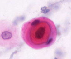
pyknosis
nuclear degenration - karyorrhexis
nuclear degenration - karyolysis
nuclear degenration - karyolysis
nuclear and cytoplasmic degenration
regenerative activity
macrophages
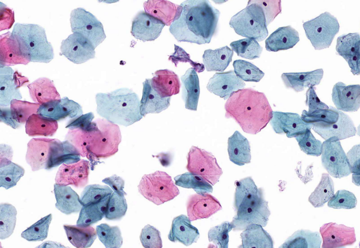
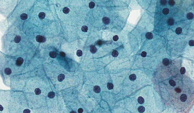
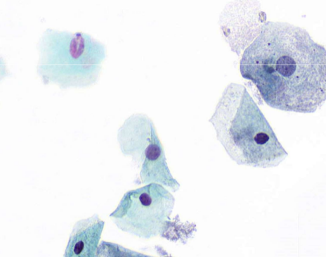
squamous cells
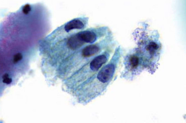
bronchial cells
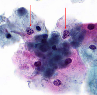
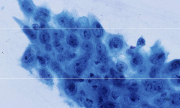
metaplastic cells
parabasal cells
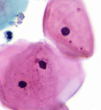
mucus producing cells - goblet cells
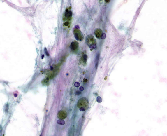
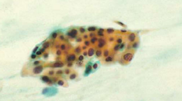
alveolar macrophages
squamous metaplasia
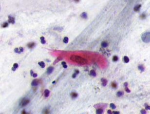
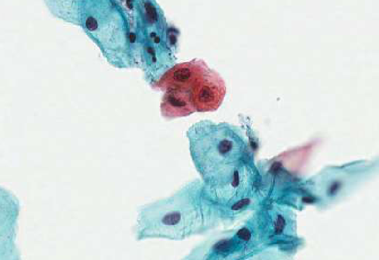
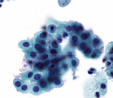
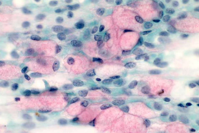
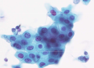
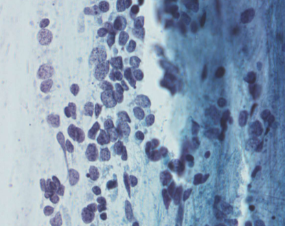
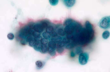
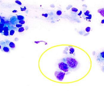
squamous cell carcinoma
squamous cell carcinoma
squamous cell carcinoma
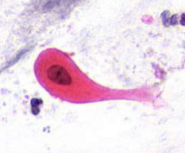
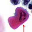
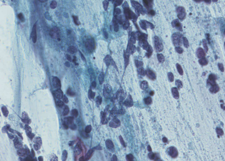
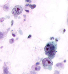
squamous cell carcinoma - tadpole cell
squamous cell carcinoma - pearl formation
SCC differential - atypical squamous metaplasia
SCC differential - reactive changes
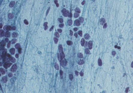
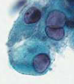
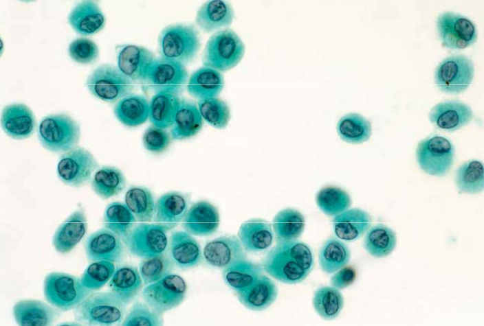
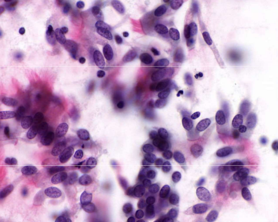
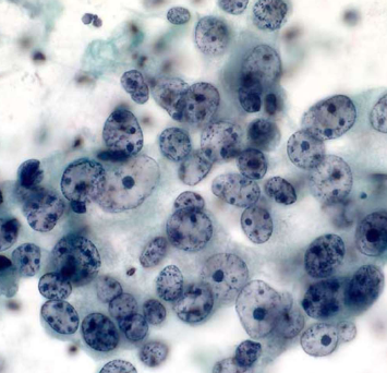
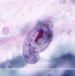
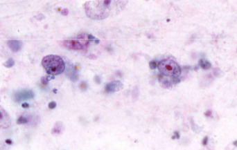
small cell carcinoma (SCLC)
small cell carcinoma (SCLC)
small cell carcinoma (SCLC)
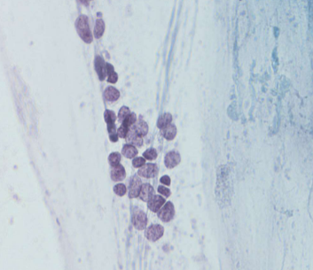
small cell carcinoma (SCLC)
small cell carcinoma (SCLC)
SCLC differential - reserve cell hyperplasia
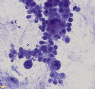
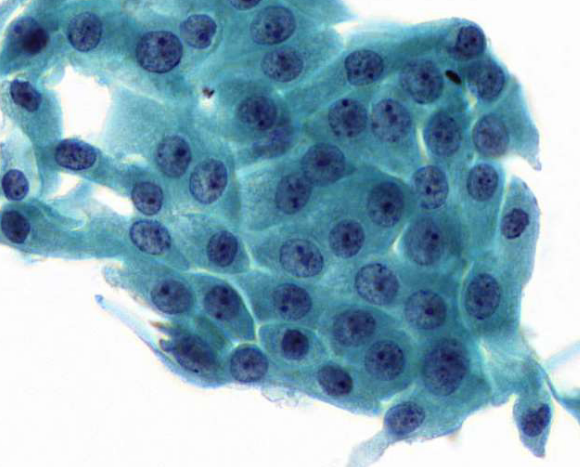
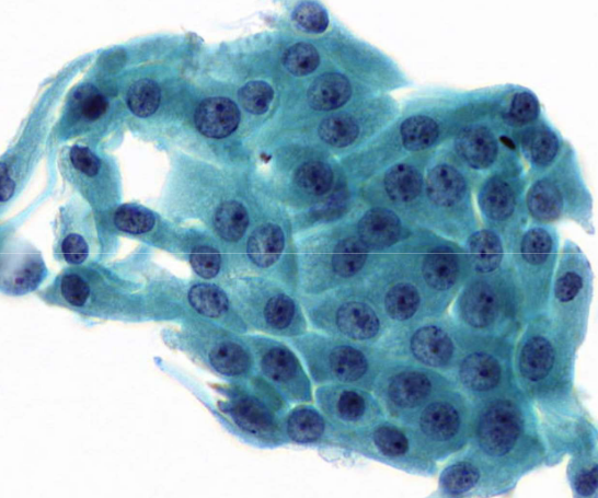
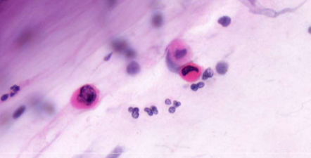
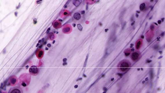
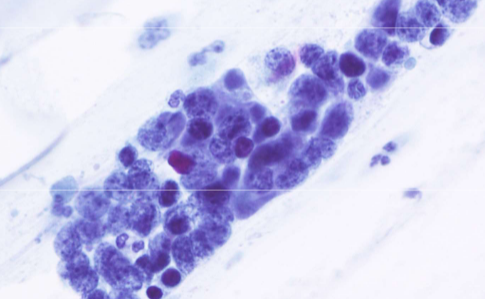
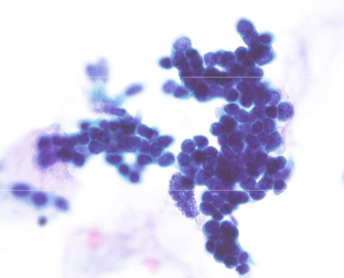
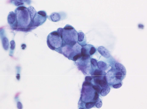
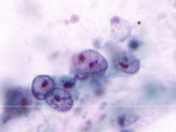
adenocarcinoma
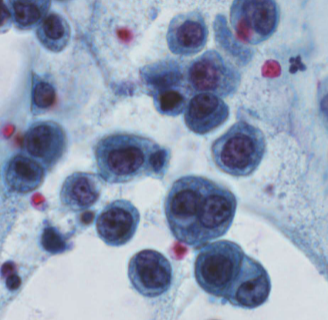
adenocarcinoma
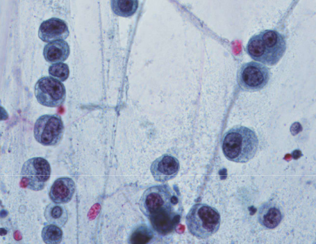
adenocarcinoma
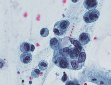
adenocarcinoma - mucin vacuole
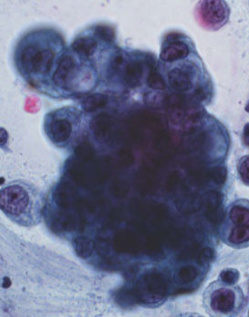
adenocarcinoma - 3D group
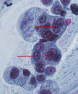
adenocarcinoma - nucleoli
adenocarcinoma differential - macrophages
adenocarcinoma differential - creola body
adenocarcinoma differential - goblet cell hyperplasia
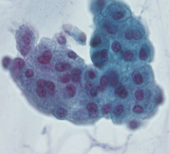
non—small cell carcinoma (NSCLC)
non—small cell carcinoma (NSCLC)
non—small cell carcinoma (NSCLC)
non—small cell carcinoma (NSCLC)
non—small cell carcinoma (NSCLC)
pap stain - airdrying artefact
giemsa stain
affinity
movement of a dye from the dye bath onto a section
Cationic dyes (+) / basic
Positive charge, binds to negatively charged tissue groups
Tissue components referred to as basophillic
Anionic dyes (-) / acid
Negative charge, bind to positively charged tissue groups
Tissue components are acidophilic
Direct dyeing
Direct attachment of dye to tissue component by ionic bonding
Opposite charge
Indirect dyeing
Attachment of dye to tissue using an intermediate substance called a mordant
Mordant: metal, has affinity for both the dye and tissue
Dye + mordant = dye lake
Metachromasia
Certain dyes which when attached to particular tissue groups, produce a colour different from the original dye
Dyeing by permeation
Difference in permeabilty of tissye structures and uses dyes of different colour and molecular size
Differential staining
Two or more of the above methods
Histochemical methods
Reagents used react with specific tissue components to produce a colour insoluble product
Metallic impregnation
Particular chemical groups in tissue have the ability to bind or reduce silver salts
Metal precipitates in or on particular tissue constituents
Argentaffin reaction
causes silver to be deposit without using a chemical reducing agent (black)