repro full review
1/665
There's no tags or description
Looks like no tags are added yet.
Name | Mastery | Learn | Test | Matching | Spaced | Call with Kai |
|---|
No analytics yet
Send a link to your students to track their progress
666 Terms
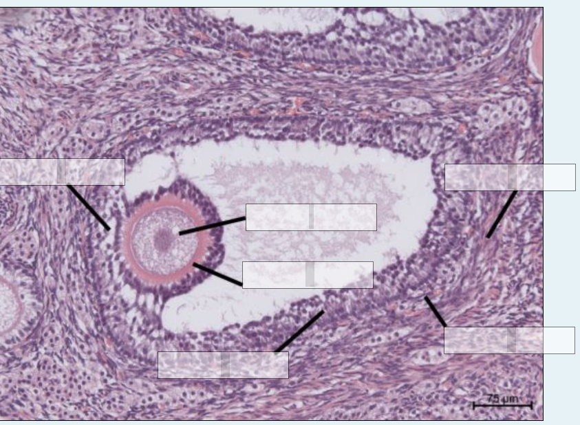
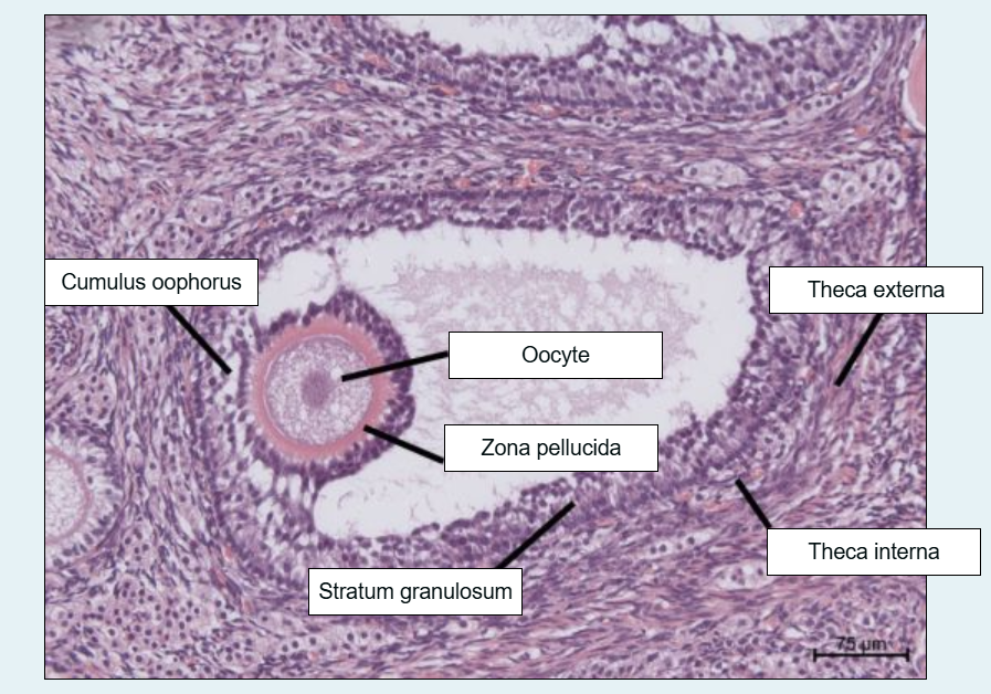
isthmus
uterine horn
fimbria
ampulla
oestrus
pregnancy
anoestrus
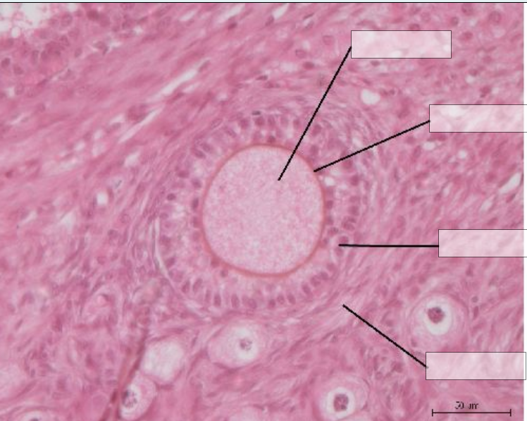
oocyte
zona pellucida
granulosa
thecal layer
this is a secondary lecture
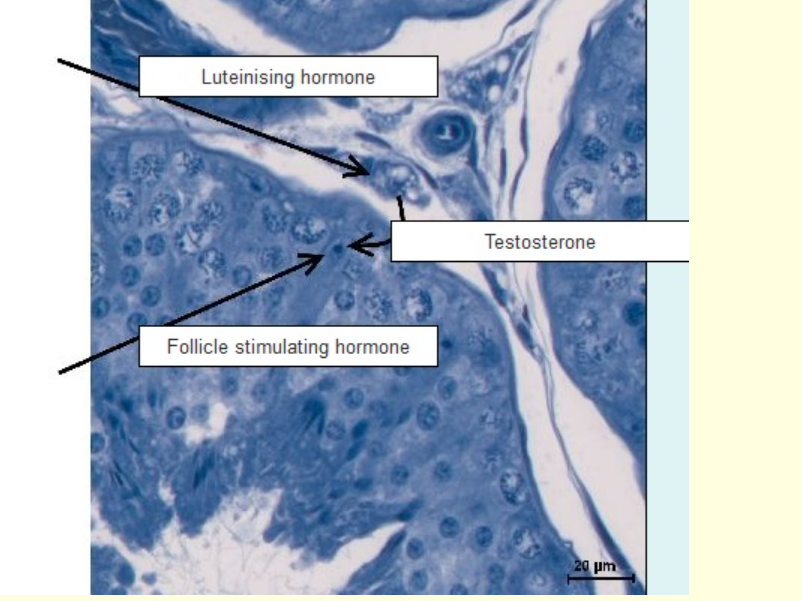
leydig cell
order at which the developing sperm cell differentiate
spermatogonia
primary spermatocytes
secondary spermatocytes
spherical spermatids
elongated spermatids
spermatozoa
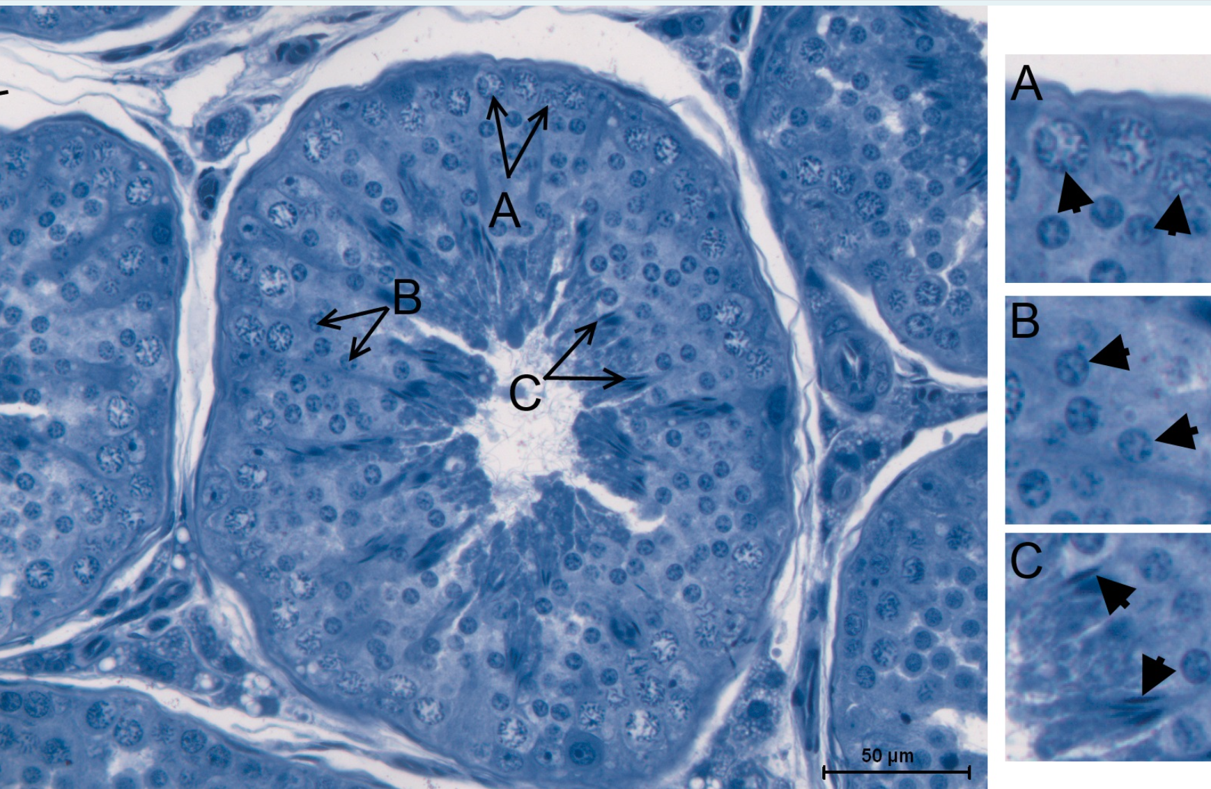
a- primary spermatocytes
b. spherical/secondary spermatocytes
c.
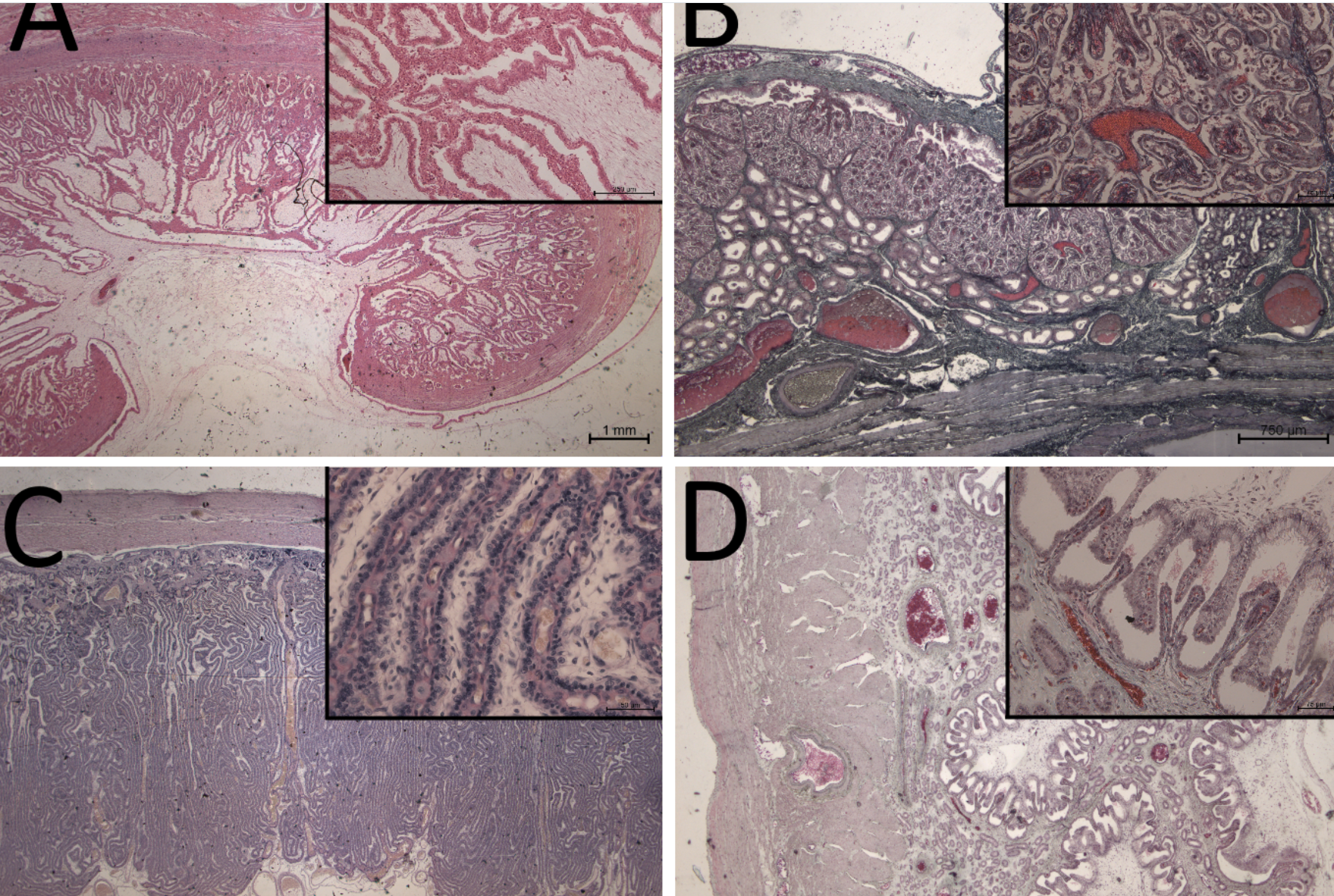
a. cotyledonary
b. diffuse, microcotyledonary
c. labyrinthine placenta
d. diffuse, folded
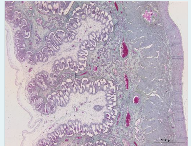
where is the foetal and maternal
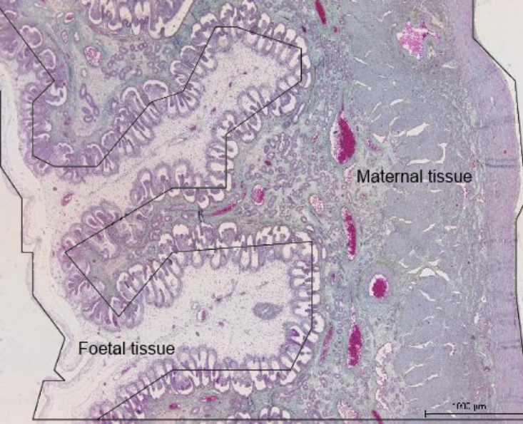
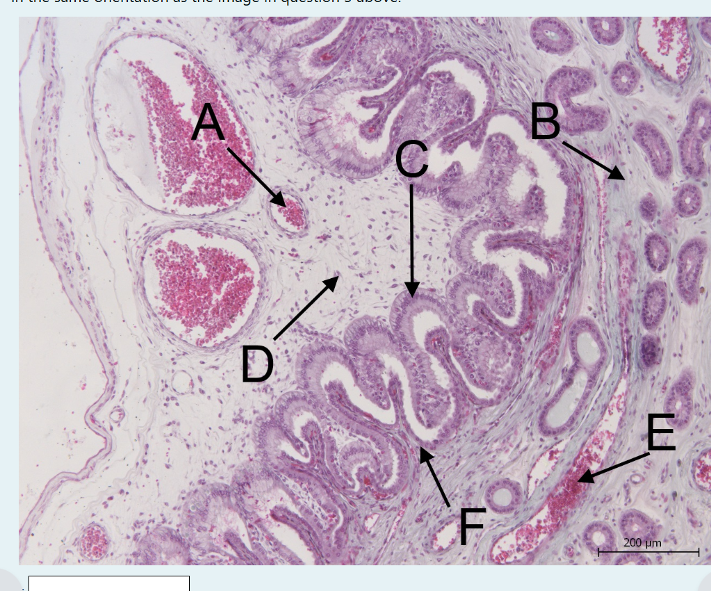
a. foetal blood vessel
b. endometrium
c. chorion
d. foetal stroma
e. maternal blood vessel
f. maternal epithelium
areolae
endometrial cup
marginal haematoma
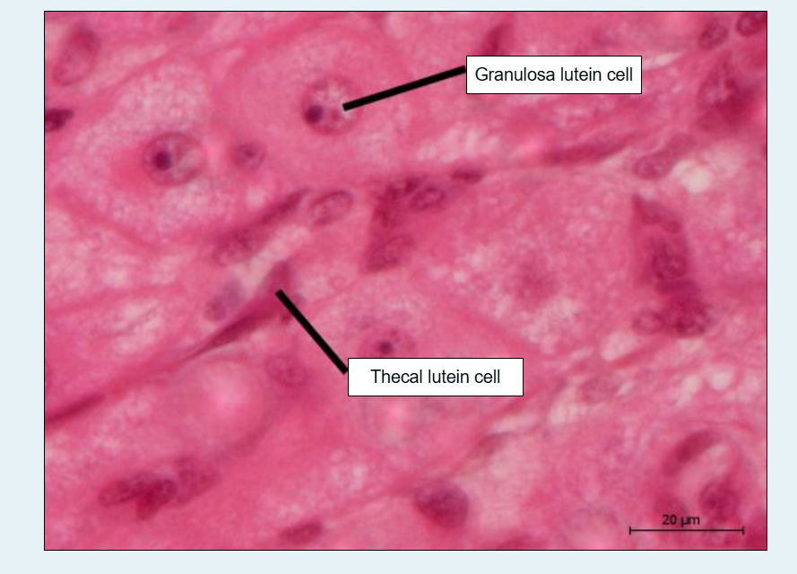
how is the cl formed
– The CL is formed from granulosa and theca cells of the ruptured follicle following ovulation. Theca and granulosa cells undergo a process termed "luteinization" to form large and small luteal cells. It is heavily vascularized.
function of the cl
what does he secrete
what does it convert
secrete progesterone under the control o
progesterone converts proliferative endometrium to secretory and inhibits smooth muscle contraction
luteolysis
the process by which the functional lifespano fht CL is terminated in the non fertile life cycle
lifespan of the cl in woman cow ewe sow mare
14
18
14
16
15
extracellular vesicles
what are they part of
where are they present
what are there important functions
what can embryonic cells able to uptake
what might embryo derived extracellular vesicles do
are part of communication between mother and embryo and carry bioactive molecules such as proteins,lipids, mrna and mirna
small ev are present in the oviducatal and uterine fluid and have important functions during fertilisation and early embryonic dev
embryonic cells are able to uptake oviductal and endometium derived small evs. conversely, embryo dervided ev may modulate oviductal and uterine function
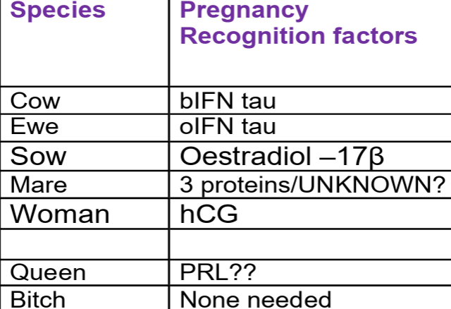
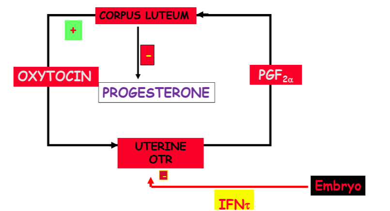
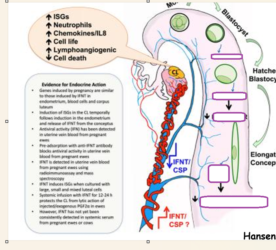
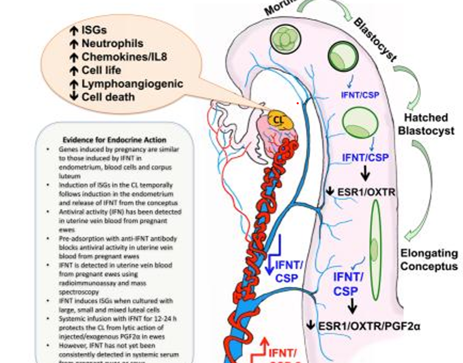
maternal recognition of pregnancy
signal from conceptus to mother
first described in sheep
takes various forms in different species
biochemical signal sent by conceptus tissues to mother
ensures maintenance of functional cl- cl continues to secrete progesterone instead of lysis and return oesrus.
pregnancy recognition in
cow
ewe
sow
mare
woman
queen
bitch
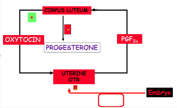
what causes luteolysis in domestic ungulates
upregulation of uterine OTR
maternal recognition of pregnancy in the sow
what hormone
why
oestrogen
blastocyst expansion means that they transform from circular to tubular
days 1/0-12 they become filamentous and reace 800-1000mm
increases surface area contact with the
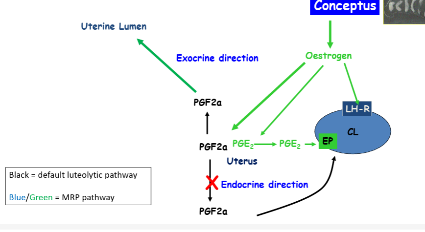
mrp signal in sows is oestradiol
secreted throughout- more secreted between days 11 and 15
impact of oestradiol in the sow
what does it induce and how
induces an overall decrease in the secretion of pgf2a by
retrograde transfer of pgf2a from the venous blood and lymph into the uterus
decrease in pgf2a from the endometrium to circulation
ability of uterine vein and artery to accumulate pgf2a
in addition, oestradiol promotes an increase in pge2 which acts as an luteotrophin to stimulate cl to secrete p4
requires 2 embryos per horn to provide sufficient signal
maternal recognition of pregnancy in the mare
signal is unknown
contunuous feto maternal dialogue
luteolysin is thought to be pg2a
endometrial otr is down regulated
ot which interacts with otr comes from uterus rather than the cl
unknown how down regulation is achieved
embryo does secrete e2 from d10 but this is not the mrp signal
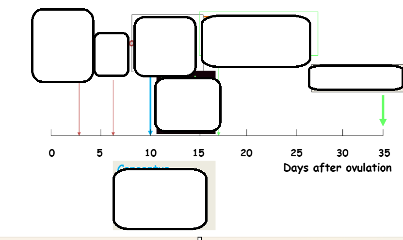
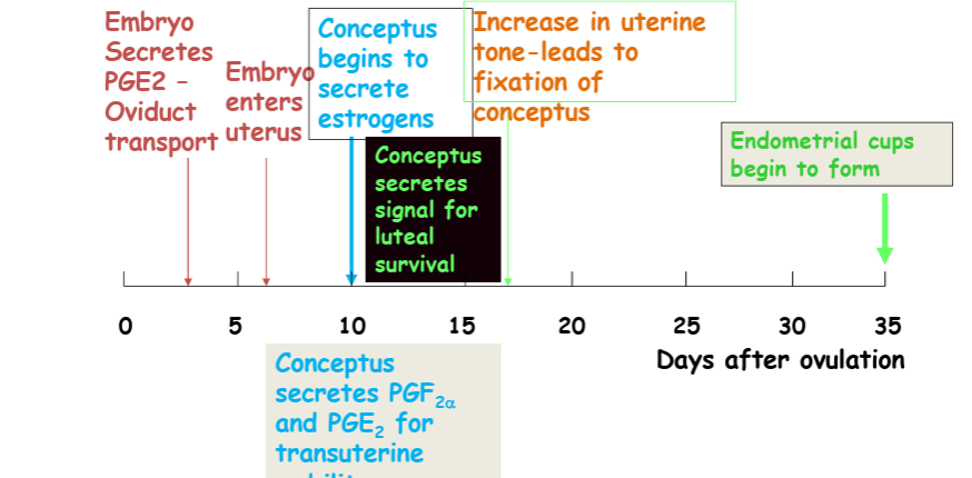
what happens when the embryo transport
how many days to transvese the oviduct
until what day does it move continuously through the lumen
what are the ciritcal days
takes 5.5-6 days to traverse the oviduct
when it finally enters uterus it remains spherical in shape and moves continuously through the uteirne lumen until day 17 afterovuation to deliver the maternal recognition of pregnancy signal to the entire endometrium
critical ays 10-14 days
placental gonadotrophins
hCG in human and some primates • eCG in the mare and other equids • Alpha chain - common to FSHA and LHA • Beta chain - unique for hCG and in common with LHB for eCG • NOT produced in other species • Bind to LH receptors - therefore main action is on the ovary to promote progesterone production • eCG: first from chorionic girdle – cells migrate into the endometrium – endometrial cups – lifespan 40 – 100/120 days – eCG related to cup presence
CG is a highly glycosylated form of LH with a very long half life (6 days) Luteotrophin is now available to ovulate or luteinise the follicles that develop in response to pituitary FSH waves
regulation of luteolysis during the oestrus cycle
proestrus: E2 increases ESR1 and OXYR and PGR
oestrus: no P4 so the PGR not active
dioestrus: P4 is up, binds to PGR
-ve feedback on ESR1, low OXYR till day 10
day 10-11 PGR downregulated by continuous P4- therefore block removed = day 11-12 ESR1 + and day 13-14 OXYR increase
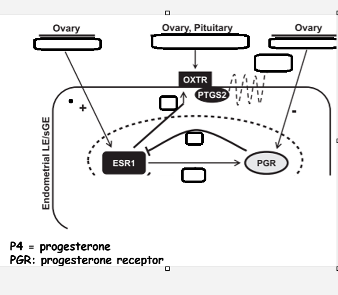
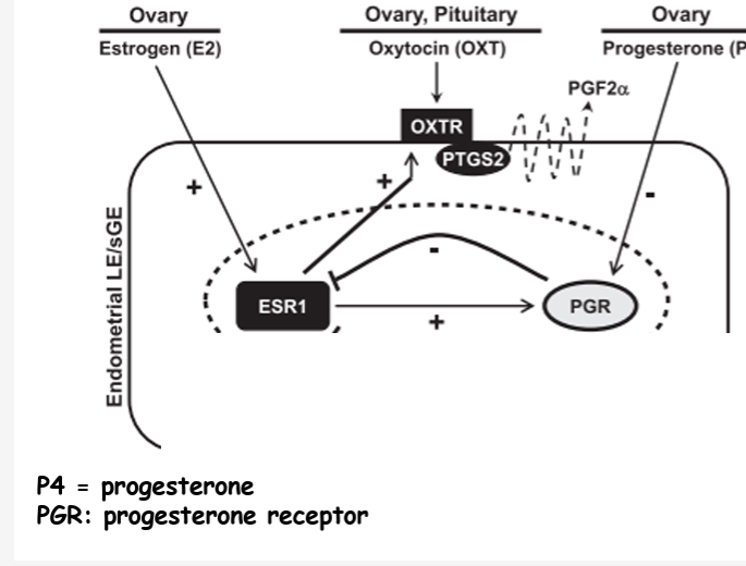
upregulation of otr in the endometrium precedes lteolysisby PGF2a mean
rising estrogen levels in the endometrium triggers the upregulation of OTRs
then oxytocin binds to them
this stimulates the endometrium to release PGF2a
this travels to the ovary, more oxytocin
patterns of PGF2a, OXYR and P4 across the cycle when there
no conceptus present
conceptus present
when there is conceptus present
pulsatile pgf2a release
p4 level drop
otr increase
luteolysis occurs
when there is conceptus present
little pgf2a secretion
little otr expression
p4 levels remain stable.
extracellular vesicle function
embryonic development
embryo migration
cell apoptosis
cell proliferation
cell fusion
immune regulation
cell adhesion
angiogenesis
MRP signal in ruminants when there is conceptus presnbent
day 10- IFN tau
binds to interferon r type i on the endometerial LE
this stimulates IRF2 signalling
blocks ESR1 (through inhibiting signalling)
blocks OXYR
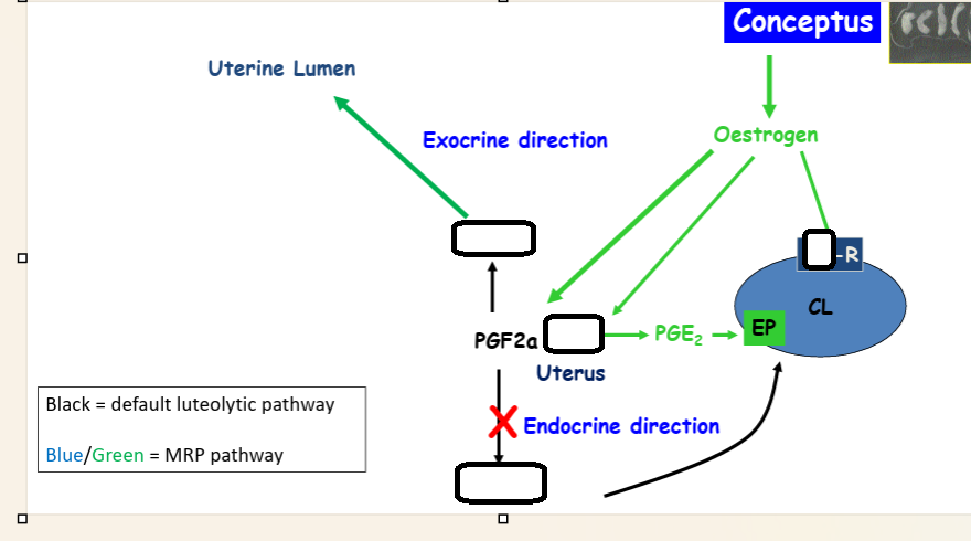
in pigs life of the corpora lutea are extended by a combination of increased secretion of estradiol and decreased secretion and redirection of PGF2a
migration of eCG
first from chorionic girdle
cells migrate into the endometrium to the endometrial cups
lifespan 40-100/120 days
eCG related to cup presence
what does luteotrophin do in response to pituitary FSH waves
ovulate or luteinise the follicles that develop
oestrus cycle in the bitch
normal lifespan of CL is at least duration of normal pregnancy
so no need for maternal recognition of rpgnancy
oestrus cycle in the queen
queen is a reflex ovulator
hence normally no luteal phase if not mated
once formed, the corpus luteum normally last for 40-45 days
however normal duration of prgnancy is 60 days
so factors must be secreted in order to ensure that the lifespan of the CL is extended from 40-45 days to 60 days
maybe PRL
also placenta provides progesterone in these latter weeks of pregnancy
transport of protein hormones from fetus to mother
what are the fetally derived proteins and what are they produced by
which type of placenta is transport easier in
transport in mares and ruminant
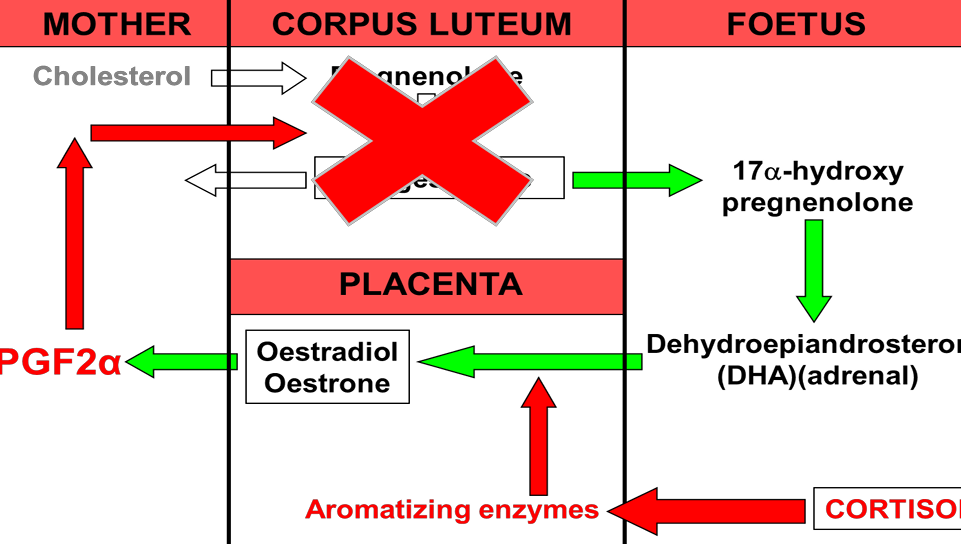
fetally derived proteins: placental gonadotrophins and placental lactogens both produced by the placental trophpblast cell in some species
in species with a haemochorial placenta it is easier as trophoblast invade and line maternal blood vessels so easy transfer
in mares the chorionic girdle cells which produce eCG detach from the epitheliochorial placenta and migrate into the endometrial stroma forming endometrial cups, and hte secretion of the cups are directly released into maternal tissue
in ruminants placental lactogen migrate into the endometrium and hten the whole cell breaks down releasing its secretory grnaules close to the basement membrane and so enar the maternal capillaries
iron transfer
some species have a haemophagous region where maternal blood cells are phagocytosed by fetal chorionic epithelium
the trophoblast cells express genes eg heem oxidase which opens up the ring structure of heme and release ferric iron creating biliverdin which is reduced to bilirubin
may form crystals of hematoidin
other species produce special proteins with a high iron content
secreted by uterine glads and taken up y opposing allantochorion
may be arranged in areolae
eg uteroferrin reaches the blood and is cleared by the fetal liver whre the iron is extracted and utilised for hemoglobin synthesis
some uteroferrin is excreted with feta urine and reaches allantoic sac
in ares ron supplied by uteroferrin
it is secreted by endometrial glands and taken up by areolar trohobalst cells trough a pinocytotic or endocytotic process
hepcidin
what is it
a hormone involved in iron homeostasis
acts of ferroportin
controls the main inflows of iron into plasma eg duodenal enterocytes and macrophages and hepatocytes
during pregnnacy it controls the placental transfer of iron from the maternal plasma to the fetal circulation
when hepcidin concs are low within fetus, iron enters blood plasma at high rate
when hepcidin concentration are high, iron is trapped in enterocytes, macrophages and hepatocytes thus not relased into plasma and not available
IgG
need for innate immunity in early life
more effcient with fewer placental layers
negligible transfer in 6 layer
may be areas of placenta specialised for this
eg in the bitch IgG taken up in the haemophagous zone is phagocytosed so does not reacht he fetal circulation intact
but there are a subpopulation of maternal blood vessels in the labyrithine zone which are specialised for this
method of synchronisation of oestrus in ewes- what to think
you need to think about the degree of syncronisation needed,
the season
and economic and market factors
the physiological method- ram
what do rams stimulate through which cues
how do you do it
when is it/not effective
what increases success
the RAM effect
rams stimulate gonadotrophin secretion and ovulation in anoestrus ewes through chemosensory cues
it involves introduction of rams to ewes that have been previously isolated from the males for3-4 weeks
only effective at certain times of the year (before the natural breeding season start, not females in deep anoestrus)
majority of ewes ovuate within 6 days of being introduced to the ram
priming with progesterone (intravaginal sponges or intra muscular injections) prior to the introduction of rams increases percentage of ewes showing oestrus behaviour
pharmacological method- progestagens
how does it work
what happens when withdrawn
what must it last
what must be given in anoestrus
progestagens
works in cyclic female by suppressing release of gonadotrophins
on withdrawal of the progestagen, negative feedback is removed
this leads to increasing amouonts of gonadotrophins and therefore oestradiol
this increased E2 leads oestrus and ovulation
must last the lenght of the luteal phase
in anoestrus females the progestagen withdrawal is compemented by follicle stimulating treatments eg PMSG
progestagens can be administered by different methods and routes
eg sponges contains synthetic progestagens, FGA or MAP
pharmacological- prostaglandin
what does it induce
what does this then cause
what does this lead to
injection timing
pgf2a can synchronise eostrus in cyclic ewes
induce luteal regression
progesterone levels fall and negative feedback from progesterone is removed
level of gonadotrophin start to rise
leads to increased follicular growth an e2 production which leads to oestrus within 2-3 days and ovulation shorty after
CL is only responsive between days 5-14 of the cycle so need 2 injections 10-14 days apart for optimum synchronisation of the flock
problems with hte prostaglandin method
variability of response
need to inject cyclic animals
induced oestrus leads to poor fertility, possibly due to limited exposure of the tract to progesterone
pharmacological- melatonin
what kind of ewes given to
to be successful when does it have to be given
how long to elevate
has been used to advance the onset of oestrus in seasonally anoestrus ewes
to be successful melatonin treatment has to be initiated after a period of long day length
need to elevate melatonin for -5 weeks for this method to be effective in bringing forward the season
how to induce early luteal regression in the cow
what is given
what oes this induce
what does this lead to the start of
when is the second injection needed
pgf 2a
from day 6-16 pgf2a will induce luteal regression
leads to start of a new follicular phase which means that the animal will come into oestrus and ovulate shortly after
for synchro of animals that are all at differnt stages, one injection is not enoguh
a second injection is needed 11-13 days after because at that time all animals will have a functional corpora lutea
cow progestagens
how long to treat
what to combine
effect in non cyclic ewes
what to inject at the removal of PMSG
for negative feedback
mimic the luteal phase of the cycle
need to treat for 10-12 dys
to make sure that the natural CL has regressed by the time of progestagen withdrawal, it is customary combine progestagen treatment with a luteolytic factor
could use oestradiol at start or prostaglandin at end
oestriadiol preferred as it alo affects follicular dynamics hwich tend ot improve ertility
in non cyclic the progestagen sensitises the HPG axis which means that it can be used in cattle with inactive ovaires
injecting ith PMSG at removal of the progestgen stimulates follicular maturation and ovulation
Oestrus and ovulation after treatment with progestagens occur earlier and with a more precise timing than following prostaglandin injection alone
sheep inducing
induce with dexamethasone
PGF2a does not work well in sheep due to high placental progesterone
PGF2a also not very successful for treating ring womb even if directly applied to the cervix
cow- induction of oestrus
induce with dexamethasone followed by PGF2a
dog
induction of parturition is rare
oxytocin used to initiate uterine contraction
horses induction and what to avoid
induce with low dose oxytocin
avoid PGF2a as causes abdominal pain
corticosteroids do not work
pigs
induce with PGF2a and oxytocin
corticosteroids do not shorten gestation in pigs
what is the gestattion length of a mare
330-340 days
what is the gestation length of a cow
270-280 days
gestation length of a ewe
147-150 dats
gestation length of a sow
115
gestation length of a bitch
63-65 days
gestation length of a guinea pig
60 days
luteolysis in gthe cow and ewe
uterine pulsatile secretion of PGF2a
diffuses from uterine vein to ovarian artery via the utero ovarian plexus in cows
in sheep to utero ovarian vein to ovarian artery
travels to CL
luteolysis in sows
pusatile PGF2a release
diffuses from uterus onto adjacent ovry luteal cells
luteolysis in the mare
pulsatile PGF2a release from the uterus
travel to ovary and destroy CL
luteolysis in the bitch
relies on withdrawal of pituitary luteotrophic support (prolactin) in late diestrus
dropping of prgesterone leads to the end of diestrus
CL regression
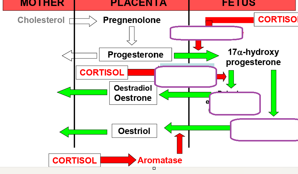
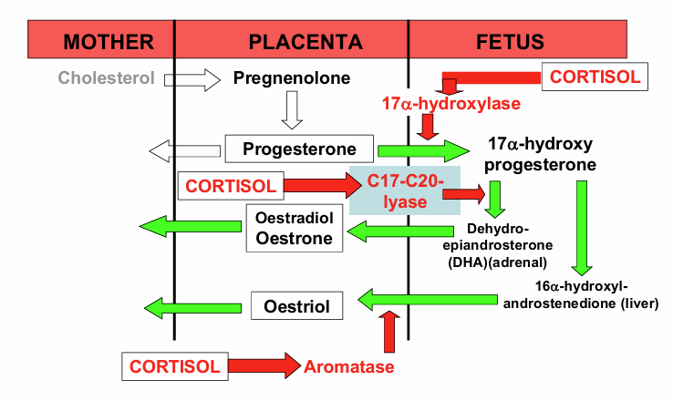
what happens to fetal glucocorticoid levels during parturition
increase
effects of glucocorticoids in the fetus
lungs
kidney
liver
gut
lungs- surfactant production, eta adrenergic receptors
kidney- glomerular filtration rate, tubular na reabsorption
liver- glycogen, gluconeogenic enzymes, igf gene expression, b adrenergic and gh receptor
gut acid secretion,digestive enzyme, mucosal growth
in goats when does the corpus luteum secrete progesterone
at the end of pregnancy
what is the luteolytic hormone in goats
prostalgandin f2 alpha
onset of parturition in goats
what must happen in order to start parturition
what is it controlled by
what is released
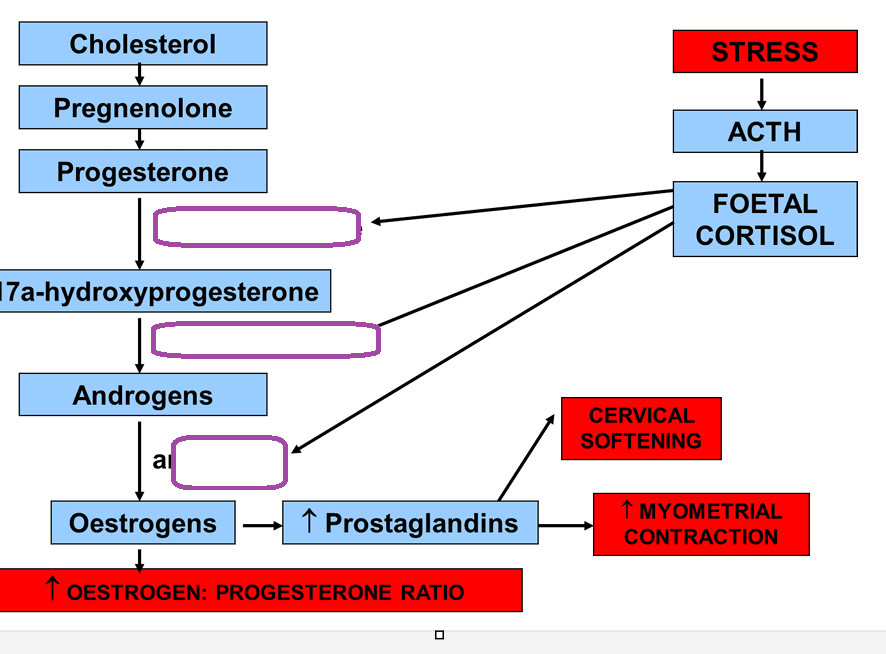
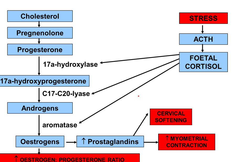
fetus controls- must stop progesterone production by the CL to start parturition
controlled by the fetal pituitary adrenal axis
fetus becomes stressed, fetal ACTH is released and cortisol increases
rise in fetal cortsol leads to an increase in oestrogen
increase in oestrogen leads to an increase in PGF 2 alpha
this increases luteoysis and decrease progesterone
increased prostaglandin causes an increase in contraction and cervical softening
how does rise in fetal cortisol incease oestradiol
leads to an increase in aromatizing enzymes
artificial induction of parturition
dexamethasone
potent synthetic corticosteroid
works across speces
takes 2-3 days to act
induction of parturition in pigs cows and sheep
pgf 2 a
induction of parturition in human and horse
oxytocin
induction of parturition in cows
when should it not be induced before
risk of using dexamethasone
what can be used to remove a mummified fetus
should not be induced before day 269 of gestation
can use dexamethasone
parturition in 48h
high incidence of retained placenta
PGF2a
work near term
used to remove mummified fetus
induction of parturiition in sows
how long does farrowing usually take
farowing takes 2-8h
PGF2a- parturition in 24-48h
oxytocin- can be given if delay in expulsionn
PGF in association with OXT 24h later
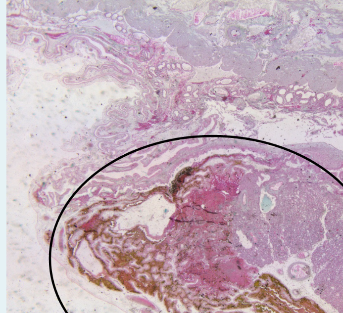
placenta
what does it interact with and what does this form
how does it connect to the embryo
any intimate apposition of fusion of the fetal tissue to the maternal tissues for physiological exchange
discarded at birth
it will possess some maternal cells although these will be in the minority
interacts with the maternal endometerium to establish the maternal fetal interface
connets to the body of the embryo by vascular supply and latter the umbilical cord
functions of placenta
exchange of nutrients and waste
changes metabolism of mother
protection from trauma and teratogens
immunological protection
hormone secretion
influence development vital organs
non invasive implantation
epithelial integrity retained ( it may be breeched locally , transiently or in very late gestation)
invasive implantation and 2 types
conceptus breaks through the surface endometrium
interstitial- very deep invasion and surface epithelium restores
eccentric- stroma only partially invaded and conceptus continues to project into lumen
ewe implantation
what does it require
begins and ends at how many days
type
what happens just before implantation
requires the endometrium to be receptive
occurs beginning at 15 days and is complete by 28 days
non invasive- epithelial integrity retained
just before implantation- blastocyst elongation
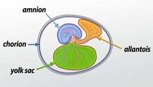
what 4 placental membranes originate from the trophoectoderm
yolk sac
amnion
chorion
allantois
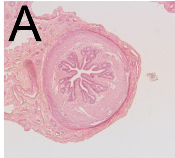
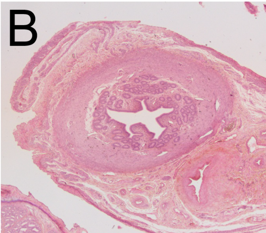
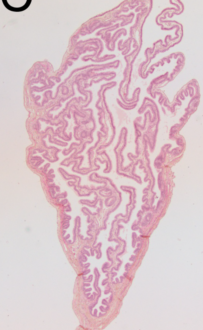
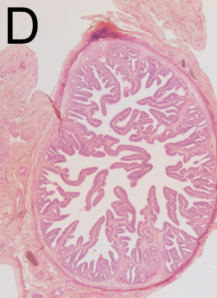
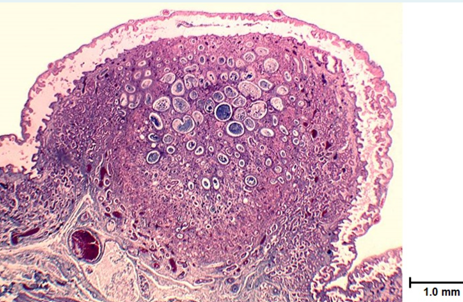
chorionic villus
function
what does it define
how many per placenta
what s it called in ewes and when
what do trhey attach to and what does this form
exchange of nutrients and gases
chorionic villous distributed in different ways and defines the type of placenta
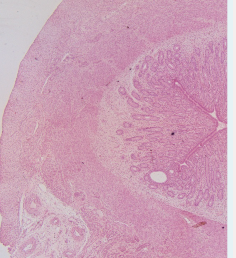
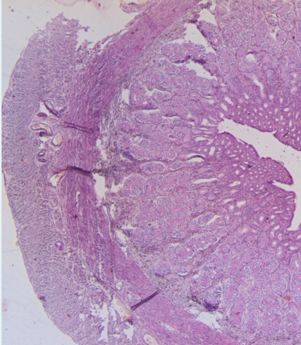
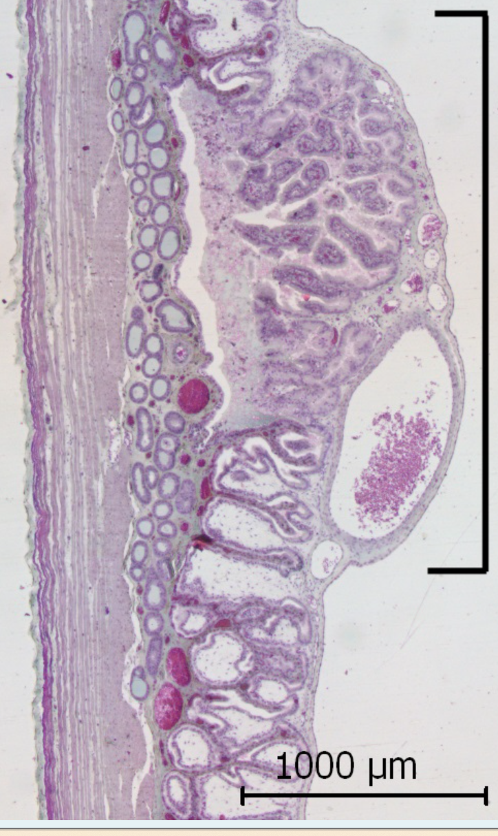
ewe at distinct contact points called cotyledons
70-100 per placenta
the cotyledons attach to uterine caruncles on the maternal side
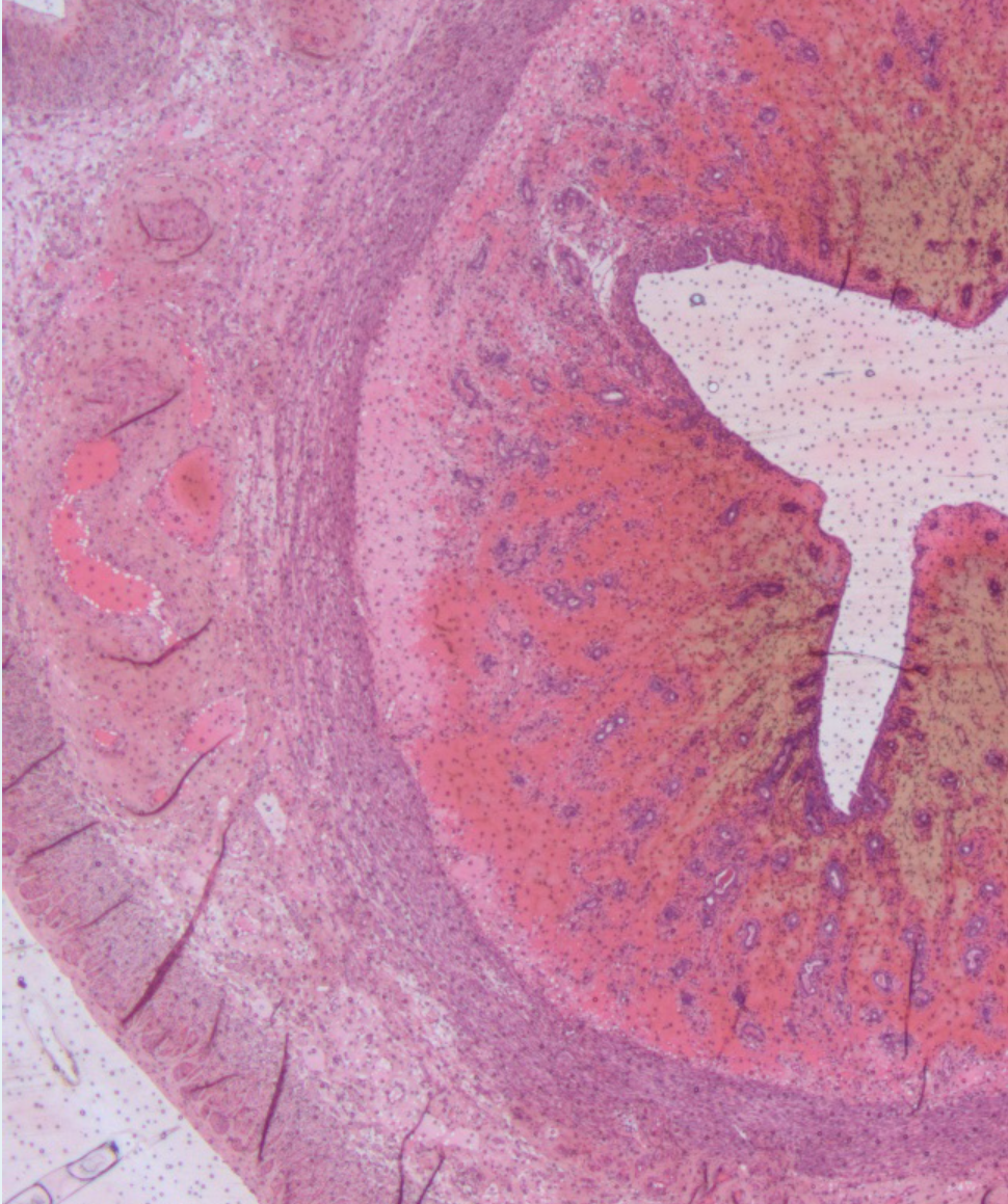
the term placentome refers to the combined caruncle and cotyledon
the tissue between the codtyledons is more simple intercotyledonary chorion- simple trophoblast
discoid / cotyledonary
the 6 layer intact- epitheliochorial
fetal endothelium
fetal connective tissue
chorionic epithelium
uterine epithelium
maternal connective tissue
maternal endothelium
umbilical cord
connects the placenta to fetal circulation - ie provies lumen to channel the nutrients and gaes
two arteries and 2 veins
normal to have numerous plawues- foci of squamous metaplasia with keratin
maternal- fecal circulation
what takes blood from the placenta to the foetus
what takes blood from the foetus to the placenta
what is thecapillary of the foetus very close to
vein takes blood from placenta to foetus
artery vice versa
exchange takes place at a placental level- lungs not needed
capillary of the foetus is very close to the cpaillary of the endometrium