PL3102 CA1 + Finals
1/133
There's no tags or description
Looks like no tags are added yet.
Name | Mastery | Learn | Test | Matching | Spaced |
|---|
No study sessions yet.
134 Terms
What are examples of glia cells?
Oligodendrocytes
Astrocytes
Microglia
Schwann cells
How do neurons create a functional circuit?
Brain neuron
Intrinsic neuron
Sensory neuron/Motor neuron
Muscle
Vice Versa
What is an afferent and efferent signal respectively? What is an interneuron?
Afferent signal: Carrying information towards the brain
Efferent signal: Carrying information away from the brain
Interneuron: Form local connections between neurons within a single structure of the brain
How do you measure the electrical activity of a neuron?
Intracellular/single cell recording
What are these electrical signals within a neuron? What are they a result of?
They are changes in the voltage of the cell membrane which travel down the axon
They are a result of the movement of ions between the inside and outsidet of the cell
What is the cell membrane of a neuron made up of? What is its function?
Phospholipid bilayer
Function: To regulate what kind of molecules can enter the cell
At rest, is the inside of the neuron more negatively or positively charged? Are there more Na+/K+ inside/out the cell?
Negatively charged
More Na+ outside the cell
More K+ inside the cell
What is the resting potential of a neuron?
It is the electrical gradient of the cell membrane at rest: the difference in charge between the inside and outside of the cell
This electrical gradient is related to the concentration of ions inside vs outside the cell
-70mV
What causes ions to move across the cell membrane?
Electrical gradient
At rest, the inside of the cell is more negatively charged
Sodium and potassium are positively charged. Opposite electrical charges attract, so the electrical gradient at rest compels these ions to move into the cell
Concentration gradients
At rest, there are more Na+ ions outside the cell and more K+ ions inside the cell
Ions will tend to diffuse from an area of higher concentration to an area of lower concentration
What maintains the resting potential? (Why doesn’t the cell membrane neutralise itself?")
Sodium channels are closed at rest
Therefore, Na+ ions are unable to follow the electrical and concentration gradient to enter the cell
K+ channels are open but there is no nett movement as K+ ions leaving the cell due to the chemical gradient are balanced by ions entering the cell due to the electrical gradient
Sodium-potassium pump
Actively transports sodium out of the cell and potassium into the cell
1 ATP = 3 Na+, 2 K+
Actively transport involves ATP because it is moving the ions against its concentration gradient
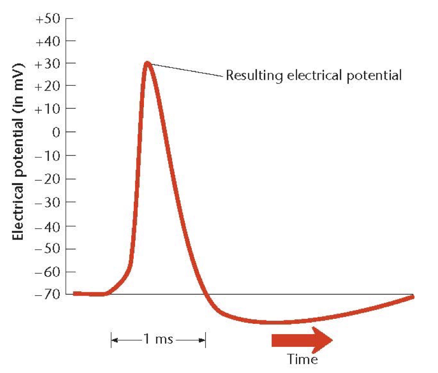
Explain the phases of the electrical potential graph of an AP
Baseline electrical state of the cell (resting membrane potential) (-70mV)
Initial, weak depolarisation of the cell membrane (caused by synaptic input from other neurons)
Triggers a rapid large depolarisation of the cell membrane
Due to the opening of voltage-gated Na+ channels
The channels will only open when the membrane potential crosses a certain threshold
Sodium rushes into the cell once channels open
This makes the inside more positively charged
Followed by rapid repolarisation of the cell membrane (overshooting the baseline state)
Due to inactivation and closure of Na+ channels and the opening of voltage-gated K+ channels
Potassium exits the cell once channels open
This makes the inside negatively-charged again
Gradual return to baseline state
Due to closure of K+ channels and sodium-potassium pump acting to restore the resting potential
How does the signal (AP) travel?
The AP propagates down the axon due to the diffusion of ions that enter during the depolarisation phase
Adjacent channels open in reaction to the increased intracellular charge
This leads to the chain reaction of the channels opening one after another
What is the absolute refractory period?
It is a period following an AP in which the neuron cannot fire another AP
This is due to the closure and inactivation of voltage-gated sodium channels
APs rely on the opening of voltage-gated Na+ channels for the rapid depolarisation of the cell
What is the relative refractory period?
It is a period following an AP in which it is less likely for the neuron to fire another AP
Because the neuron’s electrical potential overshot the baseline state of the cell following the rapid repolarisation of the cell
The cell membrane is now hyperpolarised
Therefore, stronger input is needed to reach the threshold to trigger a new AP
What is the importance of the refractory period?
It is to prevent the AP moving backwards along the axon
What is Myelin?
It is made up of the membrane of glia cells e.g. oligodendrocyte (it wraps around the axon body)
It is an insulating sheath around an axon
It increases the speed at which the AP travels down the axon
APs jump between the gaps in the myelin sheath (called Nodes of Ranvier)
Why is the synpase important?
Allows communications between neurons
Allows neurons to integrate signals from different sources and process information
Forms the basis of cognition
Mediates the plasticity of our brain
The ability of our brain to change over time from experience
Allows us to learn new information and form new patterns of behaviour
Mediates the effects of many drugs on the mind
Mediates the effect of genes on behaviour
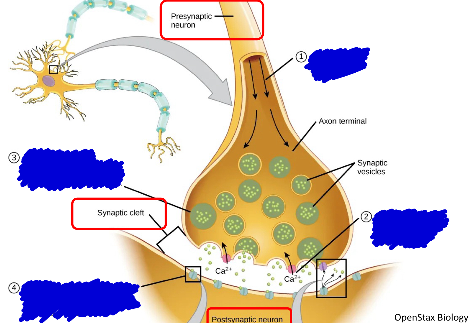
Describe the chemical events that occur at the Synapse
The AP arrives at the axon terminal
The arrival of the AP opens voltage-gated Ca²+ channels causing Ca²+ ions to enter the axon terminal
The Ca²+ ions triggers the synaptic vesicles to fuse with the cell membrane, releasing neurotransmitters into the synaptic cleft
The neurotransmitters diffuses across the synaptic cleft and bind to the ligand-gated ion channels on the postsynaptic membrane
Binding of neurotransmitter opens the ligand-gated ion channels, resulting in graded potentials → Which can ultimately lead to an AP in the postsynaptic neuron
Neurotransmitter levels reduce over time, leading to the closure of the ion channels
What are the different type of receptors that can be found on a neuron cell membrane? What are their differences?
Ligand-gated ion channels (ionotropic receptors)
Ligand: a molecule that binds to something
Triggered by binding of a neurotransmitter
Open or close in response to the binding of specific neurotransmitters to their extracellular domains
When neurotransmitters bind to these receptors, they induce conformational changes that directly alter the permeability of the channel to specific ions, allowing ions to flow across the cell membrane.
Voltage-gated ion channels
Activated by changes in the membrane potential
Open or close in response to alterations in the electric potential across the cell membrane
These channels have specific voltage-sensing domains that respond to changes in membrane potential, causing conformational changes that regulate the opening or closing of the channel pore
Metabotropic receptors
Indirectly coupled to ion channels via intracellular signaling cascades
When neurotransmitters bind to metabotropic receptors, they activate associated G proteins, which then initiate intracellular signaling pathways.
The activated G protein will bind to the effector protein
This leads to second messenger molecules being produced, activating enzymes that open ion channels
These pathways modulate ion channels or other cellular processes through the action of second messenger molecules, leading to slower and longer-lasting effects compared to ionotropic receptors
What is Serotonin and how does an SSRI work?
Serotonin is a neurotransmitter involved in regulating mood amongst other things
SSRIs block the reuptake of serotonin into the presynaptic neuron, so that it remains in the synaptic cleft for longer, enacting its effects
What is Dopamine and what drugs can affect its effects?
Dopamine is a neurotransmitter involved in reward, motor function, and other things
Dopa is a precursor for Dopamine
Levadopa is a precursor for dopa
Antipsychotic medications block the D2 receptor of the postsynaptic neuron preventing dopamine from entering the postsynaptic neuron
MAOIs inhibit degradation of dopamine to DOPAC
Cocaine blocks reuptake of dopamine by the presynaptic neuron (so does methylphenidate and many antidepressants but less strongly)
Where does specificity come from?
Lock-and-Key Mechanism: The interaction between a receptor and its ligand is often described using the "lock-and-key" analogy. In this model, the receptor's binding site (the lock) has a specific shape that only fits the particular neurotransmitter or ligand molecule (the key) that matches its shape and chemical properties. This ensures that only the appropriate ligand can bind to and activate the receptor
What is a temporal summation of signals?
Several impulses from one neuron over time
What is a spatial summation of signals?
Impulses from several neurons at the same time
Where do graded potentials and APs tend to happen in the neuron?
Graded potentials: Dendrites/soma
AP: Axon hillock
What is the Excitatory Postsynaptic Potential (EPSP)?
A temporary depolarization of the postsynaptic membrane potential caused by the release of neurotransmitters from a presynaptic neuron
EPSPs occur at chemical synapses where the neurotransmitter released by the presynaptic neuron binds to receptors on the postsynaptic neuron's membrane
What is the Inhibitory Postsynaptic Potential (IPSP)?
Voltage of the cell membrane becoming hyperpolarised (more negative than the baseline state of a neuron)
Less likely for an AP to occur
What is the difference between excitatory synapse and inhibitory synapse?
Excitatory synapse:
An AP in the presynaptic cell that depolarises the membrane of the postsynaptic cell (makes it more likely to fire an AP)
The depolarisation is caused by sodium ions entering the neuron
Inhibitory synapse:
An AP in the presynaptic cell that hyperpolarises the membrane of the postsynaptic cell (makes it less likely to fire an AP)
The hyperpolarisation is caused by chloride ions entering the neuron
What determines whether a neuron fires an AP?
The dendrites of a given neuron may synapse with axons of many other neurons, some excitatory and others inhibitory
It depends on the mixture of excitatory and inhibitory signals it currently receives
Neurons are wired together into circuits/networks that can process signals in complex ways
Excitation and inhibition interact in complex ways
What does the “all-or-nothing” law mean for APs?
Synaptic inputs to a neuron can vary in magnitude
However, there is not such thing as a “weak” AP, it either happens fully or not at all
“Greater brain activity” = Increased rate of firing (not increased magnitude of AP)
However, greater excitatory input to a neuron can increase its firing rate (frequency of APs)
What are neurotransmitters?
They are chemicals released by neurons that affect other neurons
Different neurons use different neurotransmitters
What are examples of neurotransmitters? What are their functions?
Glutamate: commonly works as an excitatory neurotransmitter, opening ligand-gated sodium ion channels to depolarise the postsynaptic cell
GABA: commonly works as an inhibitory neurotransmitter, opening ligand-gated chloride ion channels to hyperpolarise the cell
Made up of amino acids
What are neuromodulators?
Some neurotransmitters are often referred to as neuromodulators
Unlike the 1-to-1 type of synaptic transmission that neurotransmitters like Glutamate and GABA exhibit, neuromodulators can influence brain function in a more diffuse manner
They often act via metabotropic receptors rather than ligand-gated ion channels, with slower but more long-lasting effects
Made up of monoamines (also modified from amino acids)
What are examples of neuromodulators? Where are they produced and what are their functions?
Noradrenaline
Produced by neurons in the Locus Coeruleus
Function: Increase arousal and alertness
Serotonin
Produced by neurons in the Raphe Nuclei
Function: Regulate mood, sleep, etc.
Dopamine
Produced by neurons in the Substantia Niagra and Ventral Tegmental Area
Function: Reward system, motor function, cognition
What neurotransmitters are made up of Neuropeptides?
Endorphins
Substacne P
Neuropeptide Y
How are neurotransmitters synthesised?
Neurotransmitters are synthesised within the neurons that release them
Precursors are derived from our diet
E.g. Phenylaline → Tyrosine → Dopa → Dopamine → Norepinephrine → Epinephrine
E.g. Tryptophan → Serotonin
How is the anatomy of the brain studied?
Cellular level
Sub-cellular level
Whole-brain anatomy
What are methods to the study brain anatomy on a cellular level?
The neuron through a microscope
Golgi stain
A set of chemicals applied to a piece of brain tissue
What are methods to the study brain anatomy on a sub-cellular level?
Electron microscopy
Passing an electron beam through a tissue sample
What are methods to the study brain anatomy on a whole brain level?
Post-morterm samples
CT (computed tomography)
“X-ray” for the brain
Can visualise denser structures in the body (e.g. bone) but not soft tissue (e.g brain)
Therefore, the imaging is a bit low quality and grainy
CT with contrast dye in bloodstream
Able to visualise vascular network in the brain
MRI (magnetic resonance imaging)
Produces images of the brain with high spatial resolution
Able to see growth structures of the brain
MRI tractography
Diffusion-weighted imaging
Observing the movement of water molecules in the brain
Water travels down the length of the axon
This technique allows us to visualise how axons connect to different parts of the brain
How is the function of a brain determined?
Effects of damage to the brain
Effects of brain stimulation
Recording brain activity during behaviour
What are examples of how studying the effects of brain damage has allowed us to understand the function of a brain region?
GSW during WW1
Loss of vision due to GWS to the occipital cortex
Post-morterm examination of people who were unable to speak
Damage to the left prefrontal cortex discovered post-mortem in people with impaired speech
Broca’s Area: language production
Wernicke’s area: language comprehension
Stroke patients scanned with MRI/CT
Location of damage correlates with difficulty performing a social cognition task (recognising the emotions of people when presented with a part of their facial expression)
What are the limitations of studying clinical cases pertaining to the effects of damage to the brain?
Limited to what occurs naturally
Location of damage varies across cases and is unlikely to be confined to a single functional area
Researchers sometimes cause damage to non-human animals to study effects on behaviour, what are examples of techniques they used?
Ablation: Surgical removal of a brain area
Lesion: Localised damage (e.g. chemical injection)
What are examples of how stimulating the brain has allowed us to understand the function of a brain region?
Invasive methods:
Deep Brain Stimulation
Inserting microelectrodes into the brain of Ps
Stimulating specific brain regions and observing their response
Non-invasive methods:
Transcranial Magnetic Stimulation (TMS)
Limitations: Limited to superficial areas of the brain
Doesn’t have good target specificity (Instead of stimulating a single neuron, you end up stimulating the whole area)
More diffuse
Transcranial Direct Current Stimulation (tDCS)
What are examples of how recording brain activity during behaviour has allowed us to understand the function of a brain region?
Intracellular/single cell recording
Invasive
Used with lab animals, rarely with humans
EEG
Records from scalp
Measures changes by ms
Low resolution of location of the signal (Poor spatial resolution)
Good temporal resolution (time)
PET
Measures changes over both time and location
But requires exposing brain to radiation
Subject is injected with glucose labelled with radioactive atoms
PET machine detects gamma rays emitted by the radioactive glucose to track metabolism in the brain
fMRI
Measures changes over about 1 second
Identifies location within 1 to 2 mm
Good spatial resolution (location)
Poor temporal resolution (electrical recordings not as good)
BOLD signal (Blood-Oxygenation Level Dependent signal) measures the level of oxygenated haemoglobin as a proxy of neural activity
Based on the principle that when neurons in a region of the brain are active, blood supply increases
What is the CNS made up of?
Brain
Grey matter
White matter
Nuclei: clusters of cell bodies in the CNS (e.g. Raphe Nuclei)
Tracts: bundles of axons in the CNS
Caudate Nucleus
Corpus Callosum
Spinal Cord
Grey matter: made up of cell bodies & dendrites
White matter: Majority of axons
What is the PNS made up of?
Nerves (bundles of axons in the PNS)
Motor nerves
Sensory nerves
Ganglia (bundles of cell bodies in the PNS)
Autonomic Nervous System
Sympathetic Nervous System (fight or flight)
Parasympathetic Nervous System (rest and digest)
What are the different anatomical planes?
Horizontal
Sagittal
Coronal
What are the different anatomical landmarks?
Left/Right
Dorsal/Ventral
Anterior/Posterior
Lateral/Medial
What is the Cerebral Cortex?
It is the largest part of the brain in mammals
Folded sheets of grey matter with axons extending inwards
At the microscopic level, cells are organised into layers and columns
Consists of:
Gyrus: Peak/bump of the cortex surface
Sulcus: Depression/groove in the cortex surface
Fissue: a long/deep sulcus
It is involved in sensory, motor and cognitive processing
How many lobes is the cerebral cortex divided into? What are their names and respective functions? What are other notable structures in the cerebral cortex?
Frontal Lobe
Executive functions
Planning of movements
Recent memory
Some aspects of emotion
Parietal Lobe
Body sensations
Visuospatial processing
Temporal Lobe
Hearing
Advanced visual processing
Occipital Lobe
Vision
Precentral Gyrus
Primary Motor Cortex
Prefrontal Cortex
Executive functions
Working memory
Thoughts, actions and emotions
What are the broad divisions of the brain?
Forebrain
Midbrain
Hindbrain
What are the notable structures of the Hindbrain?
Brain Stem
Medulla and Pons extend from the spinal cord
Involved in autonomic functions that are critical for survival
E.g. control of breathing, HR, salivation, swallowing, sleep, etc.
Cerebellum
Control of movement (e.g. posture, coordination)
Perhaps cognitive function
What are the notable structures of the Midbrain?
Superior and Inferior colliculi
Nuclei that process sensory signals from the ears and the eyes
Substantia Nigra
Contains dopaminergic neurons important to motor control that degenerate in Parkinson’s disease
What are other notable subcortical structures?
Thalamus
Main source of sensory input to the cortex
“Sensory relay point”
Hippocampus
Memory consolidation
Spatial navigation
Amygdala
Evaluating emotional information (e.g. fear)
Ventricles
Lateral ventricles (anterior and posterior) contain cerebrospinal fluid
What is perception?
Your experience of the environment around you provided by your senses
What you perceive is your own model of the world constructed by your brain
How does the perceptual process work?
Environmental stimulus
Light is reflected and transformed
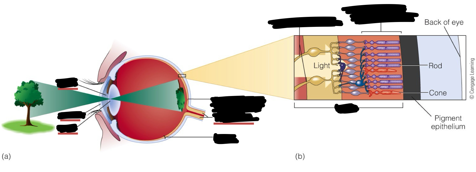
Receptor processes
Rod and cone cells line the back of the eye
They convert the light energy into electrical energy and influence what we perceive
Neural processing
Takes place in the interconnected circuits of neurons like the retina and in much more complex circuits within the brain
Each sense sends signals to different areas of the brain
Perception
“I see something”
Recognition
“It’s an oak tree”
Action
“Let’s have a closer look” walks towards tree
Perception doesn’t only rely on sensory input (bottom-up processing) but top-down processes as well (existing knowledge, expectations of the world around us, etc.)
Visual perception occurs in the brain, not the eyes
Identify these structures
Pupil
Cornea
Lens
Fovea
Optic Nerve
Retina
Optic Nerve Fibers
Retina
Photoreceptors (Rod and cones)
Note: The cornea and lens focus light onto the retina
Identify these structures
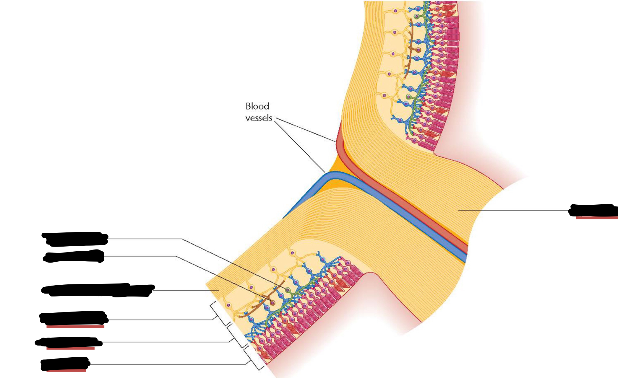
Horizontal cells
Amacrine cells
Axons of the Ganglion cells
Ganglion cells
Bipolar cells
Photoreceptors
Optic Nerve
How does the retina detect light?
Light strikes the photoreceptors
Message is transmitted to the bipolar cells
Message is transmitted to the ganglion cells
Message is transmitted to the brain via the optic nerve
The Fovea is the part of our retina that underlies the center of our visual field
Predominantly via cone cells
The rest of the retina is the periphery
Predominantly via rod cells
What are the medical conditions that result in deficits in the visual field?
Macular degeneration
Retina degenerates typically int eh Fovea
As a result, patient experiences blindspot at the centre of their visual field
Retinitis pigmentosa
Retina degenerates typically in the peripheral parts of the retina (initially)
As a result, patient experiences blindspot in the periphery of their visual field
Akinetopsia
Inability to see motion following damage to the cortex
Explain the process of transduction and adaptation to a dark environment
When light strikes the photoreceptor cells, retinal absorbs the light and it changes shape
This inactivates retinal, becoming unresponsive (‘bleached’)
This also triggers chemical events in the cell that change the electrical state of the photoreceptor thereby generating electrical signals in the cell
The photopigment needs to regenerate before it can detect light again
When we step into a darker environment, the concentration of regenerated photopigment will increase over time
The concentration of responsive photopigment determines how sensitive we are to light
Both cones and rods adapt to the dark (increasing sensitivity to light)
Once adapted, rods are much more sensitive than cones
As such, most of our visual perception in the dark arises from rod cells
How do cone cells vary?
They vary in their sensitivity to wavelength:
Short-wavelength cones (S) - Blue
Medium-wavelength cones (M) - Green
Long-wavelength cones (L) - Red
They all detect different colours (wavelengths) and differ from rods
How are the cells in the Fovea wired? What is the vision they provide like?
Packed with cone cells
Low convergence of cells (each cone excited a single ganglion cell)
Highly detailed vision (high spatial resolution)
Distinguishes among bright lights; responds poorly to dim light
Good colour vision
How are the cells in the periphery of the retina wired? What is the vision they provide like?
Packed with rod cells
High convergence of cells (multiple tods converge on a single ganglion cell)
Less detailed vision (less spatial resolution)
Responds to dim lights; poor for distinguishing among bright lights
Greater sensitivity to faint light
Poor resolution in the periphery
Poor colour vision
What is additive colour mixing?
Mixing the three primary colours (red, green, blue) to produce other colours
What is the trichromatic theory of colour vision?
Colour vision is based on the presence of three types of cone cells in the retina, each sensitive to a different range of wavelengths of light
These cone cells are most sensitive to short (blue), medium (green), and long (red) wavelengths of light, respectively
The trichromatic theory explains how our visual system perceives a wide range of colours by combining the signals from these three types of cones in various proportions
Colour perception depends on the relative response of the three cone types
Why is the relative response of all three cone types important for colour perception?
The response of a single cone is uninformative about the wavelength, partly because it confounds wavelength with intensity
The ratio of responses across the three cones to a given wavelength of light remains similar across different intensities
How does context affect our colour perception?
There is actually no 1-to-1 relationship between wavelength and colour. It depends on the context
Prior expectations about object colour affect what we perceive (e.g. colour of the strawberry in blue light, red colour is still identifiable)
Adaptation (seeing faint residues of colours we’ve seen before, “artefacts of our vision”)
Function: to normalise our environment as the scenery around us changes
Makes our perception more consistent across different environments
It depends on the current state of our visual system (e.g. adaptation), the visual context (e.g. illumination), and prior expectations
What is colour constancy in our colour perception?
The perceived colour of an object can be relatively stable across different illumination conditions, despite differences in the wavelengths of light entering our eye
This requires our visual system to somehow separate the colour of the light source from the colour of objects
How can our visual system separate the colour of the object from the colour of the light source?
Contextual cues to the lighting conditions
Adapting to the dominant colour in the environment
Past experience with familiar obejcts
What are the important features of perception?
Location
Depth
Motion
Colour
Form
What is the path of the visual pathway?
Eyes
Optic Nerve
Lateral geniculate nucleus in the thalamus
Primary visual cortex
Other cortical areas
Signals from retinal ganglion cells are relayed to the primary visual cortex via the thalamus
Pathways extending further into the cortex enable more complex processing of visual patterns
How does feature selectivity work in the retina and lateral geniculate nucleus (in the thalamus)?
Ganglion and LGN cells respond to dots of light
“center-surround” receptive field
When light falls within receptive field, it increases excitatory APs in the neuron
If the light falls outside of the receptive field, it sends inhibitory signals, thereby decreasing the signals received by the ganglion cells
How does feature selectivity work in V1 (primary visual cortex)?
Cortical cells respond to bars
Simple cells:
“Simple cells” in V1 respond to edges of a specific orientation
Some simple cells respond best to vertical edges while other simple cells can prefer horizontal or slanted orientations
Multiple ganglions’ “receptive field” (circles) make up the “bars/lines” of the simple cell
Building a “line detector” with nerve cells
The response of simple cells can then be put together to form more complex patterns (e.g. objects, faces, etc.)
Complex cells:
“Complex cells” in V1 respond to both orientation and movement direction
E.g. when the light detected is moving from R to L (not L to R)
What are location columns and orientation columns?
They are specialised arrangements of neurons that are perpendicular to the surface of the cortex
Location columns
Characterised by their preference for input from one eye over the other
Each location column receives input predominantly from one eye, with adjacent columns processing input from the other eye
Orientation columns
They are organized within the location column and based on the orientation preference of the neurons within each column
Neurons within an orientation column have similar orientation tuning properties, meaning they respond most strongly to visual stimuli oriented in a specific direction (e.g., vertical, horizontal, or diagonal)
What is tiling in regards to vision?
Wiring between neurons makes them respond to particular visual features
It is the arrangement of receptive fields of retinal ganglion cells (RGCs) in the retina, which ensures comprehensive coverage of the visual field
The retina contains different types of RGCs, each with specific receptive field properties, such as size, shape, and sensitivity to light.
Aside from V1, what are the other important parts of our cortex for visual processing?
V2: detects more complex patterns and receives signals from V1
V3 (largely interior)
V4
V5: key role in our perception of motion (in the temporal lobe)
Inferotemporal cortex
What are the dorsal and ventral pathways for vision?
Dorsal:
Towards the parietal lobe
WHERE pathway
HOW/ACTION pathway
Damage to the parietal visual areas can produce spatial neglect
Processing of visual information related to the location of objects
Ventral:
Towards the temporal lobe
WHAT pathway
Advanced visual processing
Processing of visual information related to object recognition and identification
Damage to the temporal visual areas can produce agnosia (inability to recognise objects) and prosopagnosia (inability to recognise faces)
There are neurons selective for face identity
What are the important modules in the ventral pathway?
Fusiform face area
Inferotemporal cortex
It is a region located in the ventral stream of the visual cortex, specifically in the temporal lobe of the brain
It is considered to be a critical area for higher-level visual processing, particularly for the recognition of complex visual stimuli such as objects, faces, and scenes
Extrastriate Body Area (EBA)
Active when we look at parts of the body/entire body
Parahippocampal Place Area (HPA)
Responds to landscapes, buildings, etc.
What is Face Pareidolia? Why do we experience this?
Face pareidolia is a phenomenon in which individuals perceive faces or facial features in unrelated stimuli, such as inanimate objects, patterns, or random arrangements of shapes
Faces vary but have a common visual structure
The same person can look different in multiple angles & lighting
Why?
Humans possess specialized neural circuitry dedicated to processing and recognizing faces e.g. FFA
Importance of faces for social interaction, communication, and survival (biological predisposition)
Pattern Recognition Mechanisms
The human brain is highly adept at detecting and interpreting patterns in sensory input. This includes not only recognizing faces but also detecting shapes, objects, and meaningful configurations in the environment
This propensity for pattern recognition can lead to the perception of faces in random or abstract stimuli, even when no actual faces are present.
What is the FFA activated by?
Real faces
Face pareidolia
Mooney faces
Imagining a face
How do sound waves vary? Describe them
Frequency
Higher frequency = higher pitch
Rapid fluctuations in the density of air molecules in our ear
Amplitude
Higher amplitude = Higher volume (louder)
Loudness is the psychological quality of sound
Amplitude is the physical quality of the sound wave
Greater change in the air pressure over time, with more intense compression of the air molecules occurring at the peak of the sound wave
Timbre
The quality that distinguishes between 2 sounds that have the same loudness, pitch, and duration, but which still sound different
Factors affecting timbre:
The relative intensities of the harmonics that are present
The time course of the sound wave (attack & decay)
Attack refers to how the sound builds up over time
Decay refers to how the sound dissipates over time
What is fundamental frequency and harmonics?
Fundamental frequency:
Most sounds that we hear are more complex sound waves and can be described as a mixture of frequencies
The waveform has a periodic structure to it
The fundamental frequency is the overall repetition rate of waveform in 1s
The fundamental frequency determines the pitch that we hear
Harmonics:
Musical instruments also tend to produce harmonics
They are components of the overall waveform that have a frequency that is a whole number multiple of the fundamental frequency
How does our ear detect sound waves?
Sound waves are changes in air pressure that propagate through the air around us
They arrive at the outer ear and travel down the auditory canal
The sound waves collide with the ear drum (tympanic membrane) which is located at the end of the auditory canal
The sound waves vibrate the tympanic membrane, which causes three tiny bones, called an anvil, hammer and stirrup, to move and pass on the vibration into the cochlea
The cochlea is a snail-shaped structure that consists of chambers filled with fluid
When the bones of the middle ear hammer on the oval window, it creates a vibration in that fluid that travels down the length of the cochlea and vibrates a structure in the centre of the cochlea called the basilar membrane
The basilar membrane is moved up and down in response to sound waves hitting our ear, this pushes the cilia against the tectorial membrane which causes the cilia to bend.
That bending of the cilia causes ion channels in the hair cell to open, depolarising the cell.
The depolarization triggers the release of neurotransmitters across the synapse that the hair cell shares with a neuron in the auditory nerve, stimulating action potentials
This mechanical bending of the cilia to produce electrical signals is the transaction
How do we detect the frequency of a soundwave?
The “place method”
One end of the basilar membrane is called the base and the other end is the apex
The base of the basilar membrane tends to be narrow and stiff while the apex tends to be wide and floppy
The frequency of the sound wave is coded in terms of which hair cells are most activated in response to it
It has been shown that higher-frequency sounds are better able to vibrate at the base of the basilar membrane while lower frequency sounds vibrate at the apex of the basilar membrane
How do you recover hearing for a person whose hair cells get damaged (where their ability to hear gets lost)?
Cochlear implants
An array of electrodes is inserted into the chambers of the cochlea to electrically stimulate auditory nerves that originate in different sections of the cochlea
A microphone is placed in the outer ear and it converts sound waves into a pattern of electrical stimulation for the electrodes in the implant
This bypasses the functions performed by the middle ear and hair cells, instead electrically stimulating the auditory nerve fibers directly
Describe the auditory pathway in the brain
Signals from the cochlea are sent to the cochlea nuclei and superior olive in the brainstem
This signal is then passed through the inferior colliculus in the midbrain to the medial geniculate nucleus in the thalamus, and finally to the primary auditory cortex in the temporal cortex
Most activity from the left ear arrives at the right auditory cortex and vice versa
But there are also ipsilateral projections from the left ear into the left auditory cortex and from the right ear into the right auditory cortex
Thus hearing is not as lateralised as vision
What are other brain areas that are involved in auditory processing?
Broca’s Area in the left prefrontal cortex
Language production
Broca’s aphasia: can understand speech but they struggle to produce speech
Wernicke’s Area in the left posterior temporal cortex
Language comprehension
Wernicke’s Aphasia: struggle to understand speech
How does our auditory system localise the source of a sound?
Interaural intensity differences
The brain compares the intensity of the sound waves arriving at each of our ears as a way of localising where the sound is coming from
The head acts as a sound barrier, attenuating the intensity of sounds coming from a location on the other side of the head
This means that the sound has greater intensity at the ear facing more directly towards the sound source
This works best for high frequency sounds as they are attenuated by the head to a greater extent
Sound waves of low frequencies are not attenuated much by the head and so arrive at both ears with the same amplitude regardless of location
High frequency sound waves are impeded more by objects that they encounter, while low frequency sound waves are impeded less
Comparing the timing of when sound waves reach each ear
The ear furthest from the sound will detect the sound later than the ear nearer to the sound source
Our brain can compare the timing of when sound waves arrive at each ear as a way of localising where the sound is coming from
A lack of interaural timing differences implies that the sound source is located in the central plane
What other systems can affect our perception of sound? What is an example of how this affects our perception of sound?
Our perception of sound can also be affected by what we see
We have strong expectations about how particular visual events should sound, such that we can’t help but experience a kind of auditory imagery of the sound that our vision suggests we should be hearing
Example: McGurk effect
Illustrates how our perception of speech can be affected by how we see a person’s lips move
We use visual cues to interpret speech, which is generally a useful ability (e.g. helping us to follow a person’s speech in noisy environments)
Our brain is automatically using the visual information to determine what we consciously hear
In the McGurk effect, what we see misleads us about the sound that we are hearing
What we hear can also affect what we see
2 balls passing each other or bouncing off each other with or without the ‘click’ sound
What we hear can affect our visual perception of the causal interaction between objects and the trajectories that objects are moving in
The effect of sound on what we see is likely to be most pronounced when the information is ambiguous
What are the functions of touch?
It contributes to our awareness of our own body; it provides part of our sense of where our body is located
The feel of objects helps us to grip them and interact with them
A social sense; different types of touches experiences different emotions
How we express and experience attachment in early development
Describe how our skin detects touch. How do these receptors differ from one another?
The dermis contains various mechanoreceptors that are responsible for detecting pressure, stretch and vibration of the skin
The main mechanoreceptors include Meissner’s corpuscles, Merkel disks, Pacinian corpuscles and Ruffini endings
Each of these are specialised receptor endings that connect to a nerve fibre that carries signals back to the spinal cord
These mechanical/physical forces on the skin trigger ion channels to open and the production of an actional potential in the nerve attached to the receptor
Difference:
Different receptors respond to different types of touch due to factors such as the physical structure of the receptor and how deep it is located in the dermis
E.g. Pacinian Copuscle
Many receptors like this are embedded in the skin throughout the body
Helps to detect high frequency vibration against the skin
Plays a role in our perception of textures → whether a surface feels rough or smooth
How are mechanoreceptors distributed throughout the body?
The density of mechanoreceptors in our skin varies across our body
High density of receptors in our fingertips compared to the palm
Correspondingly, our ability to discriminate fine details of touch is better for our fingertips compared to our palm
Tactile acuity: the fineness of the details that can be discriminated by touch
Highest number of mechanoreceptors in hands and lips
Describe the tactile pathway in the brain
The mechanoreceptors in the skin connect to nerves that send signals to the spinal cord
From there, the signals are passed up the spinal cord to the thalamus, then to the primary somatosensory cortex (S1) at the top of the brain
How are neurons in S1 organised?
They are organised into a somatosensory map of the body, with the cortical representation of body parts following receptor density and tactile acuity
More cortical space is allocated to parts of the body with greater sensitivity to touch
Different features of the body are organised in a systematic way, where the different fingers are represented near each other, and features of the face are represented near each other
Represented by a ‘homunculus’ figure
More cortical space is allocated to parts of the body with greater sensitivity to touch → reflects that much more of S1 is dedicated to processing signals from the hands and lips region
What is an example of how the somatosensory map can change with experience?
Musicians who have played string instruments for many years
The hand used to finger the strings has greater cortical representation in the primary somatosensory cortex compared to non-musician controls
The cortical activity in this study was measured with a technique called MEG, similar to EEG, which measures neural activity non-invasively through the scalp
Describe taste and the mechanisms behind it
Taste is a chemical sense: the sensation that you get when molecules that are dissolved in our saliva are detected by taste receptors on our tongue
5 basic sensations: sweet (sugar), salty (sodium), sour (hydrogen ions (acids)), bitter (alkaloids such as caffeine), umami (msg)
The overall taste of a substance can generally be described as a mix of the basic tastes
Mechanisms behind taste:
Taste begins when the chemicals that we put in our mouth interact with the taste buds
The taste buds are mostly located in structures on the surface of the tongue called papillae
The papillae are the structures of the tongue surface that produce ridges and valleys, underlying the roughness of the tongue surface
There are different types of papillae, differing in their shape and location on the togue
Each papilla contains a number of taste buds, and each taste bud is a ‘garlic-like’ structure that contains multiple taste cells
The taste cells have tips that extend into the taste pore, to contact with the saliva on the surface of the tongue
The taste cell connects to nerve fibres, which transmits electrical signals towards the brain
Transduction occurs when a chemical in our saliva makes contact with the taste cells, causing them to generate electrical signals in the nerve fibres they connect to
There are different receptor types that respond to molecules that we experience as bitter, sweet, sour and salt
Stimulation of these receptor sites triggers a number of different chemical reactions within the cell that lead to the movement of charged molecules across the membrane, which creates an electrical signal in the receptor and an action potential in the nerve fiber that the taste cell is connected to
The taste cells send electrical signals towards the brain via different nerves that connect to different parts of the tongue and mouth
These pathways connect to the spinal cord then travel up to the thalamus, then into the insula in the primary taste cortex within the lateral sulcus
Explain the individual differences in the function of taste cells
People differ in the total number of tastebuds they have → giving rise to ‘supertasters’ and ‘nontasters’
This depends on genetics, age and other things
E.g. pregnancy increases taste sensitivity → may be an adaptive thing by helping to avoid harmful foods
Another factor is the sensitivity of the taste receptors themselves
Genes may affect the function of taste receptors, influencing how readily the individual can detect compounds in the food that elicit the experience of sweetness or bitterness