LABORATORY TECHNIQUES AND METHODS
1/119
There's no tags or description
Looks like no tags are added yet.
Name | Mastery | Learn | Test | Matching | Spaced |
|---|
No study sessions yet.
120 Terms
Microscopy
use of light and electrons to magnify objects
magnification
the apparent increase in size of an object
a. thickness of the lens
b. curvature of the lends
c. speed of light
The image enlarged depends on the following
Resolution or Resolving Power
- ability to distinguish objects as close as 0.2 micrometer (200nm max)
- human eye can see objects as small as about 0.1mm (100um)
0.2 micrometer (200nm max)
ability to distinguish objects as close as
0.1mm (100um)
human eye can see objects as small as about
a. shorter wavelength
b. greater numerical aperture
Better resolution are due to
the ability to gather light
greater numerical aperture
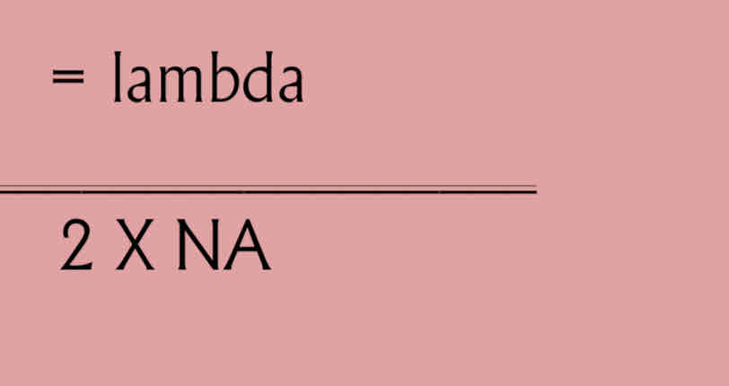
Resolution Distance
= 0.61 X wavelength
NA
Resolution or Resolving power
Contrast
different intensity between two objects or its
background
Par focal
ability of the microscope to stay in focus even you shift from different objective
Working Distance
distance between the lens and the specimen
Total magnification
the number of times the object is enlarged from its original size
objective lens x ocular lens
Total magnification formula
Gamma rays
X-days
UV light
Infrared
Microwave
Radio waves and Television
Visible light
Ocular lens
Remagnifies the image formed by the objective lens
Body
Transmits the image from the objective lens to the ocular lens using prisms
Objective lenses
Primary lenses that magnify the specimen
Stage
Holds the microscope slide in position
Condenser
Focuses light through specimen
Diaphragm
Controls the amount of light entering the condenser
Illuminator
Light source
Coarse focusing knob
Move the stage up and down to focus the image
Light microscopes
Useful magnification 1x to 2000x; resolution
to 200nm
Light microscopes
Use visible light; shorter, blue wavelengths provide better resolution
Bright-Field
Colored or clear specimen against bright background
Bright- Field
Simple to use; relatively inexpensive; stained specimens often required
Dark- Field
Bright specimens against dark background
Dark- Field
Use a special filter in the condenser that prevents light from directly passing through a specimen; only light scattered by the specimen is visible
Bright- Field
Observation of killed stained specimens and naturally colored live ones; also used to count microorganisms
Dark- Field
Observation of living, colorless, unstained organisms
Phase- Contrast
Specimens has a light and dark areas
Phase- contrast
Use a special condenser that splits a polarized light b e a m into two beams, one of which passes through the specimen, and one of which bypasses the specimen; the beams are then rejoined before entering the oculars; contrast in the image results from the interactions of the two beams
Differential Interference Contrast (Nomarski)
Image appears three dimensional
Differential Interference Contrast ( Nomarski)
Use two separate b e a m s instead of a split beam; false color and a three - dimensional effect result from interactions of light b e a m s and lenses; no staining required
Differential Interference Contrast (Nomarski)
Observation of internal structures of living microbes
Fluorescent
Brightly colored fluorescent structures against dark background
Fluorescent
An ultraviolet light source causes fluorescent natural chemicals or dyes t o emit visible light
Fluorescent
Localization of specific chemicals or structures; used as an accurate and quick diagnostic tool for detection of pathogens
Confocal
Use a laser to fluoresce only one plane of the specimen at a time
Confocal
Detailed observation of structures of cells within communities
Bright- Field
Dark- Field
Phase- Contrast
Differential Interference Contrast (Nomarski)
Fluorescent
Confocal
Light microscope
Transmission
Scanning
Electron Microscopes
Electron Microscopes
Typical magnification 1000x to 100,000x;
resolution to 0 . 0 0 1 n m
Electron microscopes
Use electrons traveling as waves with short wavelengths; require specimen to be in a vacuum, so cannot be used to examine living microbes
Transmission
Monotone, two- dimensional, highly magnifies images; may be color- enhanced
Transmission
Produce two-dimensional image of ultrastructure of cells
Transmission
Observation of internal ultrastructural detail of cells and observations of viruses and small bacteria
Scanning
Monotone, three-dimensional, surface images; may be color enhanced
Scanning
Produce three-dimensional view of the surface of microbes and cellular structures
Scanning
Observation of the surface details of structures
Scanning Tunneling
Atomic Force
Probe Microscopes
Scanning Tunneling
Individual molecules and atoms visible
Scanning tunneling
Measures the flow of electrical current between the tip of a probe and the specimen to produce an image of the surface at atomic level
Scanning tunneling
Observation of the surface of objects; provide extremely fine detail , high magnification, and great resolution
Atomic force
Individual molecules and atoms visible
Atomic force
Measure the deflection of a laser beam aimed at the tip of a probe that travels across the surface of the specimen
Atomic Force
Observation of living specimens at the molecular and atomic levels
Working areas
should be disinfected before and after every activity.
mechanical pipettes
Use mechanical
Eating, drinking and applying cosmetics
are prohibited inside the laboratory.
Wash hands
Wash _ before and after the activity
Routine disinfection
(liquid/powdered soaps)
Rapid disinfection
(50-70% alcohol for 20-30 seconds; soap scrub for 10-15 sec.)
50-70% alcohol
20-30 secs
contaminated materials
Disinfect all
chlorox or 0.5 % hypochlorite or household bleach
Decontaminate laboratory coats with
Staining
artificially coloring the organism with the use of different dyes and reagents
a. to appreciate more of the appearance and morphology of the organism
b. to differentiate one group of organism to another group of organism
c. to identify the organism by staining their special structures
Functions of staining:
simple stain
uses a single dye
Crystal violet
Methylene blue
Examples of simple stain
Uniform purple stain
Uniform blue stain
Results in simple stain
Simple stain
Reveals size, morphology, and arrangement of cells
Differential stains
(use two or more dyes to differentiate between cells or structures)
Gram stain
Ziehl-Neelsen acid- fast stain
Schaeffer-Fulton endospore stain
Examples of differential stain
Gram- positive cells are purple
Gram- negative cells are pink
Results in differential stain
Differential stains
Differentiates between Gram-positive Gram- negative bacteria, which is typically the first step in their identification
Pink tor red acid-fast cells and
blue non-acid- fast cells
Results in Ziehl-Neelsen acid- fast stain
Ziehl-Neelsen acid- fast stain
Distinguishes the genera Mycobacterium and Nocardia from other bacteria
Schaeffer-Fulton endoscope stain
Green endoscopes and pink to red vegetative cells
Schaeffer-Fulton endoscope stain
Highlights the presence of endoscopes produced by species in the genera Bacillus and Clostridium
Negative stains for capsules
Background is dark, cells unstained or stained with simple stain
Negative stain for capsules
Reveals bacterial capsule
Flagellar stain
Bacterial flagella become visible
Flagellar stain
Allows determination of number and location of bacterial flagella
Simple
(methylene blue, carbolfuchsin, crystal violet, safranin)
Simple
Aqueous or alcohol solution of a single basic dye. (Sometimes a mordant is added to intensify the stain.) Used to highlight microorganisms to determine cellular shapes a n d arrangements.
Gram
Acid- fast
Differential
Gram
React differently with different kinds of bacteria in order to distinguish among them. Classifies bacteria into two large groups: gram-positive and gram-negative. Gram-positive bacteria retain the crystal violet stain and appear purple. Gram-negative bacteria do not retain the crystal violet stain and remain colorless until counterstained with safranin and then appear pink.
Acid-fast
Used to distinguish Mycobacterium species and some species of Nocardia. Acid-fast bacteria, once stained with carbolfuchsin and treated with acid-alcohol, remain red because they retain the carbolfuchsin stain. Non-acid-fast bacteria, when stained a n d treated the same way and then stained with methylene blue, appear blue because they lose the carbolfuchsin stain and are then able to accept the methylene blue stain.
Gram-positive bacteria
retain the crystal violet stain and appear purple.
Gram-negative bacteria
do not retain the crystal violet stain and remain colorless until counterstained with safranin and then apear pink.
Acid- fast bacteria
once stained with carbolfuchsin and treated with acid-alcohol, remain red because they retain the carbolfuchsin stain.
Non-acid-fast bacteria
when stained a n d treated the same way and then stained with methylene blue, appear blue because they lose the carbolfuchsin stain and are then able to accept the methylene blue stain.
Special
Used to color and isolate various structures, such as capsules, endospores, and flagella; sometimes used as a diagnostic aid.
Negative
Used to demonstrate the presence of capsules. Because capsules do not accept most stains, the capsules apear as unstained halos around bacterial cells and stand out against a dark background.
Endospore
Used to detect the presence of endospores in bacteria. When malachite green is applied to a heat-fixed smear of bacterial cells, the stain penetrates the endospores and stains them green. When safranin (red) is then applied, it stains the remainder of the cells red or pink.
malachite green
When _ is applied to a heat-fixed smear of bacterial cells, the stain penetrates the endospores and stains them green.
safranin (red)
When _ is then applied, it stains the remainder of the cells red or pink.