Homeostasis
1/67
Earn XP
Description and Tags
Name | Mastery | Learn | Test | Matching | Spaced | Call with Kai |
|---|
No analytics yet
Send a link to your students to track their progress
68 Terms
What is the purpose of homeostasis?
To maintain a stable internal environment.
Give two factors of the internal environment that homeostasis is important in controlling.
Core temperature.
Blood pH.
Give a danger associated with core temperature being too low.
There will not be enough kinetic energy for enzymes to catalyse metabolic reactions, leading to cell death.
Give a danger associated with core temperature being too high.
Enzymes will denature and therefore not be able to catalyse metabolic reactions, leading to cell death.
Give a danger associated with blood pH being too extreme.
Enzymes in the blood will denature and therefore won’t be able to catalyse metabolic reactions, causing cell death.
Give a danger associated with blood glucose concentration being too low.
There won’t be sufficient glucose for respiration, causing cell death.
Give a danger associated with blood glucose concentration being too high.
Due to water potential of the blood being lowered, water will move out of cells via osmosis, causing the cells to shrink and eventually die.
Give a danger associated with blood water potential being too low.
Water will move out of cells via osmosis and into the blood, causing the cells to shrink and eventually die.
Give a danger associated with blood water potential being too high.
Water will move via osmosis from the blood and into cells, eventually causing the cells to burst.
Give one external factor that increases blood glucose concentration.
Ingesting food and drink containing carbohydrates.
Give two external factors that decrease blood glucose concentration.
Exercise.
Not ingesting food or drink that contains carbohydrates.
Which organ detects changes in blood glucose concentration?
The pancreas.
Which cells in the pancreas release hormones to bring blood glucose concentration back to normal?
Islets of Langerhans.
Name the two hormones that the Islets of Langerhans release to control blood glucose concentration.
Insulin.
Glucagon.
What effect does insulin have on blood glucose concentration?
Insulin lowers blood glucose concentration.
What effect does glucagon have on blood glucose concentration?
Glucagon increases blood glucose concentration.
Name the glands that secrete adrenaline.
The adrenal glands.
When is adrenaline released? What does it do and what response does this produce?
Adrenaline is released when the body senses danger. It stimulates the hydrolysis of glycogen to produce glucose, this provides more glucose for respiration meaning more movement (from muscle contraction) can take place and allow you to escape the danger.
What is glycogenesis?
The process in which excess glucose is converted into glycogen in the liver in order to lower blood glucose concentration.
What is glycogenolysis?
The hydrolysis of glycogen into glucose in the liver in order to increase blood glucose concentration.
What is gluconeogenesis?
Creating glucose from a non-carbohydrate store in the liver.
When does gluconeogenesis take place?
When all the glycogen in the liver has been hydrolysed to produce glucose.
What type of Islets of Langerhans detect increased blood glucose concentration and release insulin?
Beta cells.
What effect does insulin have on the liver and enzymes in general?
Insulin makes liver cells more permeable to glucose and activate enzymes that convert glucose into glycogen.
What type of Islets of Langerhans detect a lowered blood glucose concentration and release glucagon?
Alpha cells.
How does glucagon roughly work in the second-messenger model to restore blood glucose concentration back to normal?
Glucagon activates enzymes that hydrolyse glycogen, which produces glucose and therefore raises blood glucose concentration.
Talk me through in detail how insulin lowers blood glucose concentration.
1) Insulin binds to complementary receptors on the cell surface membranes of target cells (mainly muscle and liver cells).
2) This binding stimulates the insertion of more glucose protein channels (through vesicles containing glucose protein channels) in the cell surface membranes, making the target cells more permeable to glucose.
3) This allows more glucose to enter the target cells via facilitated diffusion.
4) Enzymes that convert glucose into glycogen are activated for storage (more glycogenesis).
What are insulin’s main target cells?
Liver cells.
Muscle cells.
Why is glucose converted into glycogen once it enters a cell?
Glucose is water soluble so would lower the water potential of the cell and cause water to move into the cell via osmosis which would eventually cause the cell to burst, however glycogen is not water soluble so would not have any effect on osmosis once in a cell.
Talk me through how glucagon increases blood glucose concentration.
1) Glucagon binds to complementary receptors on the cell surface membranes of target cells.
2) This binding causes a protein to be activated into adenylate cyclase.
3) Adenylate cyclase converts ATP into cyclic AMP (cAMP).
4) cAMP activates protein kinase (an enzyme) that hydrolyses glycogen into glucose.
5) Glucagon also activates enzymes that are involved in the conversion of glycerol and amino acids into glucose.
What are the target cells for glucagon?
Liver cells.
What is the first and second messenger in the second-messenger model of glucagon?
Glucagon is the first messenger, cAMP is the second messenger.
True or false? Adrenaline works the same way as glucagon in increasing blood glucose concentration.
True.
What is type I diabetes?
When the pancreas is unable to produce insulin.
How could an autoimmune disease provoke type I diabetes?
If the autoimmune disease attacks beta cells in the Islets of Langerhans as they are the cells that produce insulin.
What is the main treatment for type I diabetes?
Insulin injections.
What is type II diabetes?
When receptors on target cells lose their responsiveness to insulin.
What is the main cause of type II diabetes?
Poor diet alongside lack of exercise leading to obesity.
How can type II diabetes be treated?
Regulating carbohydrate intake.
Increasing exercise.
(Sometimes) insulin injections.
What does it mean if blood is hypertonic?
Blood water potential is too low.
What does it mean if blood is hypotonic?
Blood water potential is too high.
Give three things that can cause blood to be hypertonic.
Sweating.
Not drinking enough water.
Too much salt (ions) in diet.
Give two things that can cause blood to be hypotonic.
Drinking too much water.
Not enough salt in diet.
What is urine concentration like when blood is hypertonic?
Urine concentration is high.
What is urine concentration like when blood is hypotonic?
Urine concentration is low.
Where does osmoregulation occur?
In the nephrons of the kidneys.
What are nephrons?
Long tubules surrounded by capillaries.
Roughly how many nephrons are there per kidney?
Roughly 1 million per kidney.
Draw and label the structure of a nephron.
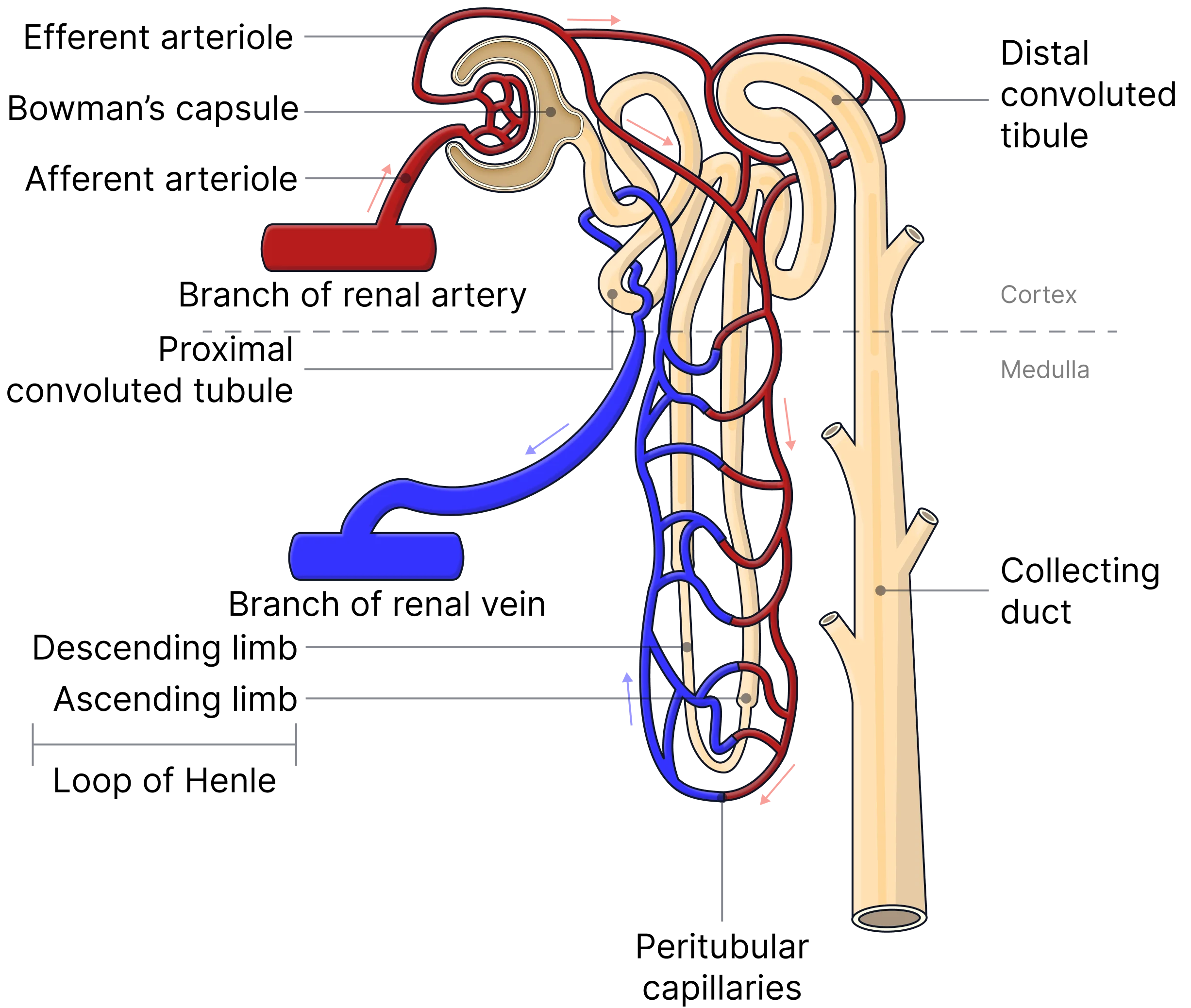
Where does ultrafiltration take place in the nephron?
In the renal capsule.
Talk me through and explain what happens during ultrafiltration.
1) Blood enters the glomerulus via the afferent arteriole.
THERE ARE THREE FILTRATION BARRIERS.
2) Plasma and dissolved substances pass through the capillary endothelium, while (larger) blood cells cannot.
3) Small molecules (such as water, glucose, ions, and urea) are able to pass through the basement membrane, while larger proteins are not.
4) Podocytes (epithelial cells of the renal capsule) ensure that only small molecules are in the glomerular filtrate.
Why is the afferent arteriole wider than the efferent arteriole?
To create a high hydrostatic pressure in the glomerular capillaries which helps ultrafiltration.
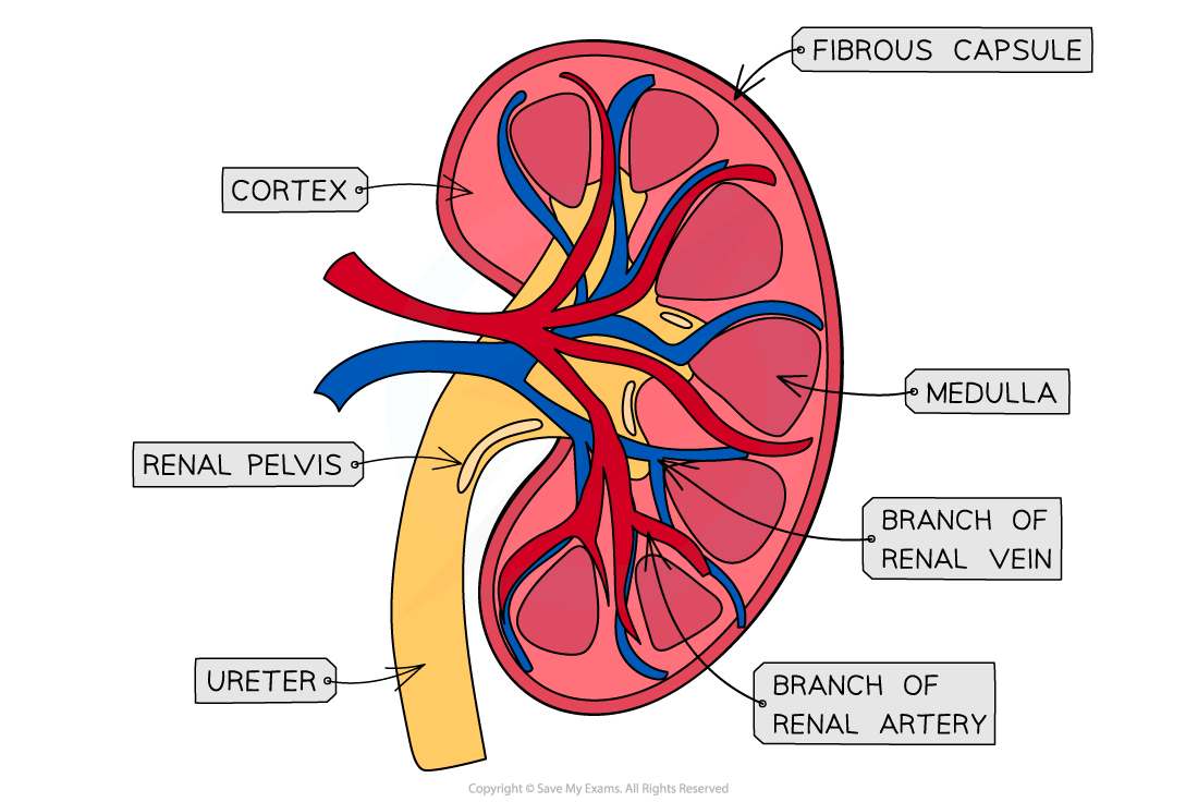
Draw and label the gross structure of the kidney.
Where in the nephron does reabsorption take place?
In the proximal convoluted tubule.
Talk me through what happens in reabsorption.
1) All glucose in the glomerular filtrate is reabsorbed by active transport and co-transport.
2) Most water in the glomerular filtrate is reabsorbed by osmosis.
How is reabsorption maximised in the proximal convoluted tubule?
The proximal convoluted tubule have many microvilli, mitochondria, and carrier proteins.
Talk me through the process of active transport (to reabsorb glucose) in the proximal convoluted tubule.
1) Epithelial cells lining the proximal convoluted tubule actively transport sodium ions into the blood using a sodium-potassium ion pump in their membranes.
2) This active transport creates a lower concentration of sodium ions in the proximal convoluted tubule than in the blood and lumen.
3) Sodium ions in the lumen (where the glomerular filtrate is) move via co-transporter proteins down their concentration gradient, carrying glucose molecules with them into the proximal convoluted tubule.
4) Glucose moves into the blood via facilitated diffusion.
Talk me through how a sodium ion gradient is maintained in the loop of Henle.
1) Water moves out of the descending limb (and into the interstitial space) via osmosis (as water potential lower in interstitial space), sodium ions do not as the descending limb is impermeable to sodium ions.
2) This osmosis causes the water potential of the filtrate to decrease as it goes further down the loop of Henle (increasing concentration of filtrate).
3) Sodium ions are actively transported into the interstitial space from the ascending limb.
4) Sodium ions diffuse out of the filtrate at the bottom of the ascending limb (where the filtrate is most concentrated).
5) This active transport lowers water potential in the interstitial space which makes water move via osmosis into the medulla from the filtrate in the descending limb (water reabsorbed).
Which limb on the loop of Henle is impermeable to sodium ions?
The descending limb.
True or false? The walls of the ascending limb are permeable to water.
False. THE WALLS OF THE ASCENDING LIMB ARE IMPERMEABLE TO WATER.
What are the protein channels that water can pass through?
Aquaporins.
Where in the body detects changes in blood water potential?
Osmoreceptors in the hypothalamus in the brain.
How does the hypothalamus detect and increase blood water potential if it is too low?
1) When blood water potential is too low, water leave osmoreceptors via osmosis.
2) This causes osmoreceptors to shrivel.
3) The stimulates the hypothalamus to produce more ADH (antidiuretic hormone).
How does the hypothalamus detect and decrease blood water potential if it is too high?
1) Due to increased blood water potential, water moves into osmoregulator cells via osmosis.
2) This stimulates the hypothalamus to produce less ADH.
Where is ADH released into the blood?
From the posterior pituitary.
What is the target organ of ADH?
The kidney.
How does ADH work in increasing water reabsorption?
1) ADH binds to complementary receptors in the cell surface membranes of cells lining the distal convoluted tubule and the collecting duct.
2) This binding activates a phosphorylase enzyme.
3) This activation stimulates vesicles containing aquaporins to move towards and fuse with the cell membrane, increasing the permeability to water.
Define negative feedback.
When an internal change stimulates action to return the condition to it’s original level (by doing the opposite).