Lab Exam 2
1/160
There's no tags or description
Looks like no tags are added yet.
Name | Mastery | Learn | Test | Matching | Spaced |
|---|
No study sessions yet.
161 Terms
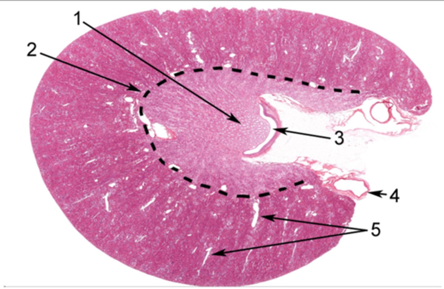
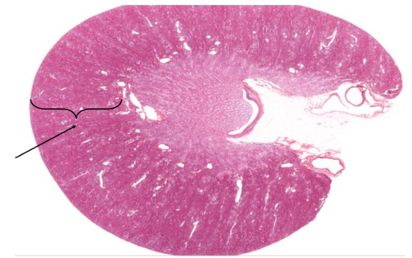
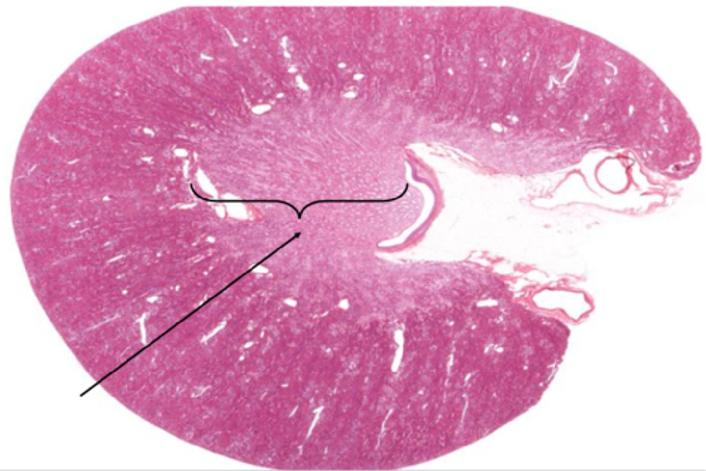
Identify the organ in cross section
kidney
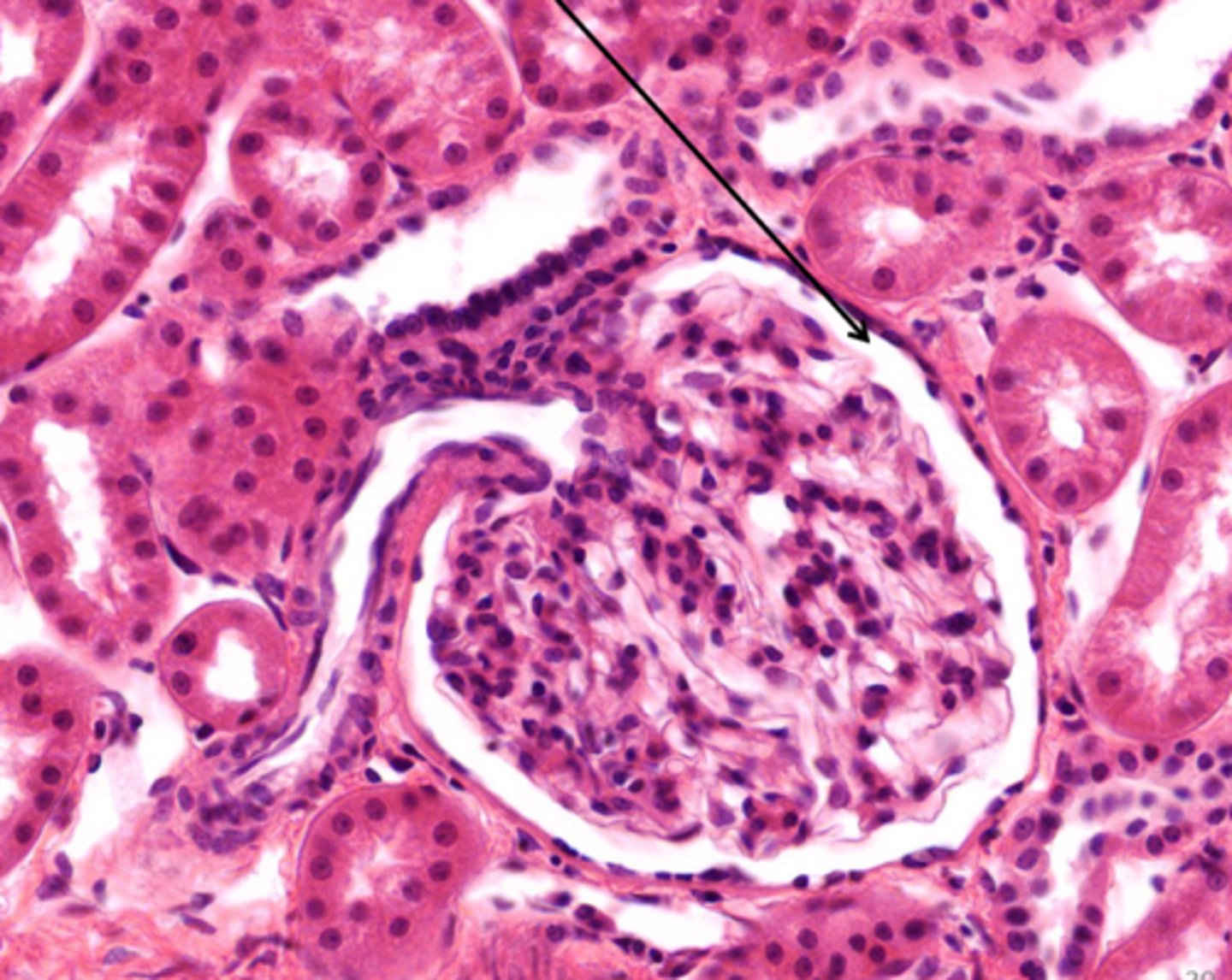
The clear space around the glomerulus is the lumen of
Bowman's capsule
Identify the structure on the kidney
cortex
Identify the structure on the kidney
medulla
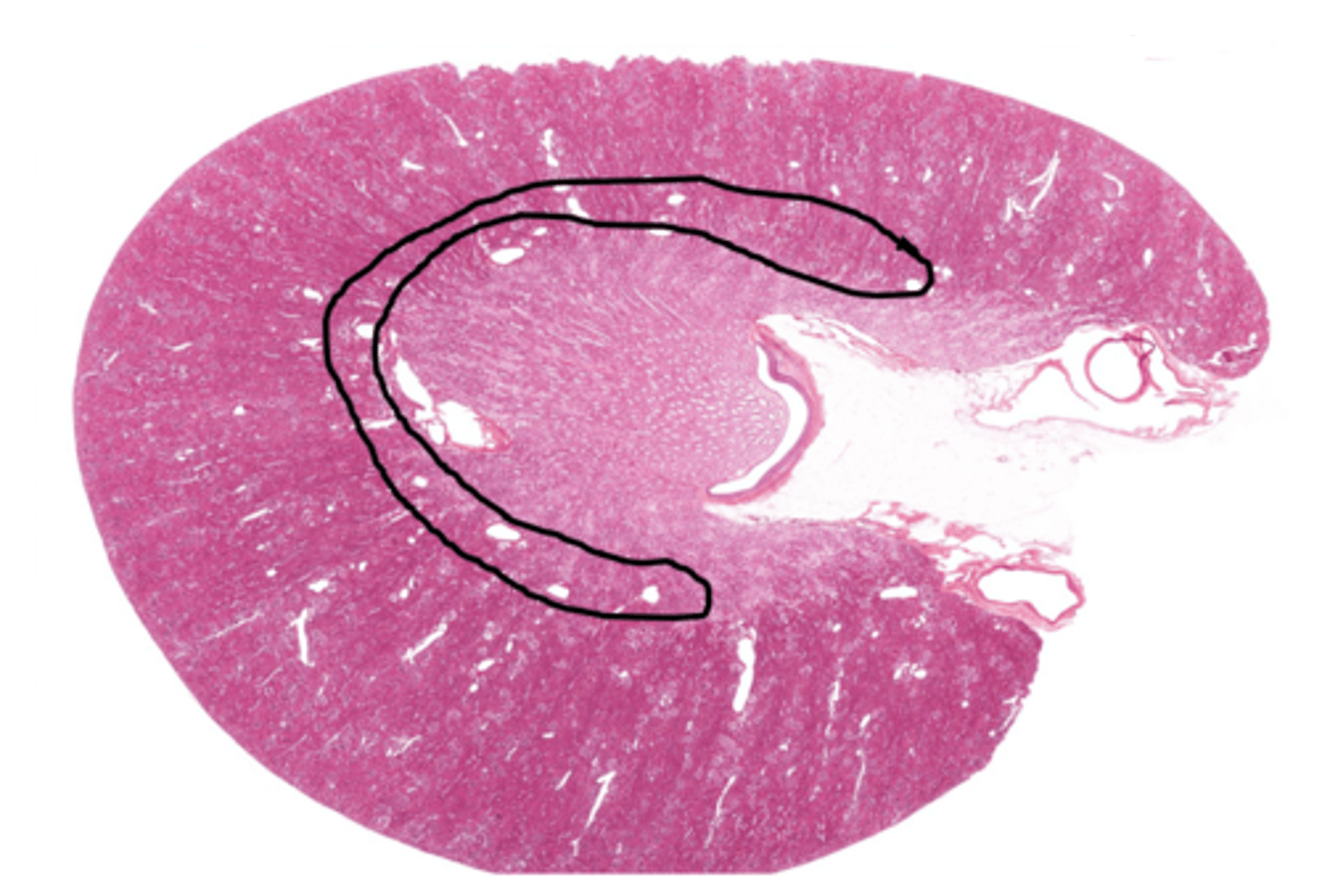
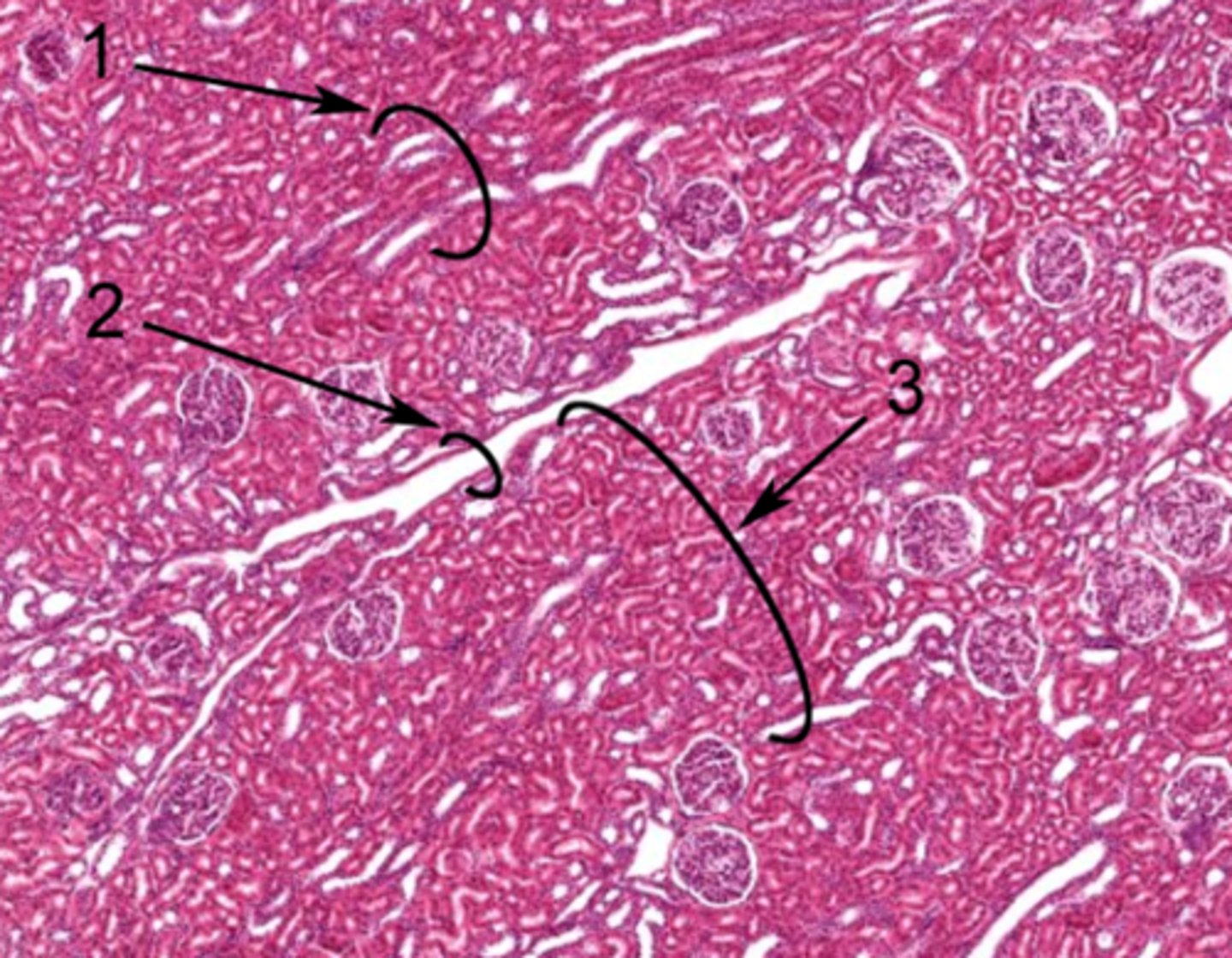
What division of blood vessels is found in the area outlined
arcuate vessels
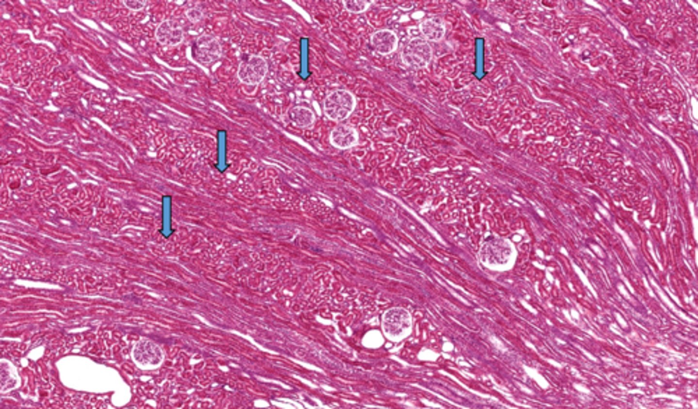
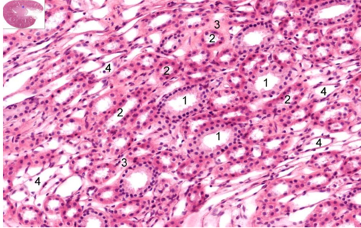
Identify these collections of convoluted structures
cortical labyrinth (pars convoluta)
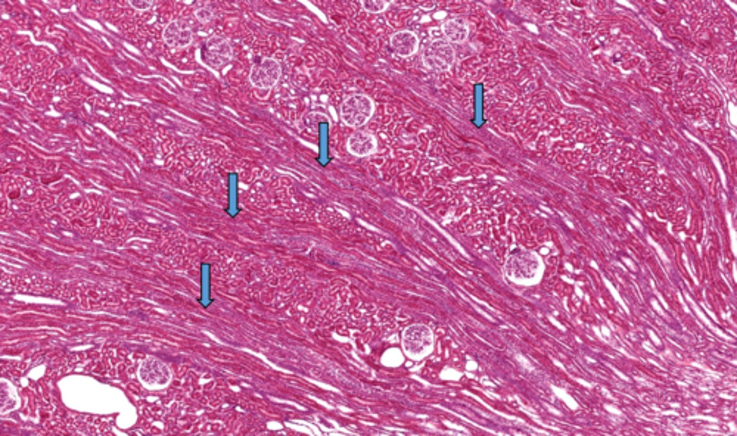
Identify the collection of straight structures in the cortex of the kidney
medullary rays
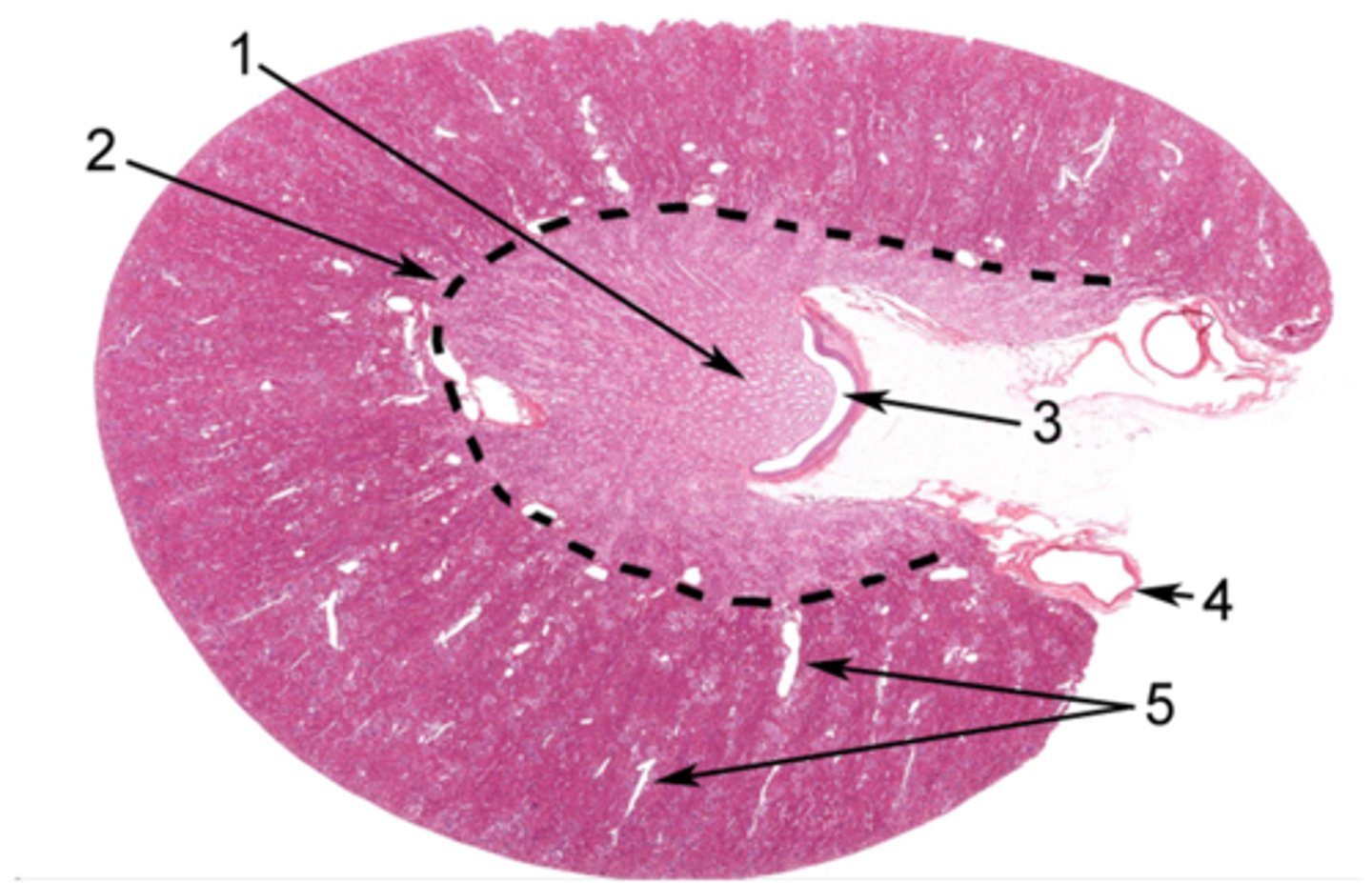
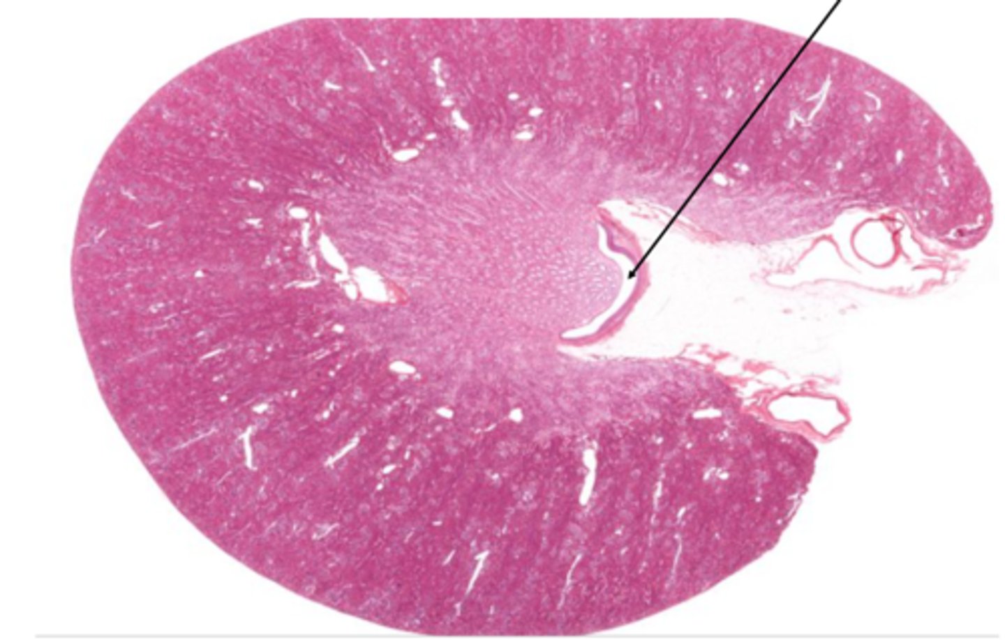
Identify the area of the kidney at 1
renal papilla
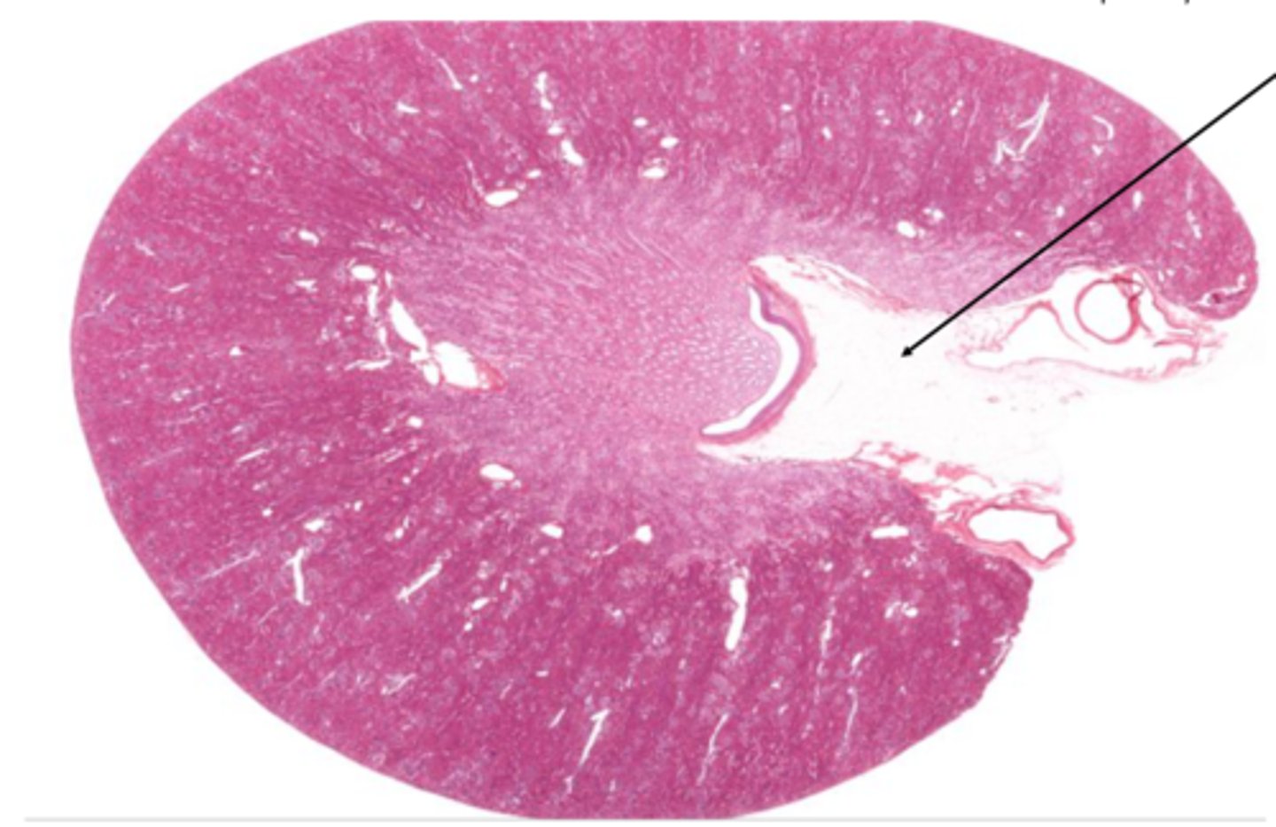
Identify the structure on the kidney
renal pelvis
Identify the structure on the kidney
renal sinus


Identify the structure of the nephron
glomerulus
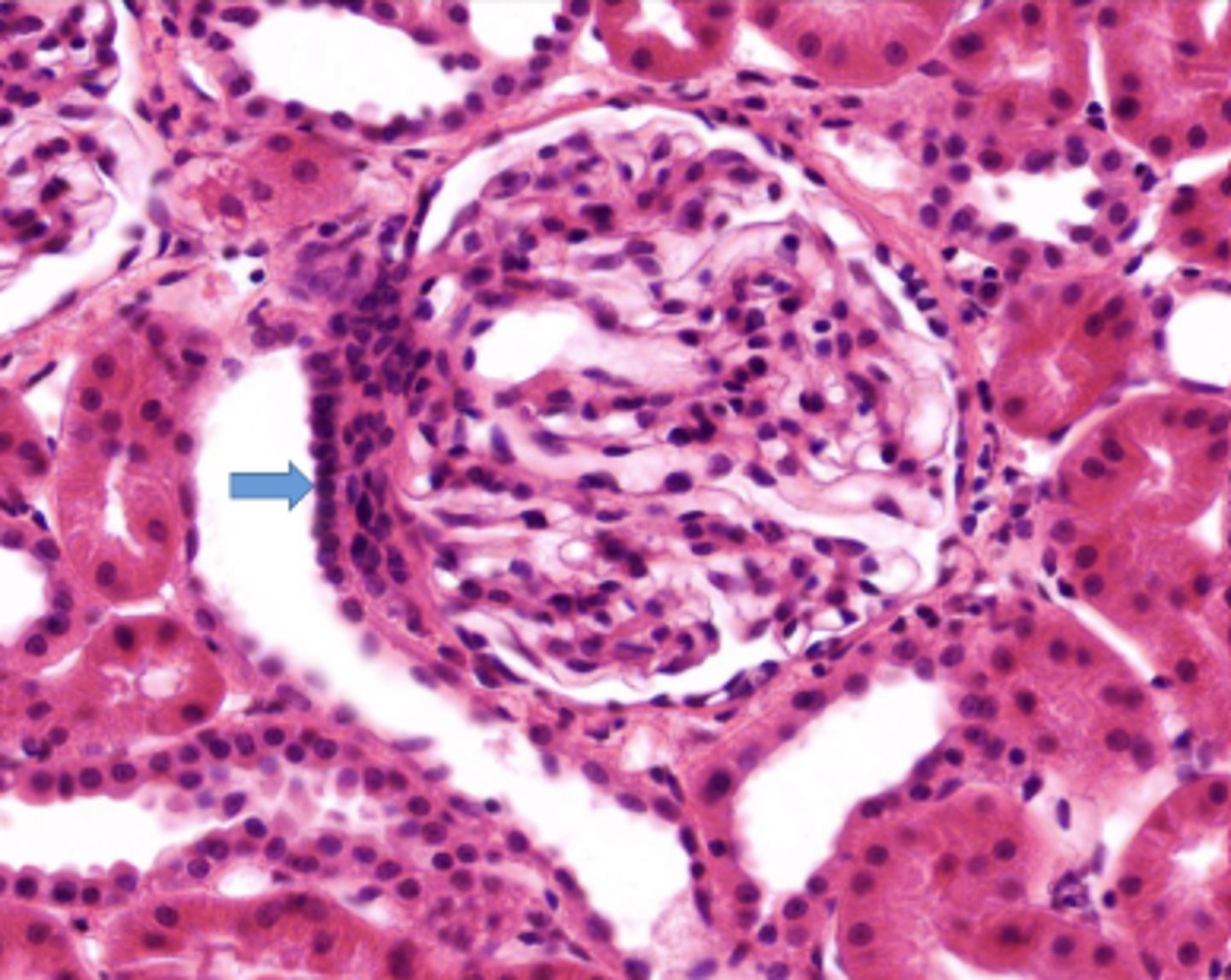
Identify this portion of the glomerulus, communicating with the efferent and afferent arterioles
vascular pole
Identify the nuclei on the distal convoluted tubule
macula densa
What is the function of the macula densa
monitor NaCl concentration
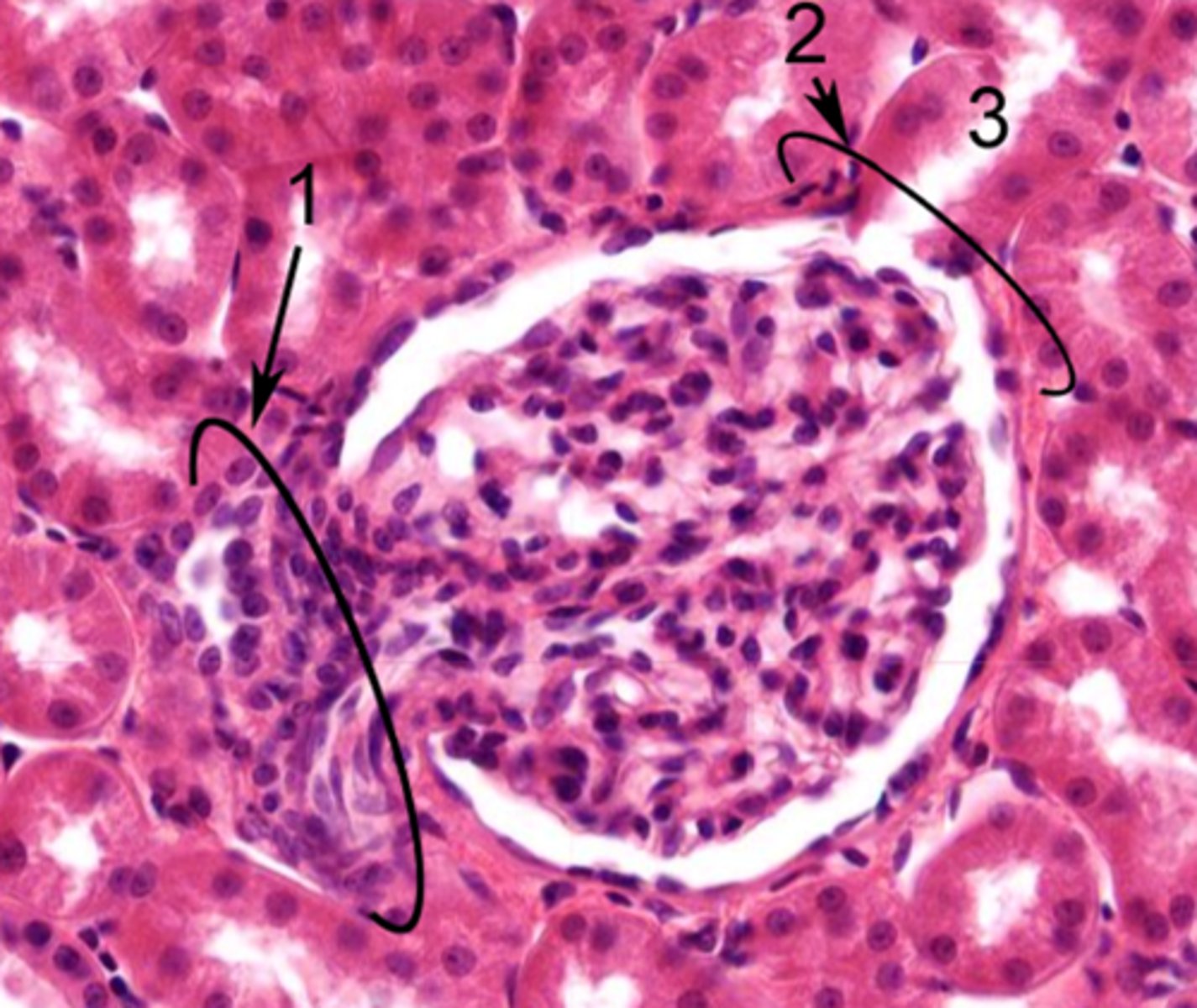
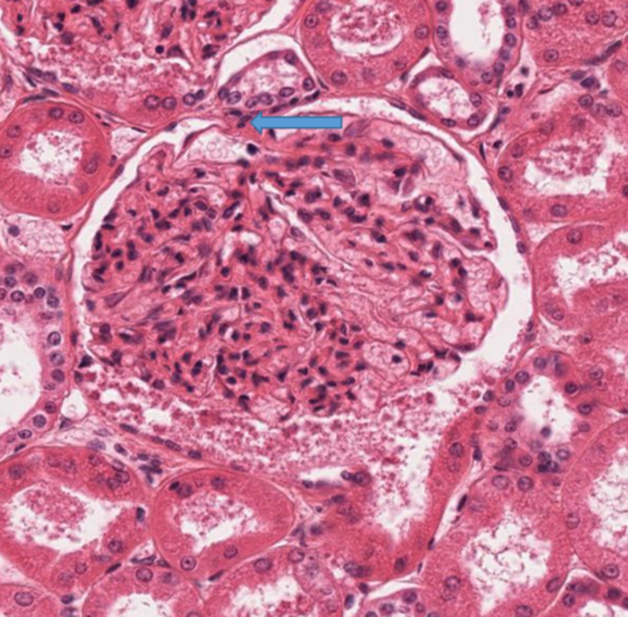
Identify the structure of the glomerulus at 2
urinary pole
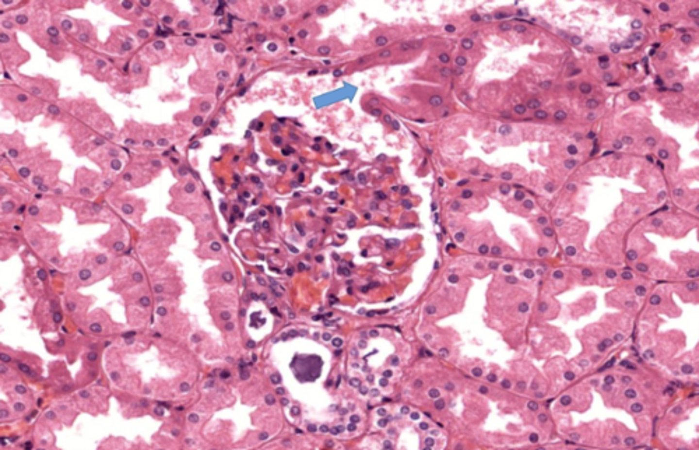
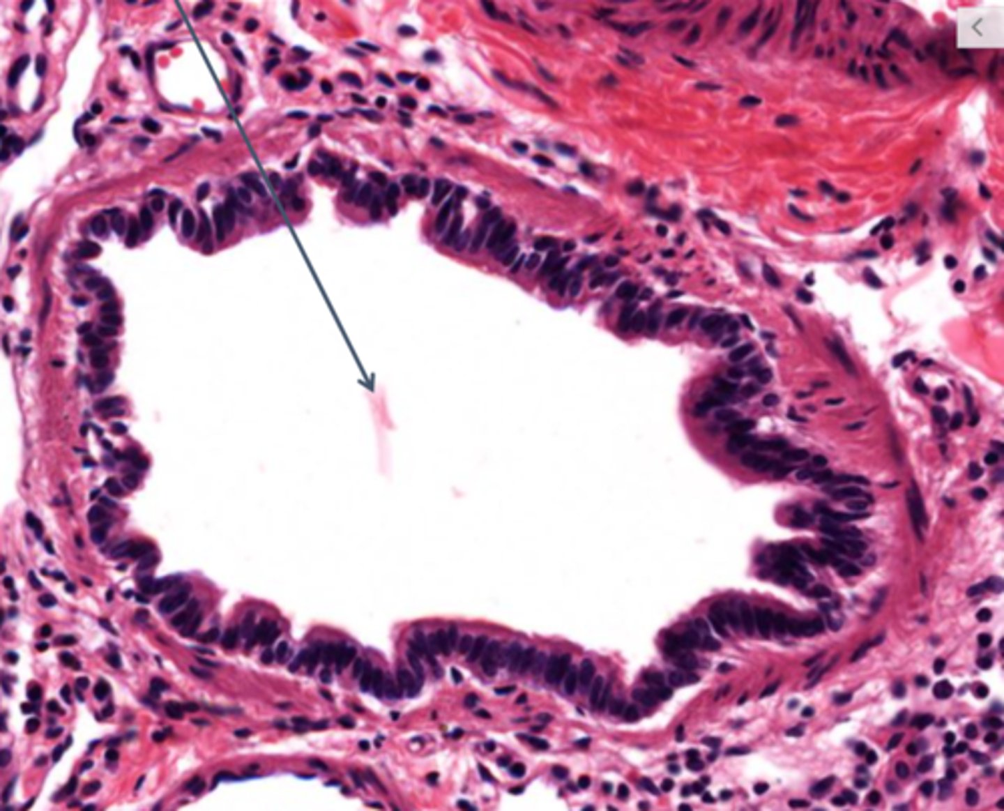
The arrow shows where filtrate enters what segment of the nephron
proximal convoluted tubule
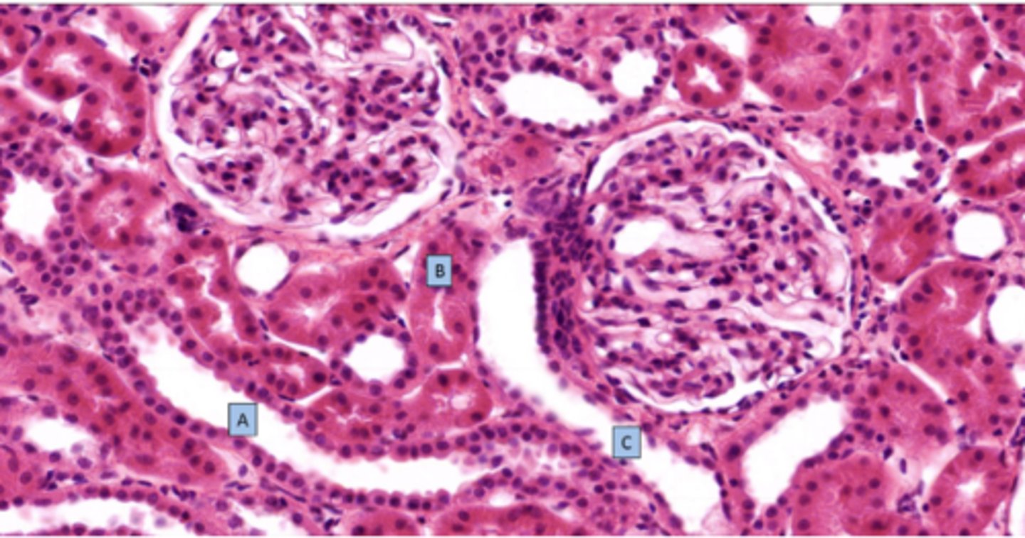
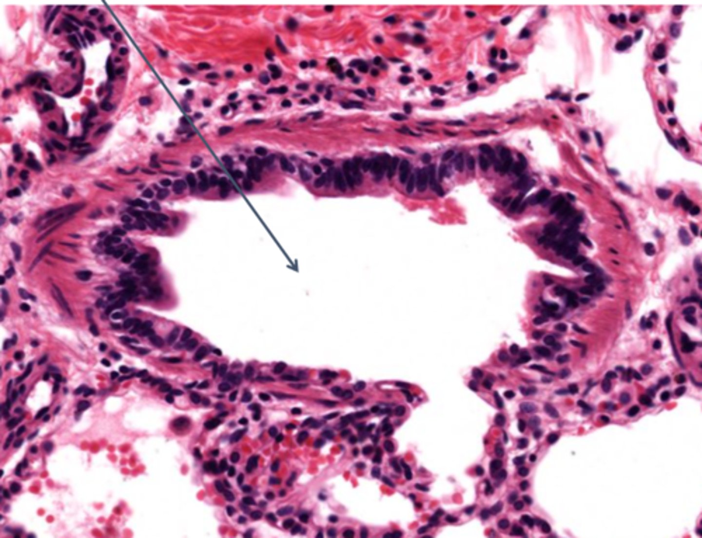
What is the structure at C
distal convoluted tubule

Identify the structure of the medulla at 2
thick loop of henle

Identify the structure of the medulla at 4
thin loop of henle

Identify the structure of the medulla at 1
collecting duct
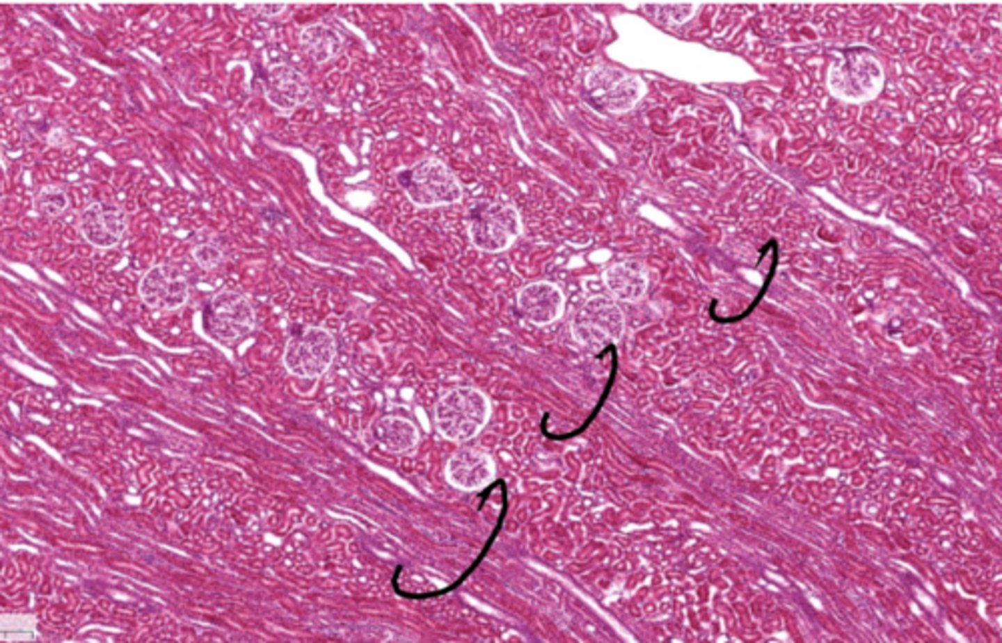
The circled structures are the centers of the
kidney lobule
Identify the structure in the cortex of the kidney at 2
interlobular arteries
What joins the glomerulus at the vascular pole of the renal corpuscle
afferent and efferent arterioles
Where is the descending loop of henle
comes out of the proximal convoluted tubule beginning in the medullary rays (resembles PCT but is straight)
Where is the ascending loop of henle
in the outer portion of the medulla and in medullary rays (resembles DCT)
What cells does the glomerulus contain
mesangial cells and podocytes
The distal convoluted tubule and vascular pole contain which cells
juxtaglomerular cells
Identify the structure of the medulla at 3
interstitium
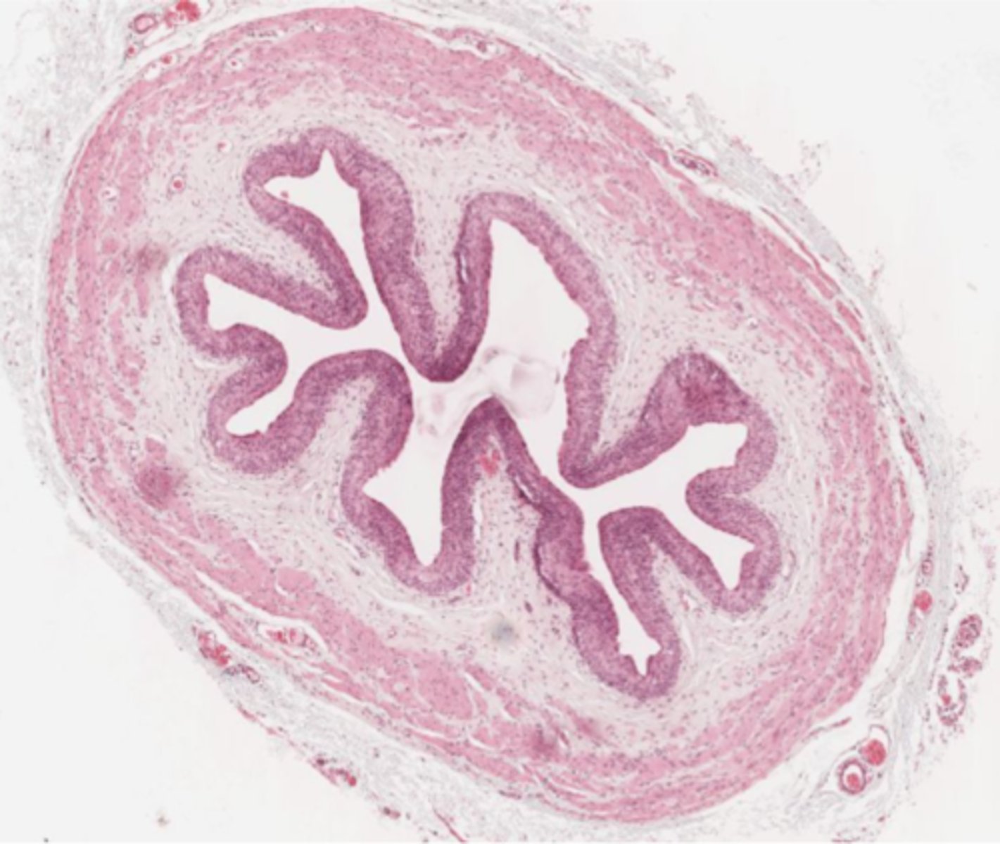
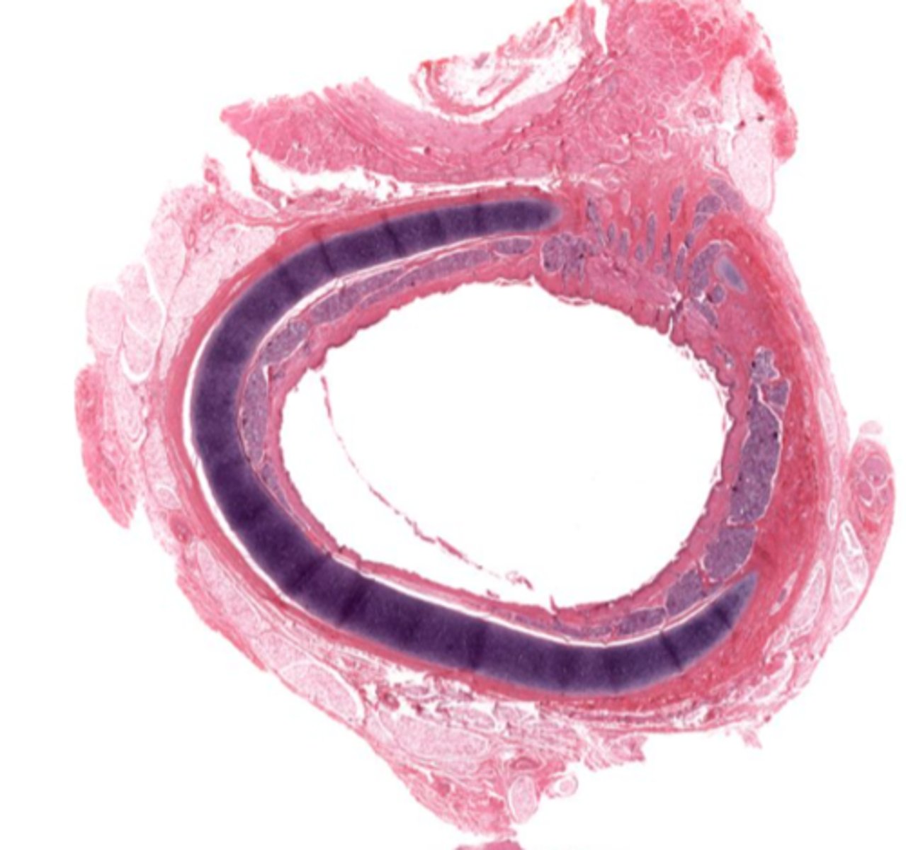
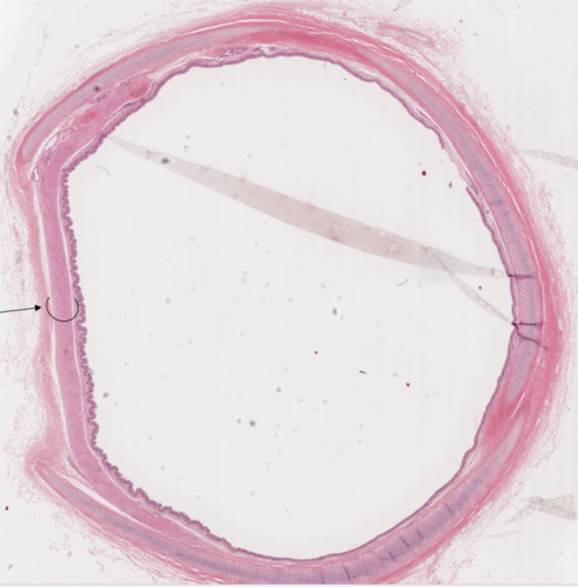
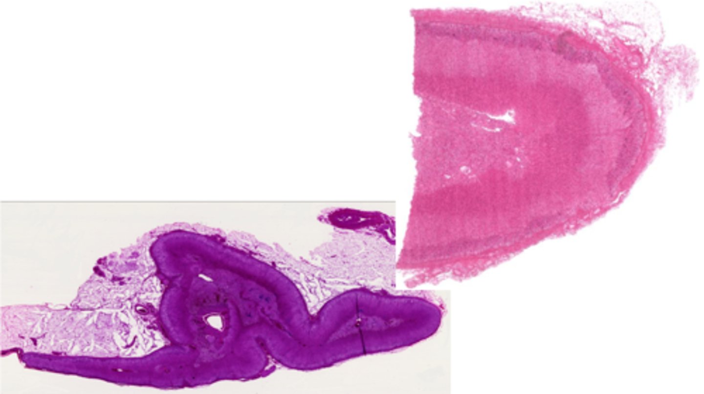
Identify the duct in cross section of the urinary system
ureter
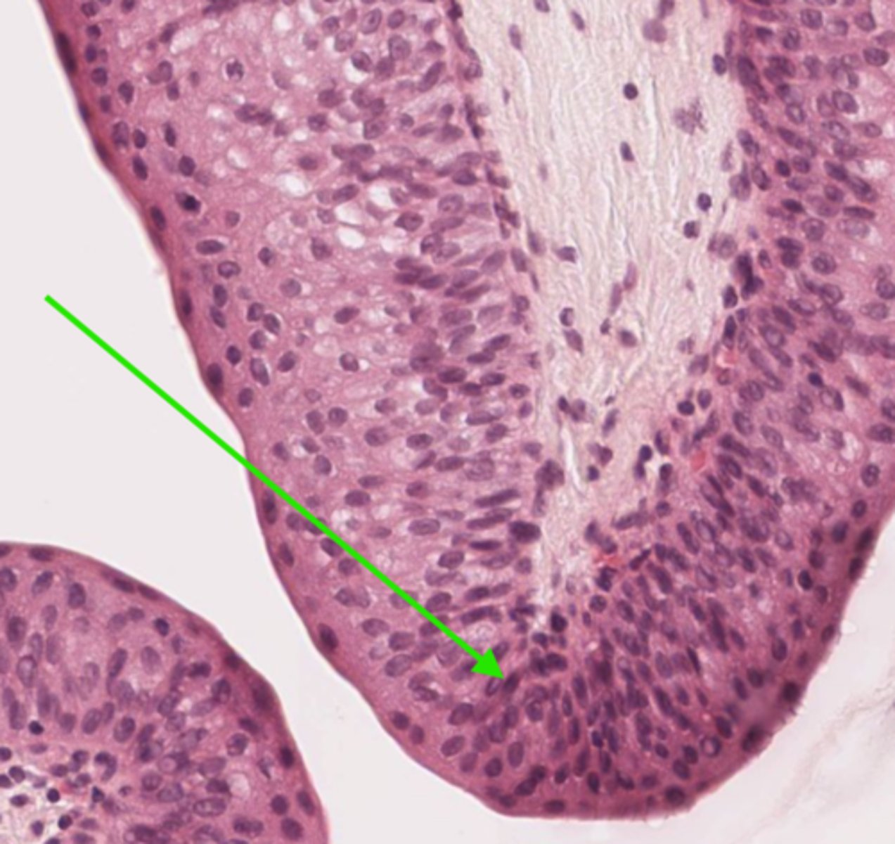
Identify the layer of the ureter
transitional epithelium
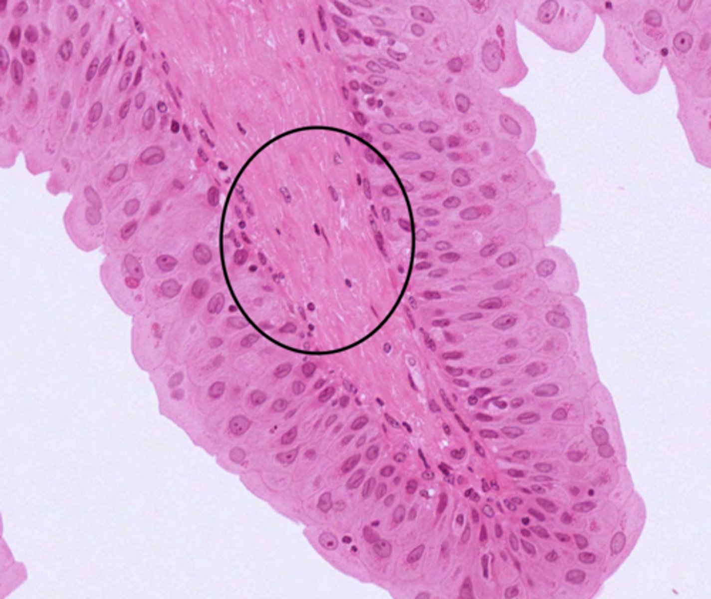
Identify the layer of the ureter
lamina propria

Identify the layer of the bladder at 4
smooth muscular wall
Identify the organ in cross section
trachea
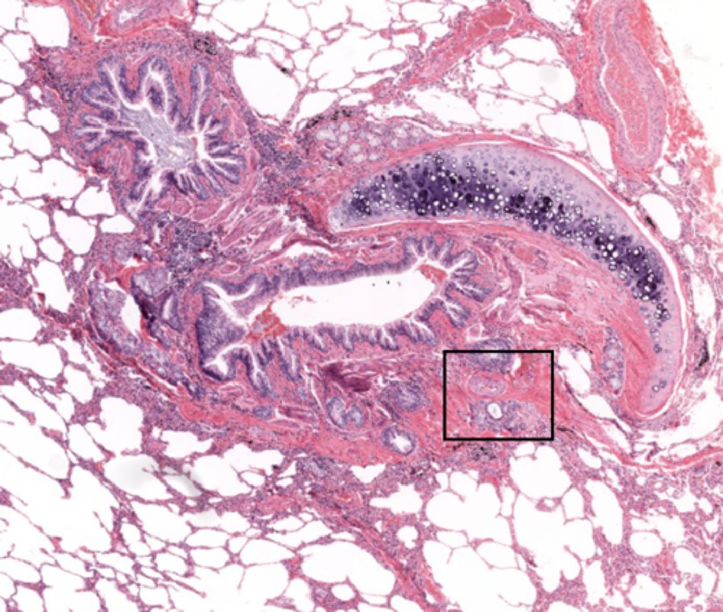
Identify the organ
lungs
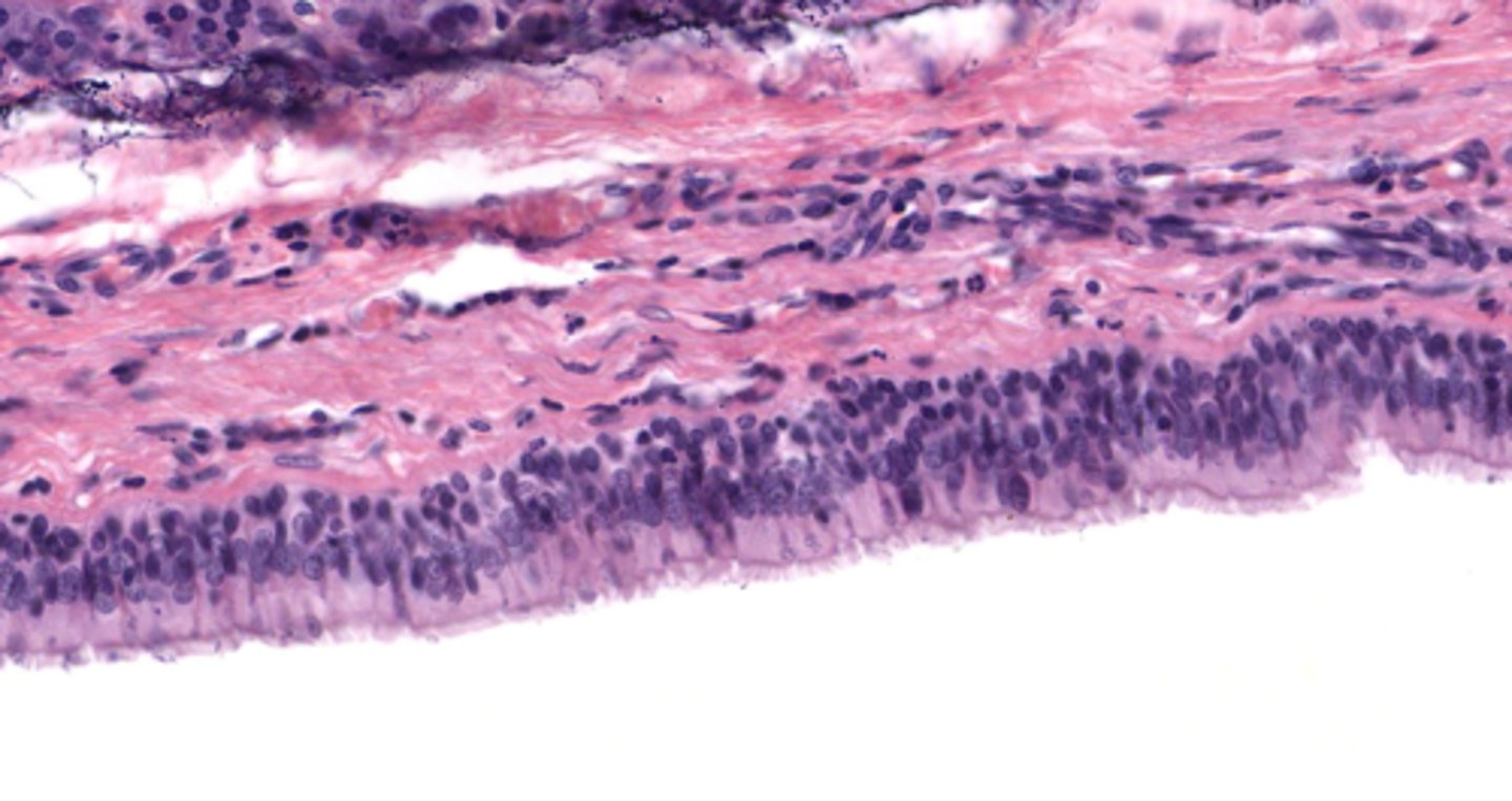
Classify the epithelium of the trachea
pseudostratified columnar ciliated with goblet cells
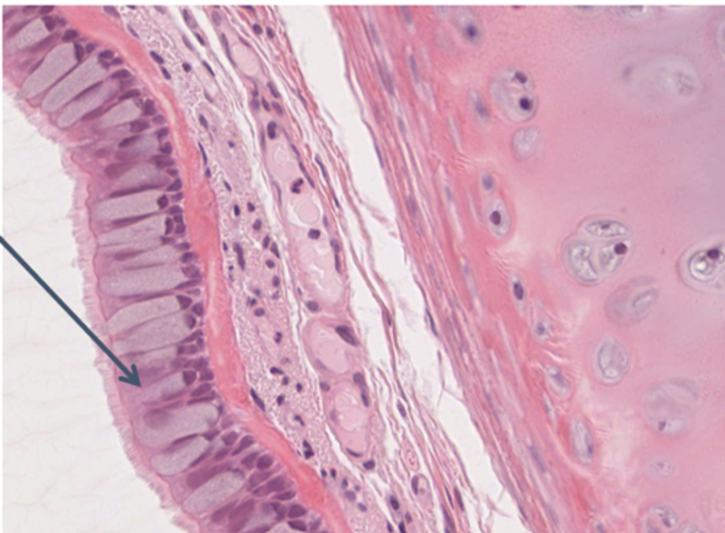
What is the name of this type of epithelium
respiratory epithelium

Identify the layer of the respiratory tract at 4
mucosa
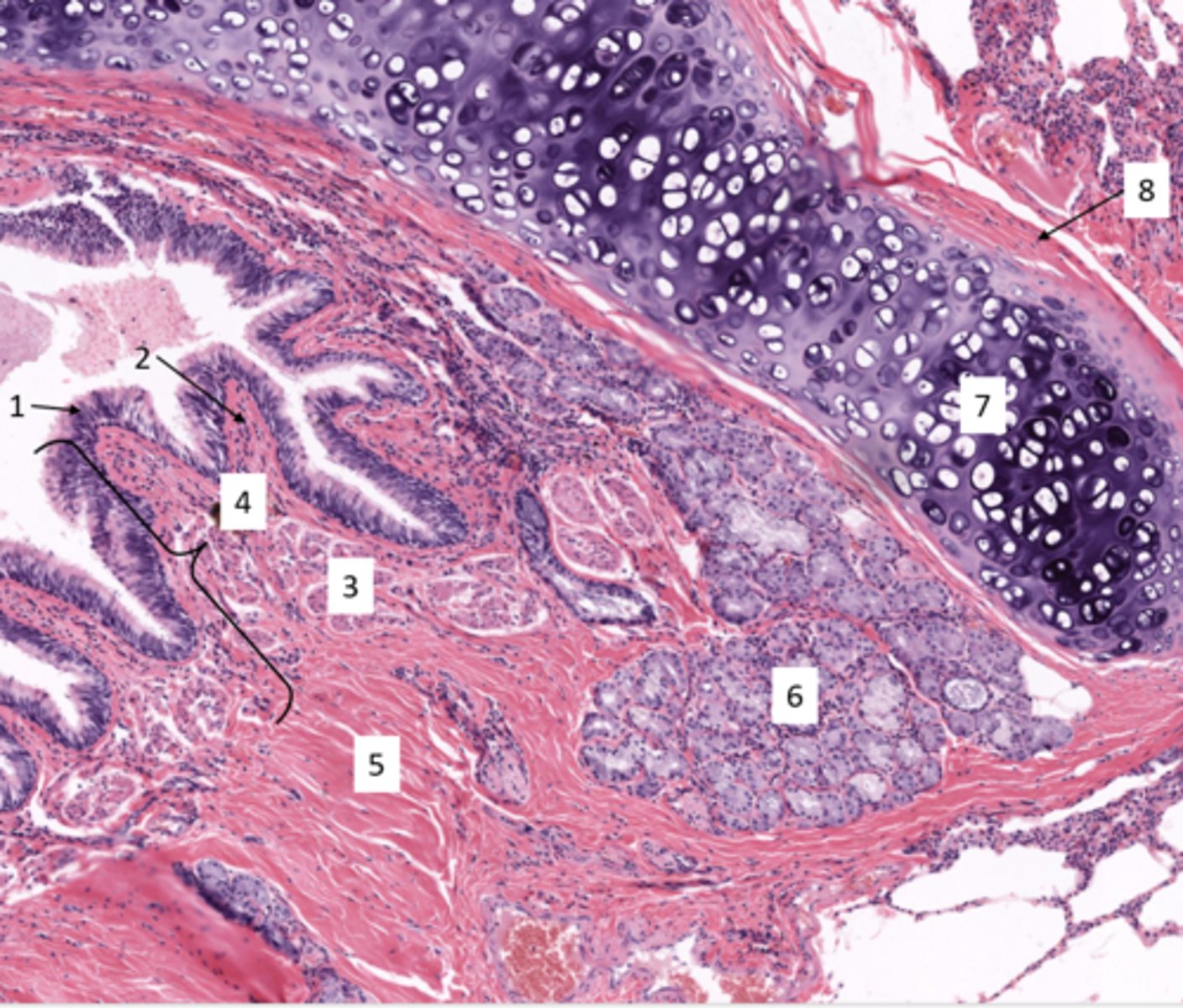
Identify the layer of the respiratory tract at 2
lamina propria
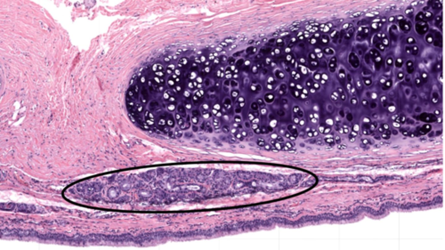
These glands are in which layer of the trachea?
submucosa
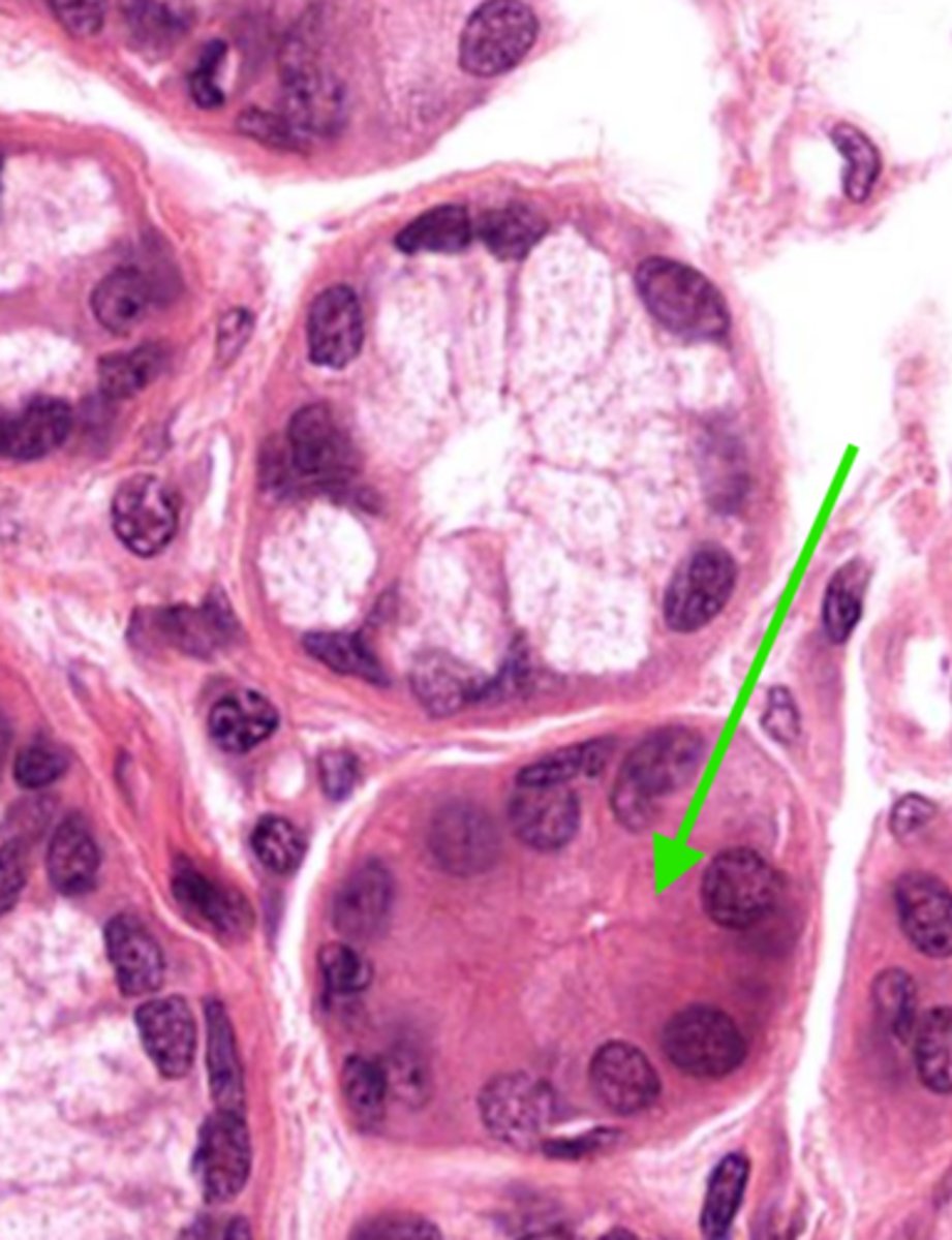
Identify the gland in the mucosa and submucosa
seromucous gland
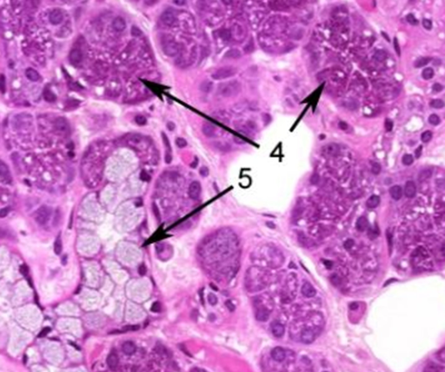
Identify the cell at 5
mucous cells

Identify the cell at 4
serous cells
Identify the muscle and organ
trachealis muscle of the trachea
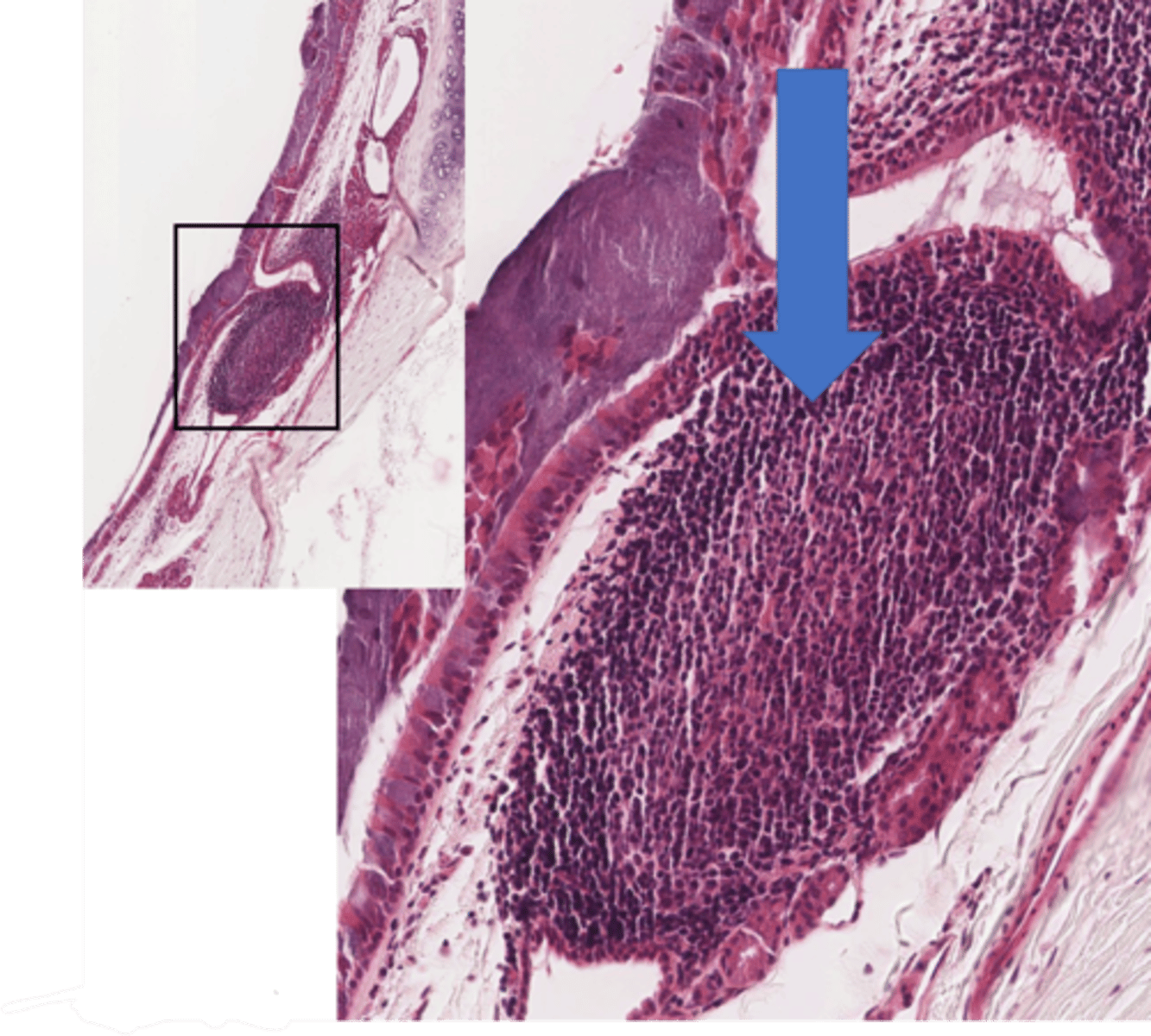
Identify the structure in the trachea
secondary lymph node
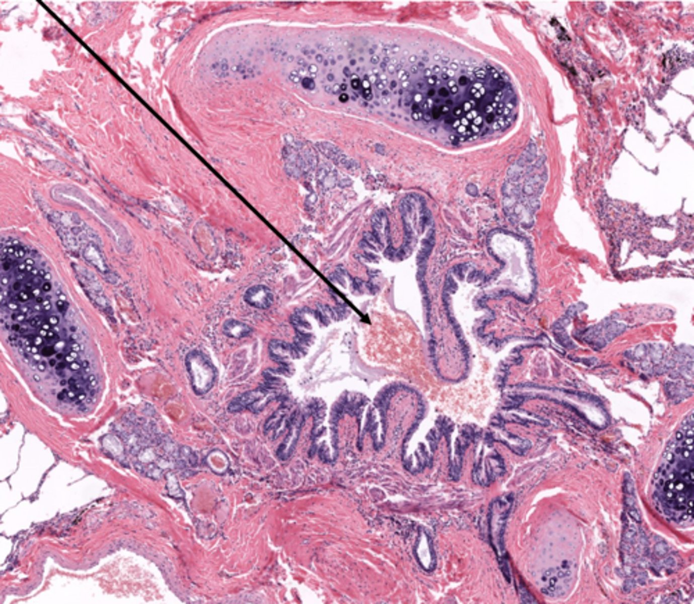
Identify the type of airway
bronchi
Identify this type of airway
bronchioles
T/F: Only bronchi have cartilage
true
Identify the type of bronchiole
terminal bronchioles
Identify the type of bronchiole
respiratory bronchioles
Identify the airway segment between the lines.
alveolar ducts

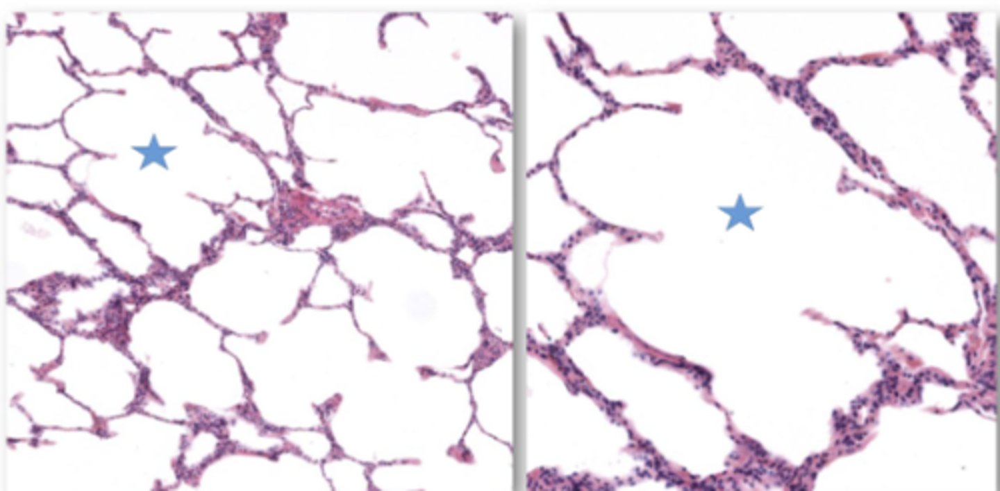
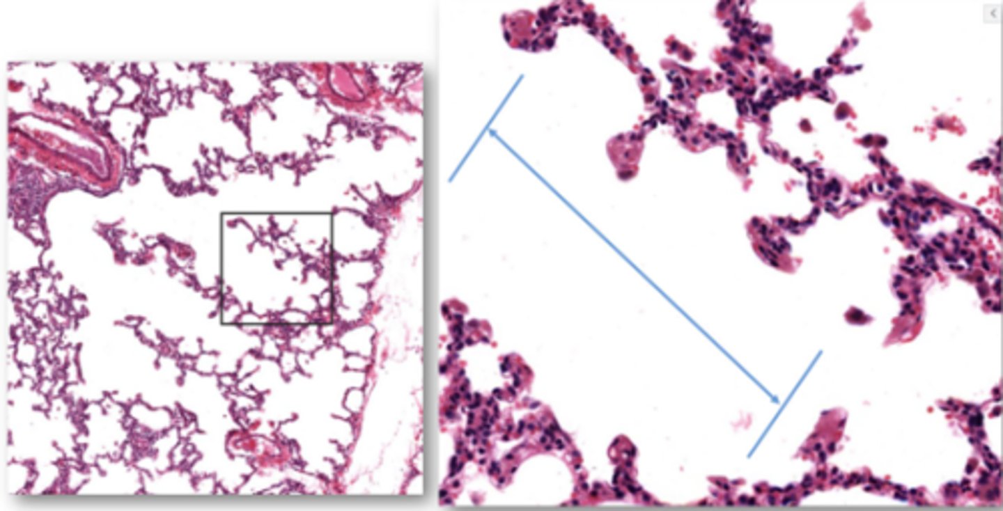
Identify the starred portion of the airway
alveolar sacs
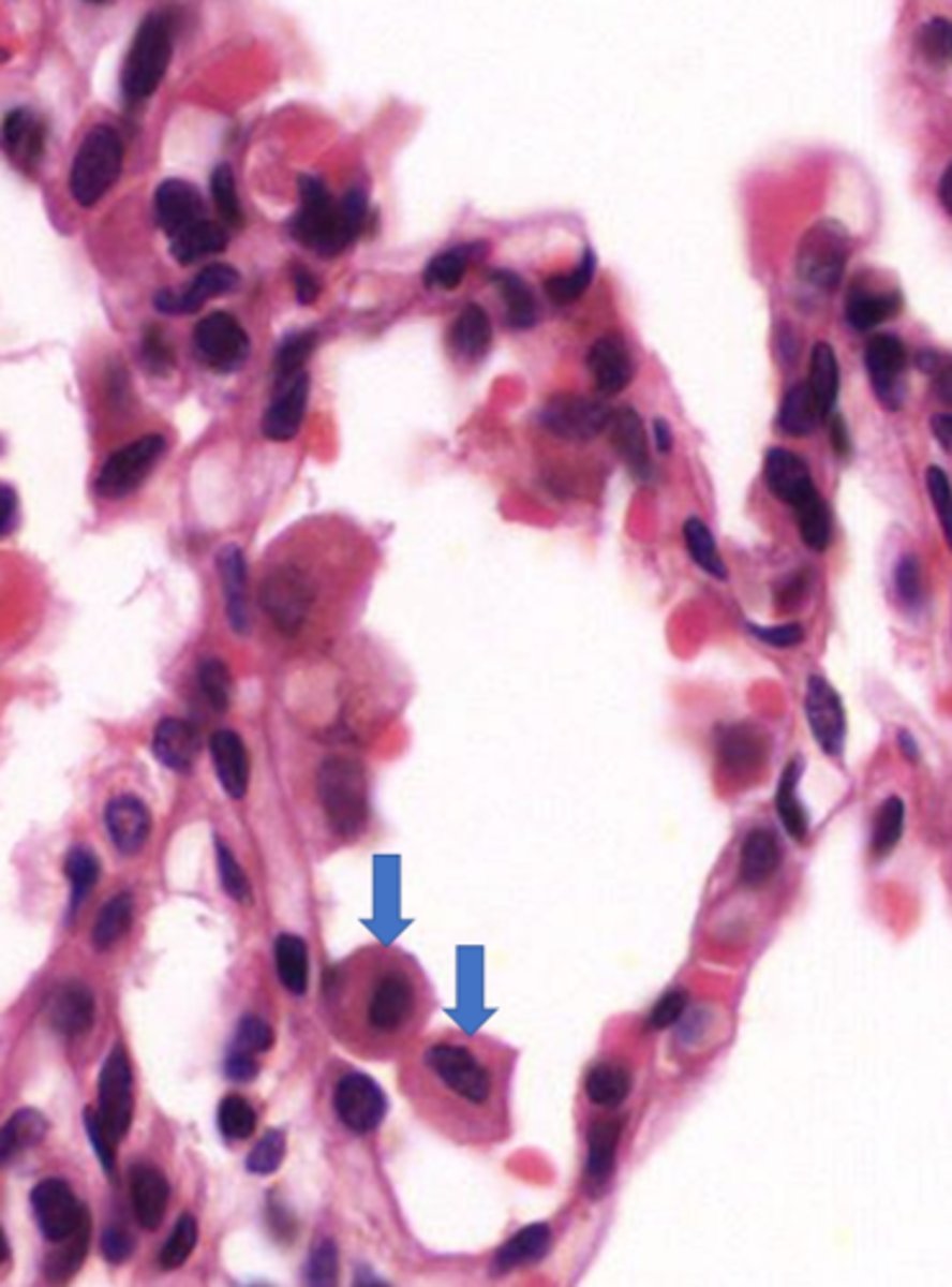
Identify these unattached cells in this alveolus
pulmonary alveolar macrophages
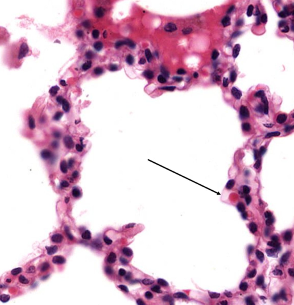
Name the barrier located here
blood air barrier
What cells make up the blood air barrier
type 1 pneumocyte and endothelial cells
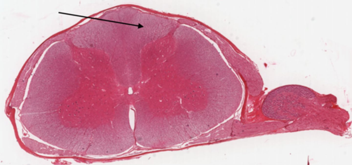
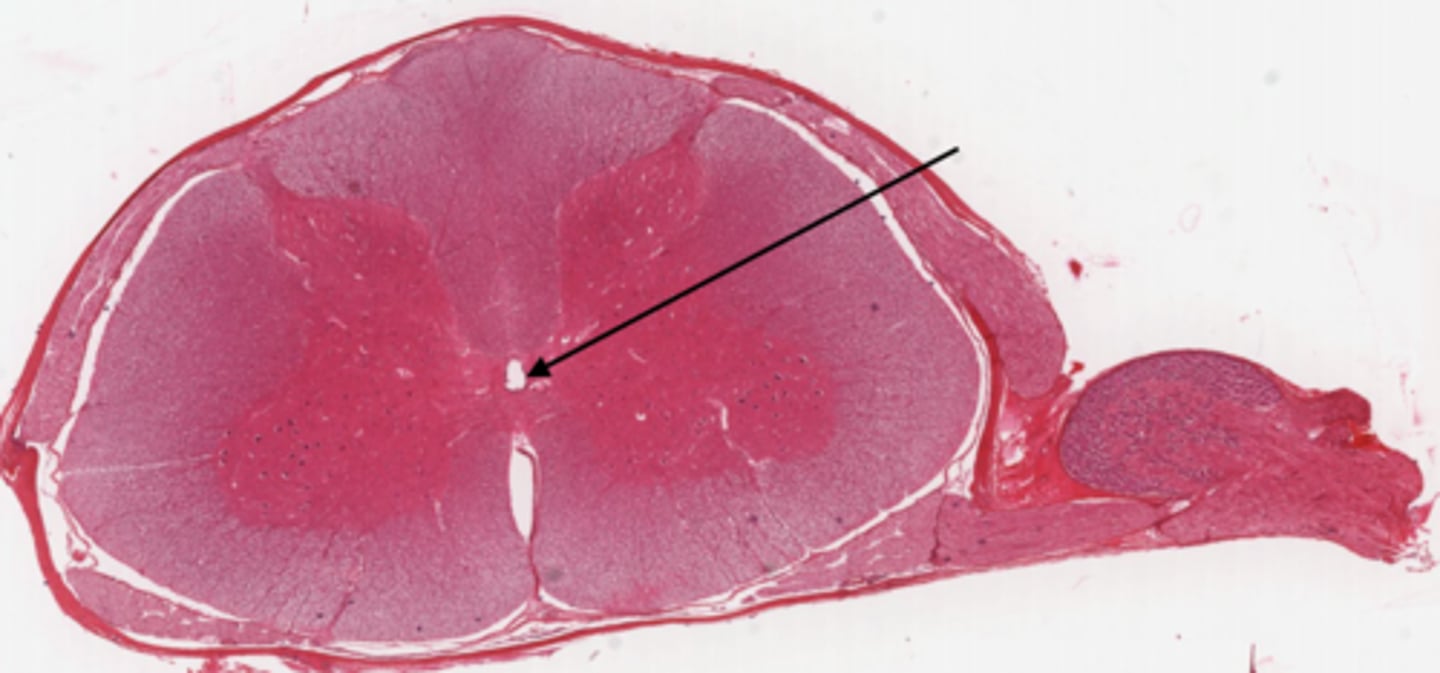
Identify the part of the spinal cord
dorsal funiculi
Identify the part of the spinal cord
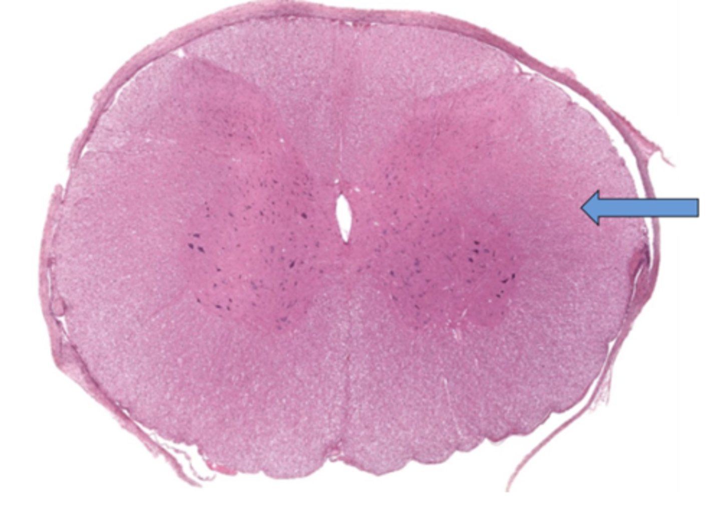
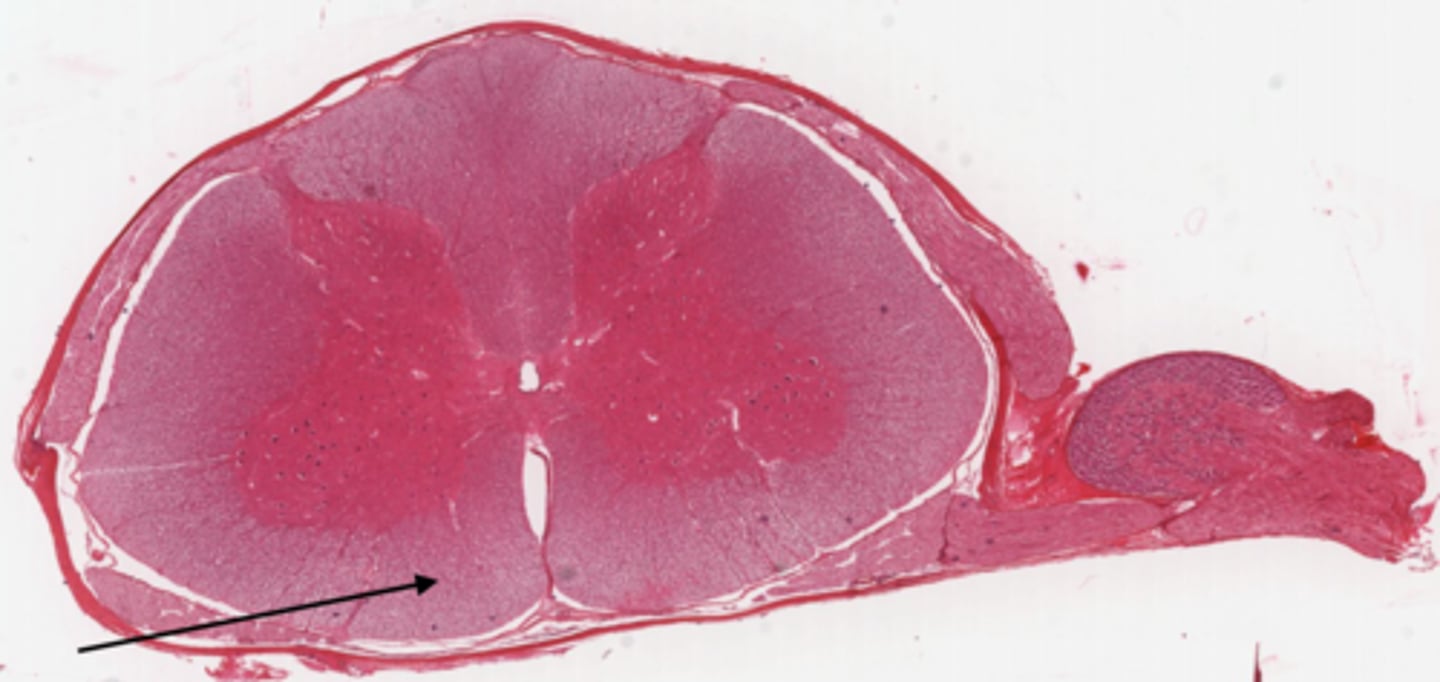
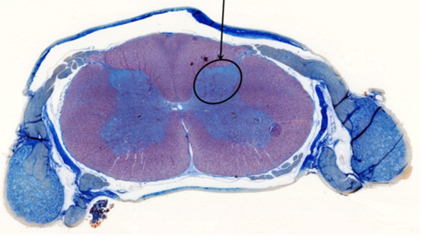
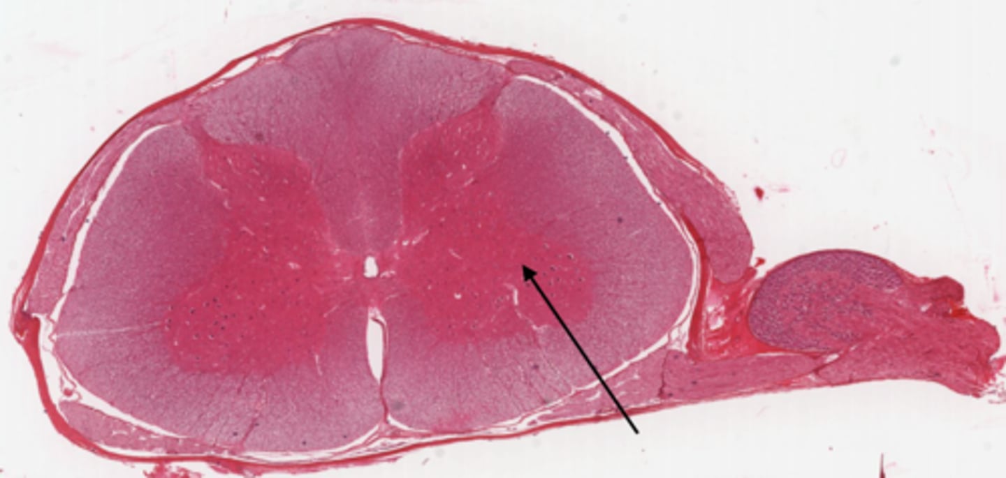
lateral funiculi
Identify the part of the spinal cord
ventral funiculi
Identify the part of the spinal cord
dorsal horn
Identify the part of the spinal cord
ventral horn
Identify the part of the spinal cord
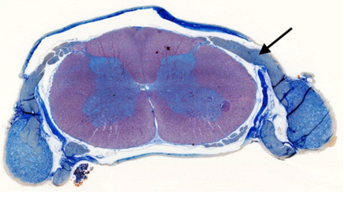
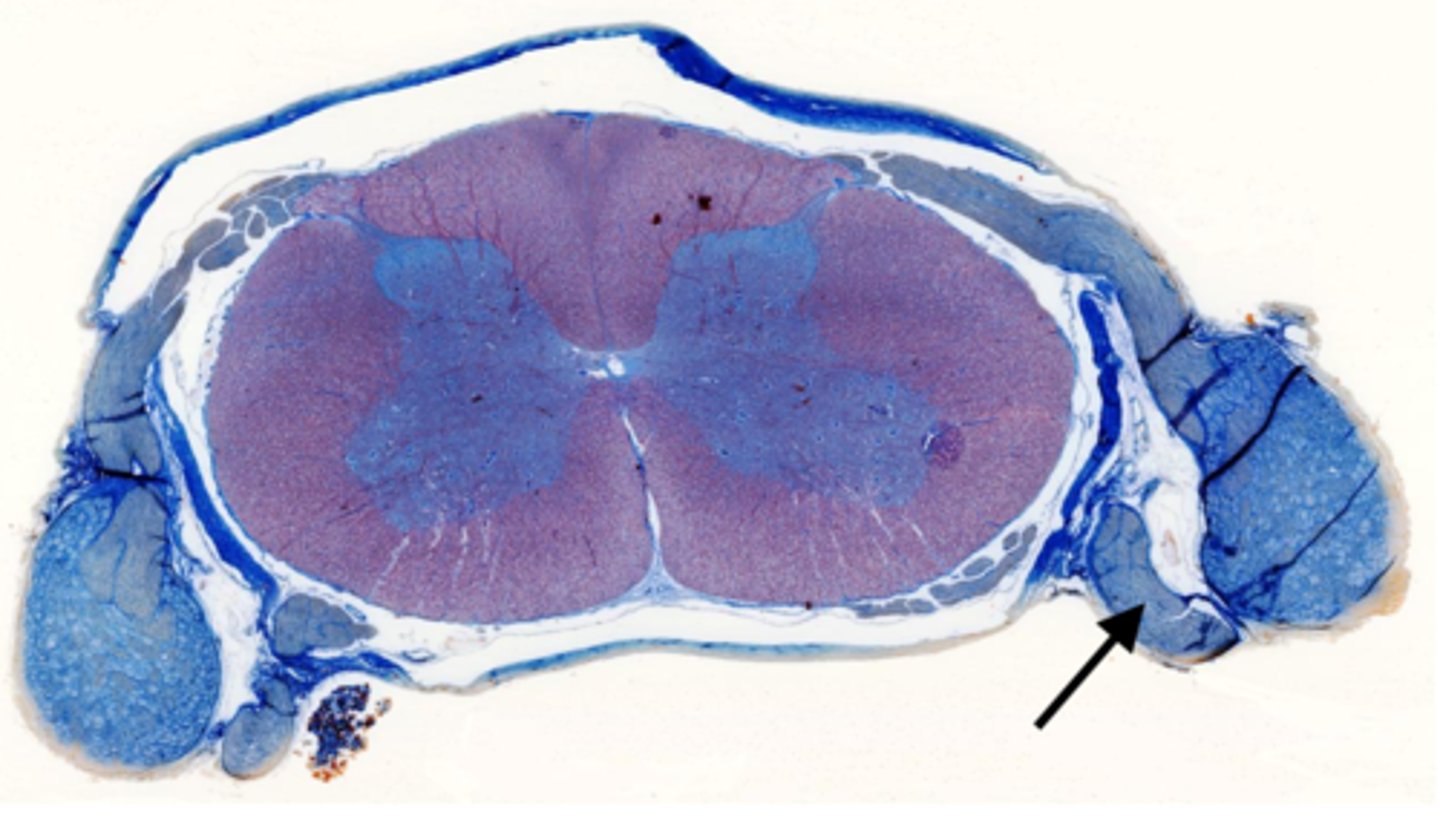
dorsal root of spinal nerve
Identify the part of the spinal cord
ventral root of spinal nerve
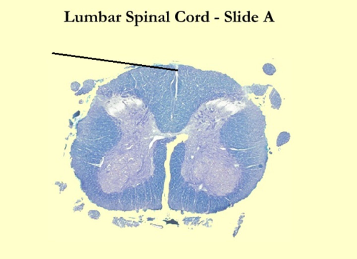
What is the midline structure that separates the dorsal left and right halves of the white matter
dorsal median septum
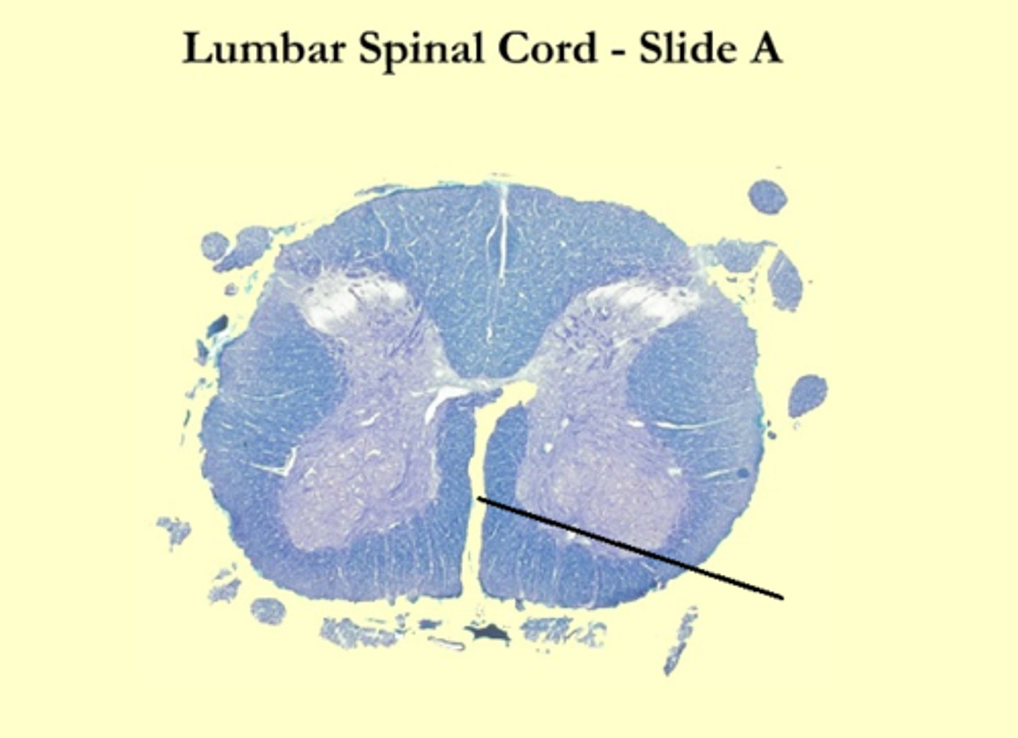
What is the midline structure that separates the ventral left and right halves of the white matter
ventral median sulcus
Identify the section of the spinal cord that contains nuclei
gray matter
Identify the section of the spinal cord that contains nerve tracts
white matter
Identify the part of the spinal cord
central canal
Identify the cells that line the ventricles of the brain and the central canal of the spinal cord
ependymal cells
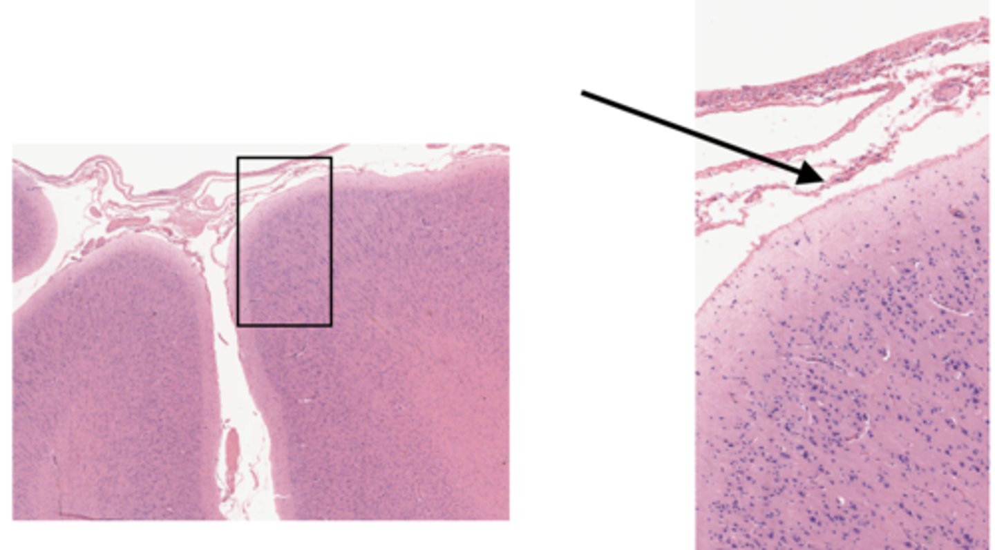
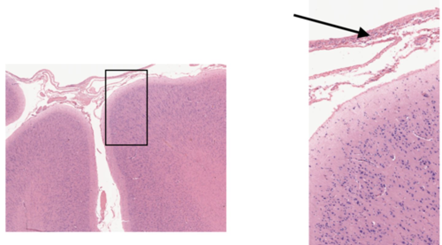
Identify the part of the meninges
pia mater
Identify the part of the meninges
arachnoid

Identify the part of the meninges
dura mater
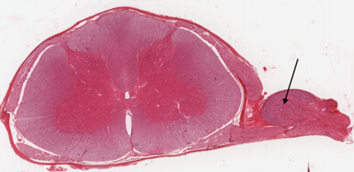
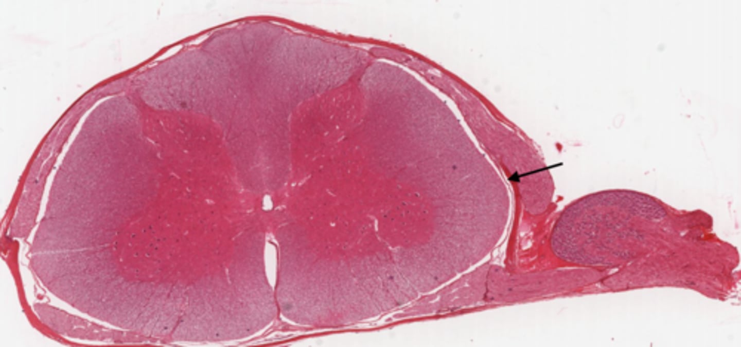
Identify the part of the spinal cord
dorsal root ganglion
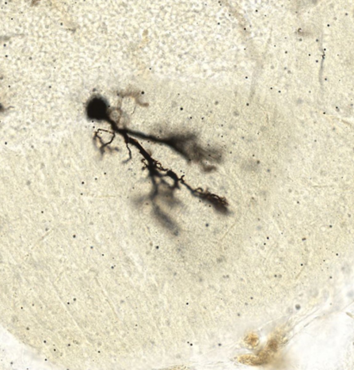
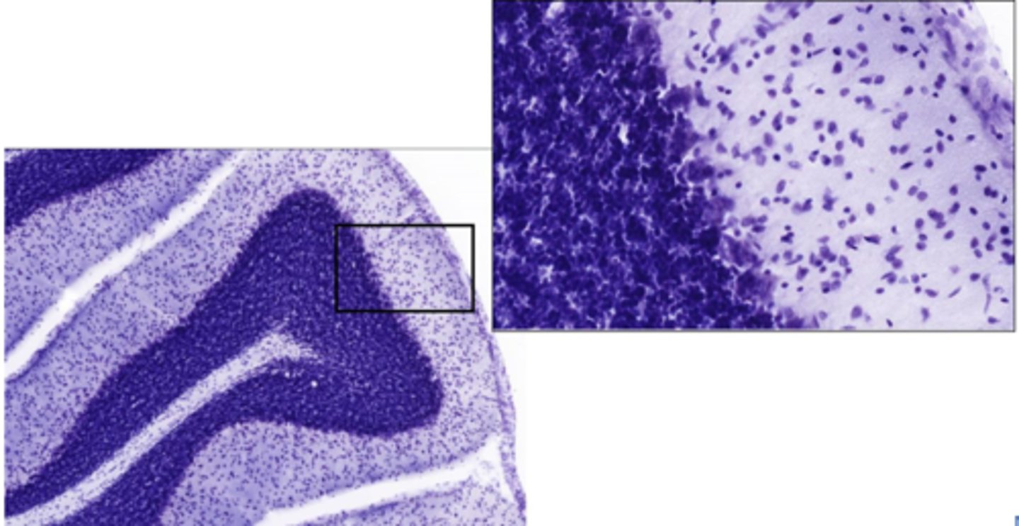
Identify this cell
purkinje cells in the brain
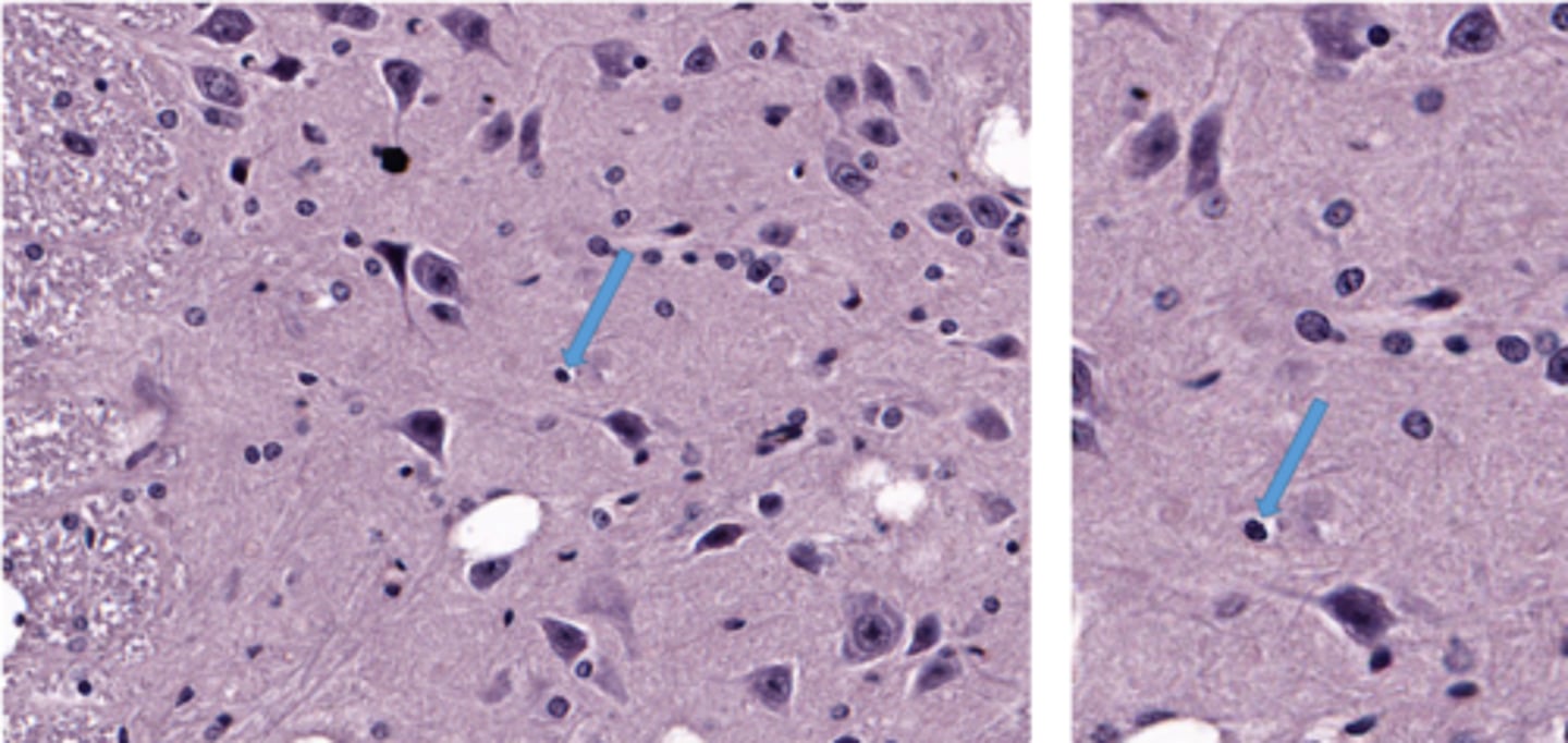
Identify the glial cell
oligodendrocyte
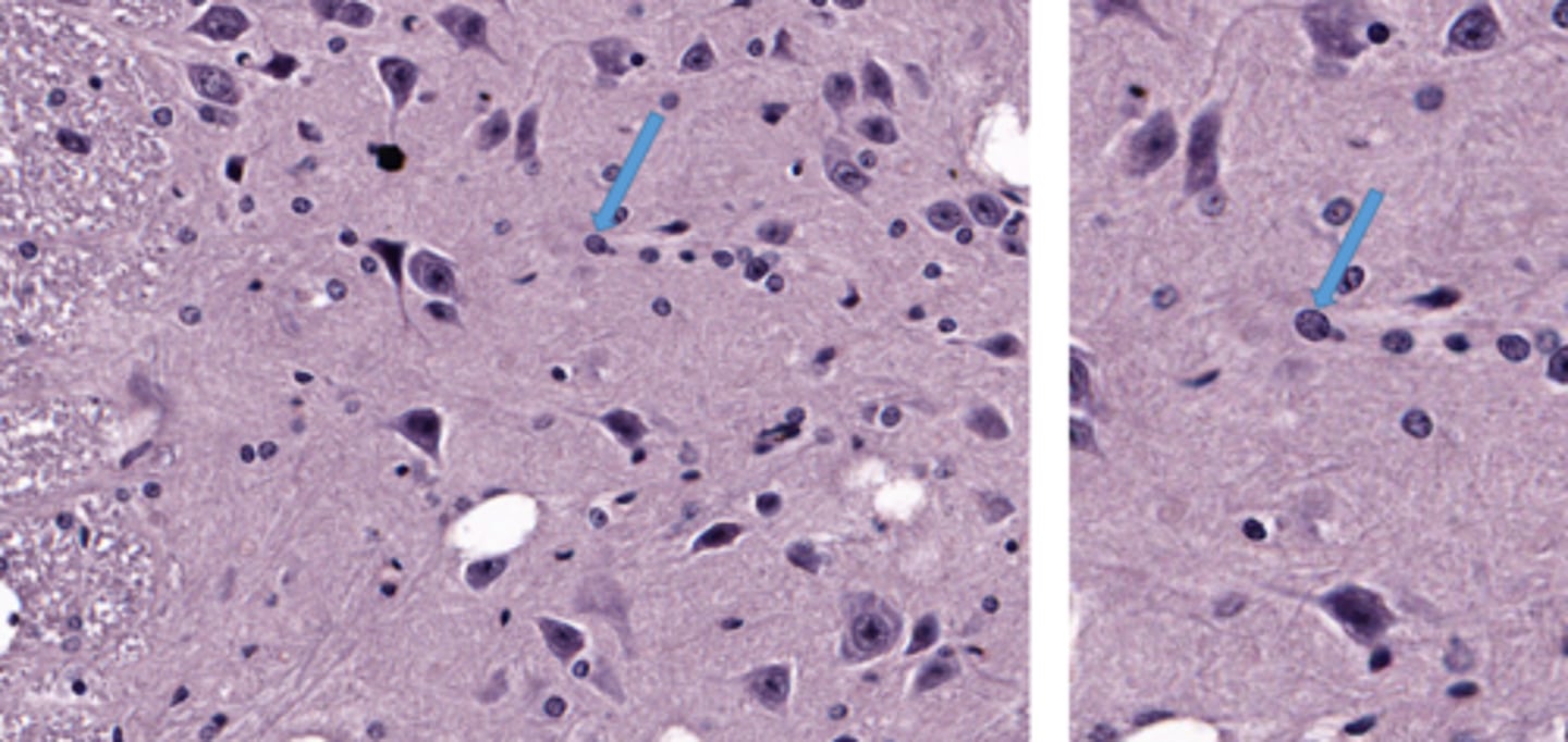
Identify the glial cell
astrocyte
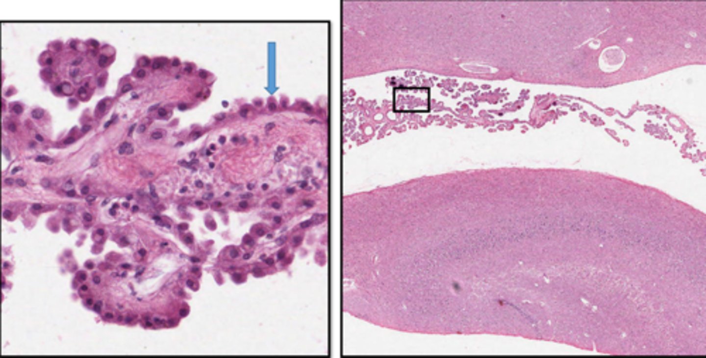
Identify the cuboidal cells on the surface of this choroid plexus
ependymal cell
Identify the glial cell
microglial cell
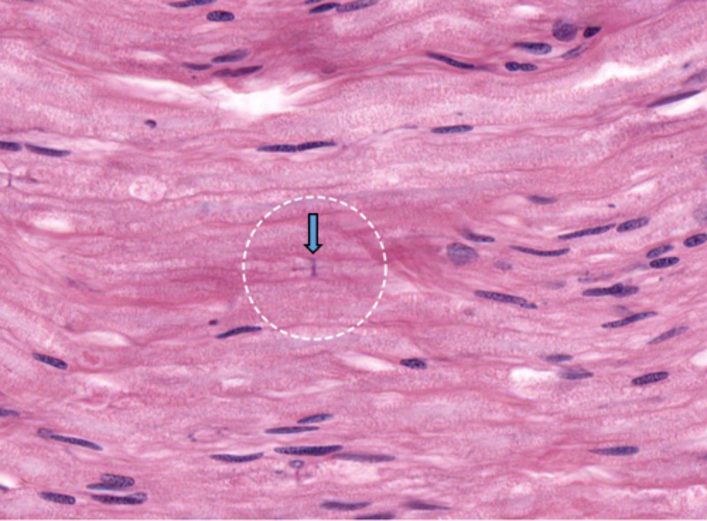
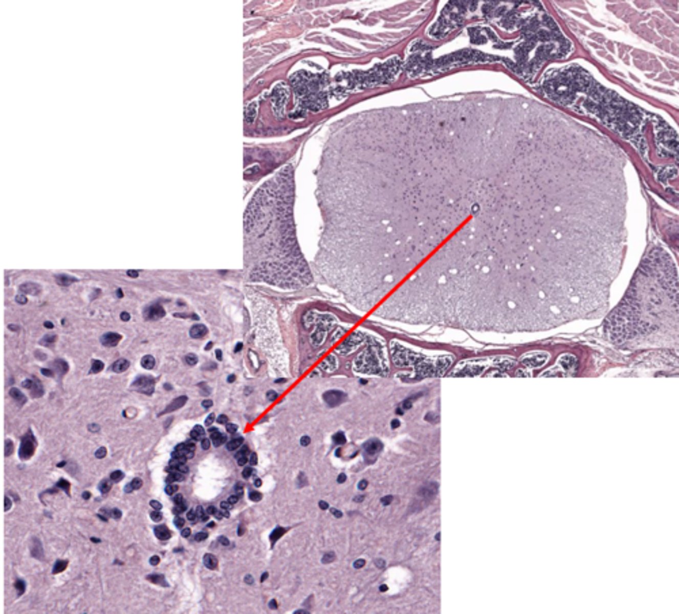
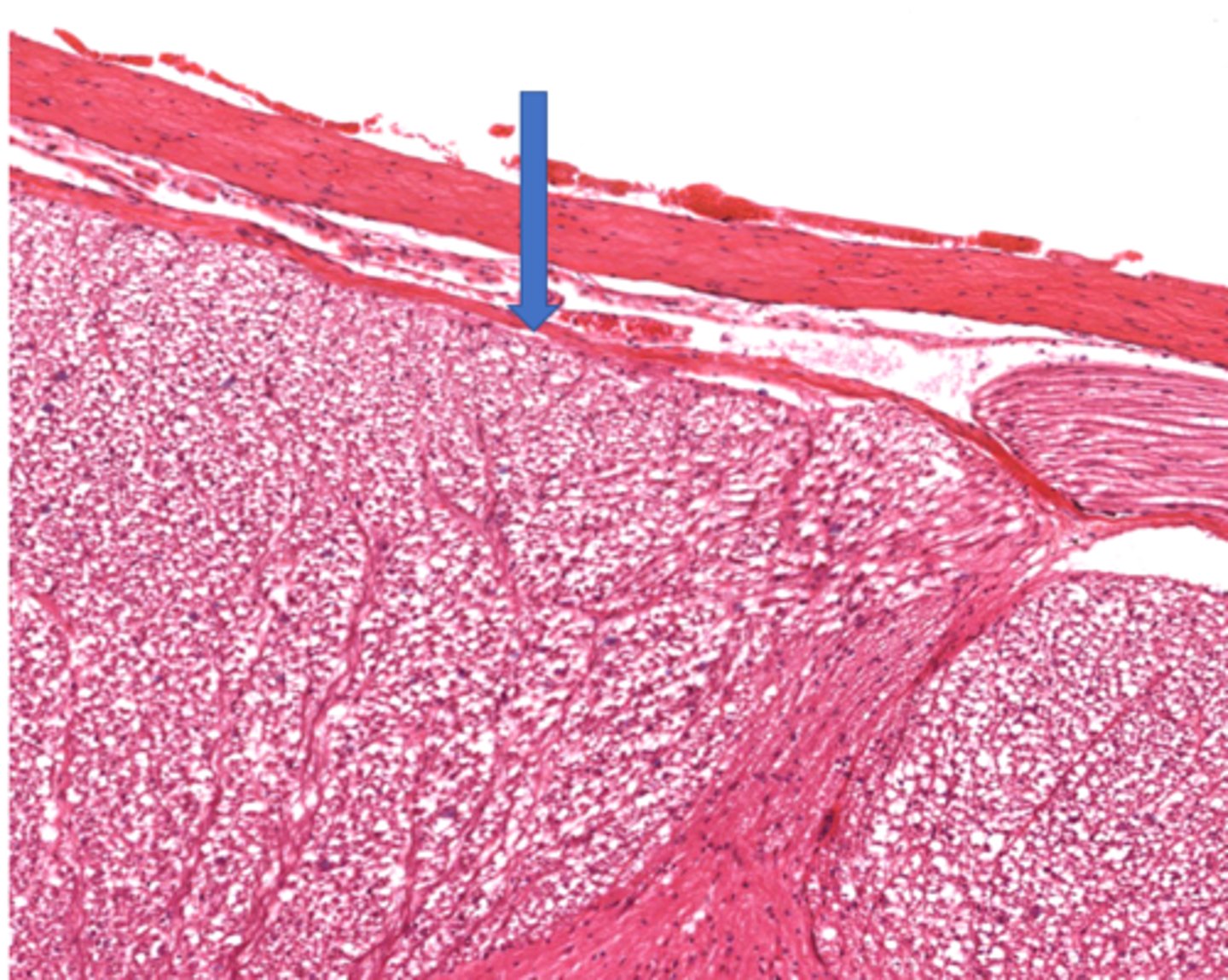
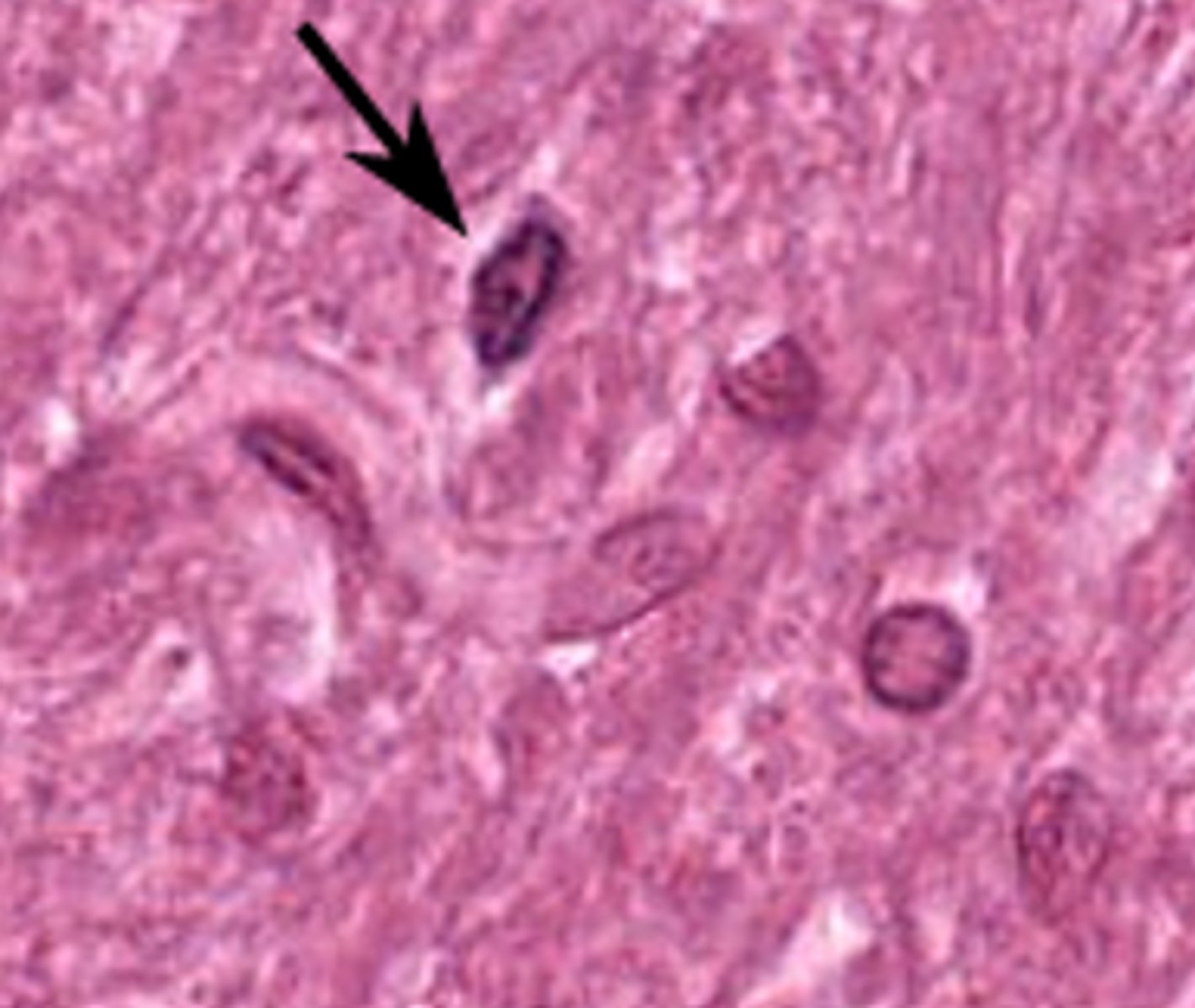
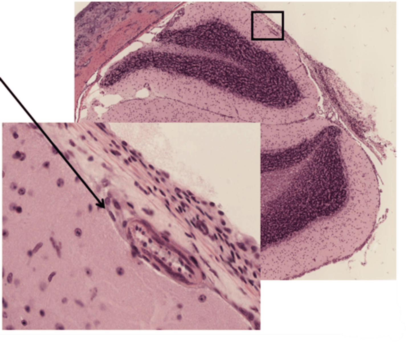
The structure at the arrow represents the junction between adjacent cells of what type
schwann cell
Identify this cell
satellite cell
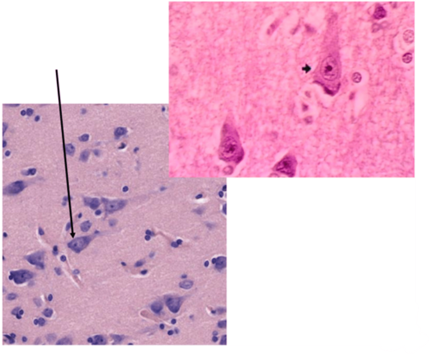
Identify this cell
pyramidal cell
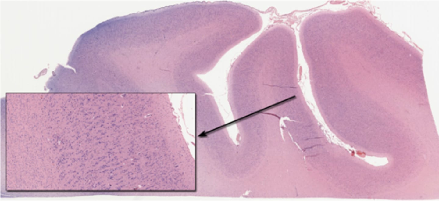
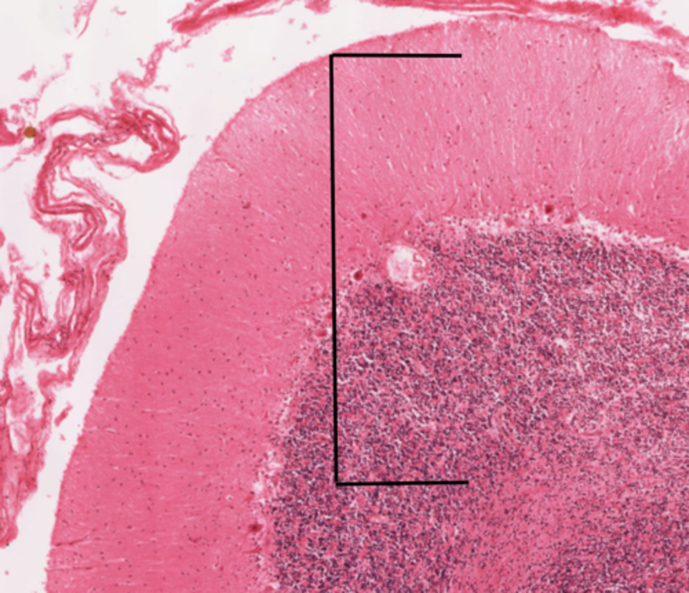
Identify the specific part of the brain
cerebrum
Identify the portion of the CNS
cerebellum
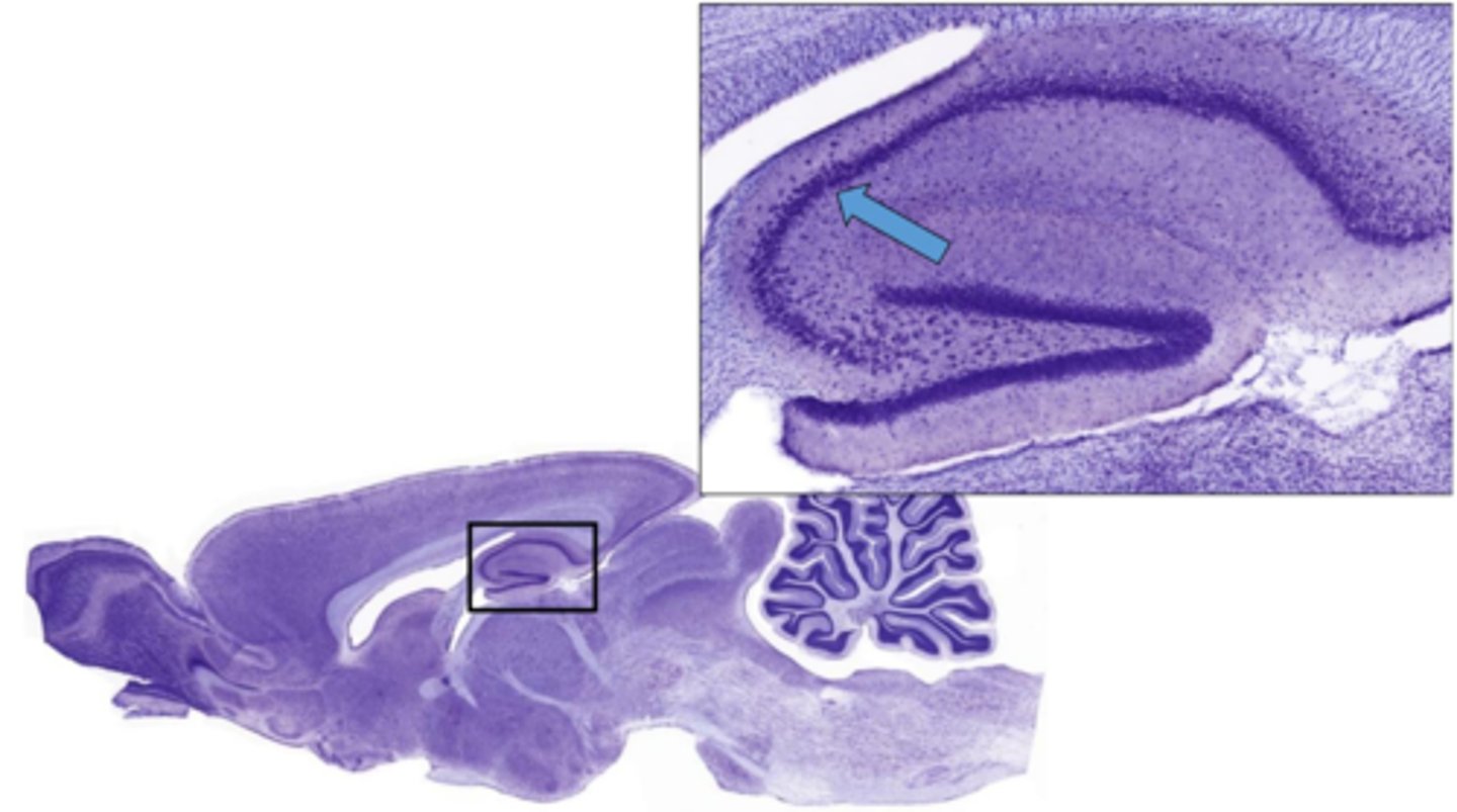
Identify the specific part of the brain in the inset
hippocampus
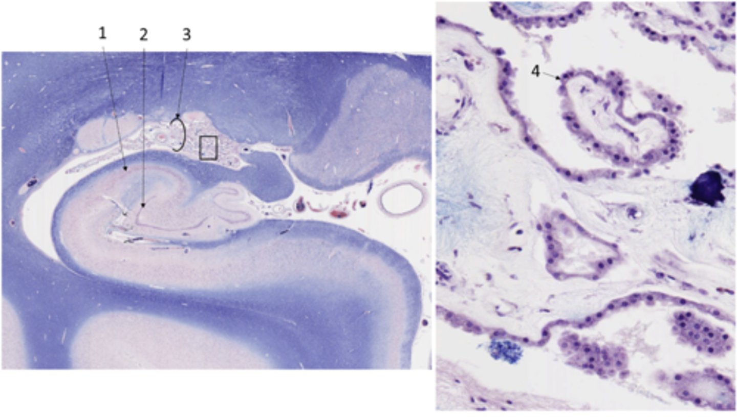
Identify the structure at 3 (enlarged at the right)
choroid plexus
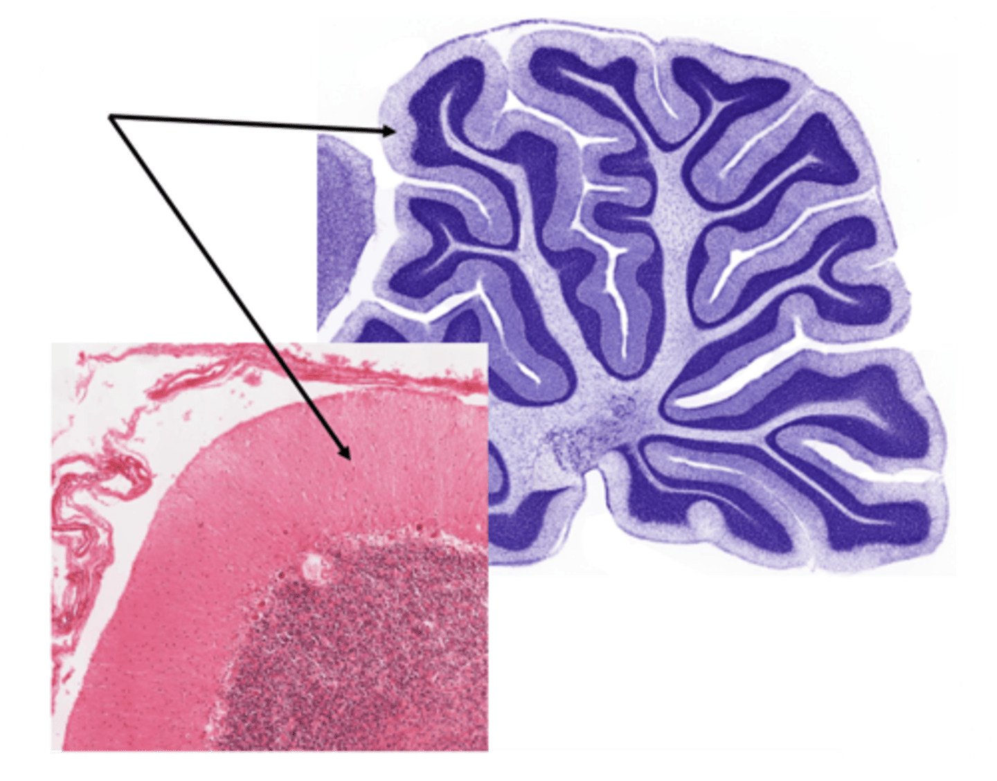
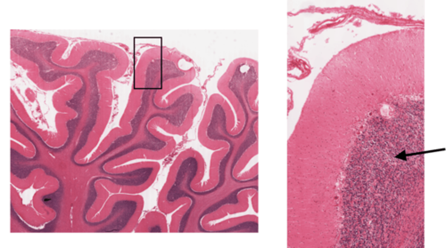
Identify the layer of the cerebellum
molecular layer
Identify the layer of the cerebellum
white matter
Identify the layer of the cerebellum
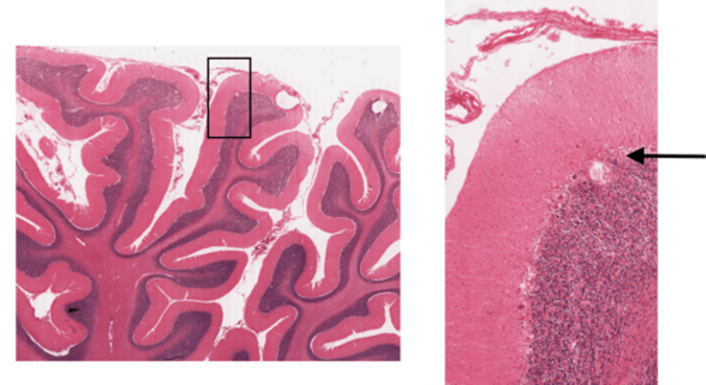
purkinje layer
Identify the layer of the cerebellum
pia mater
Identify the layer of the cerebellum
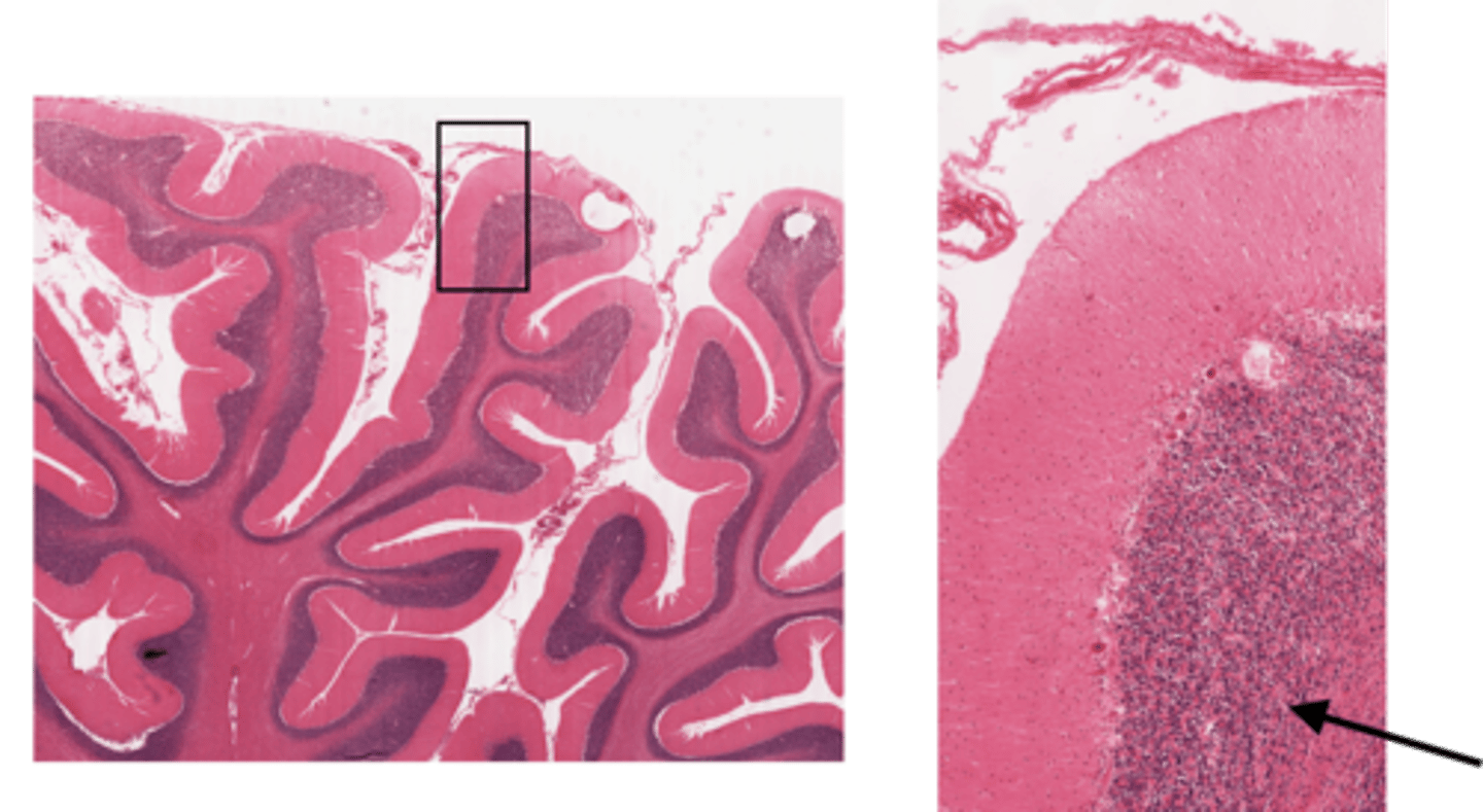
granular layer
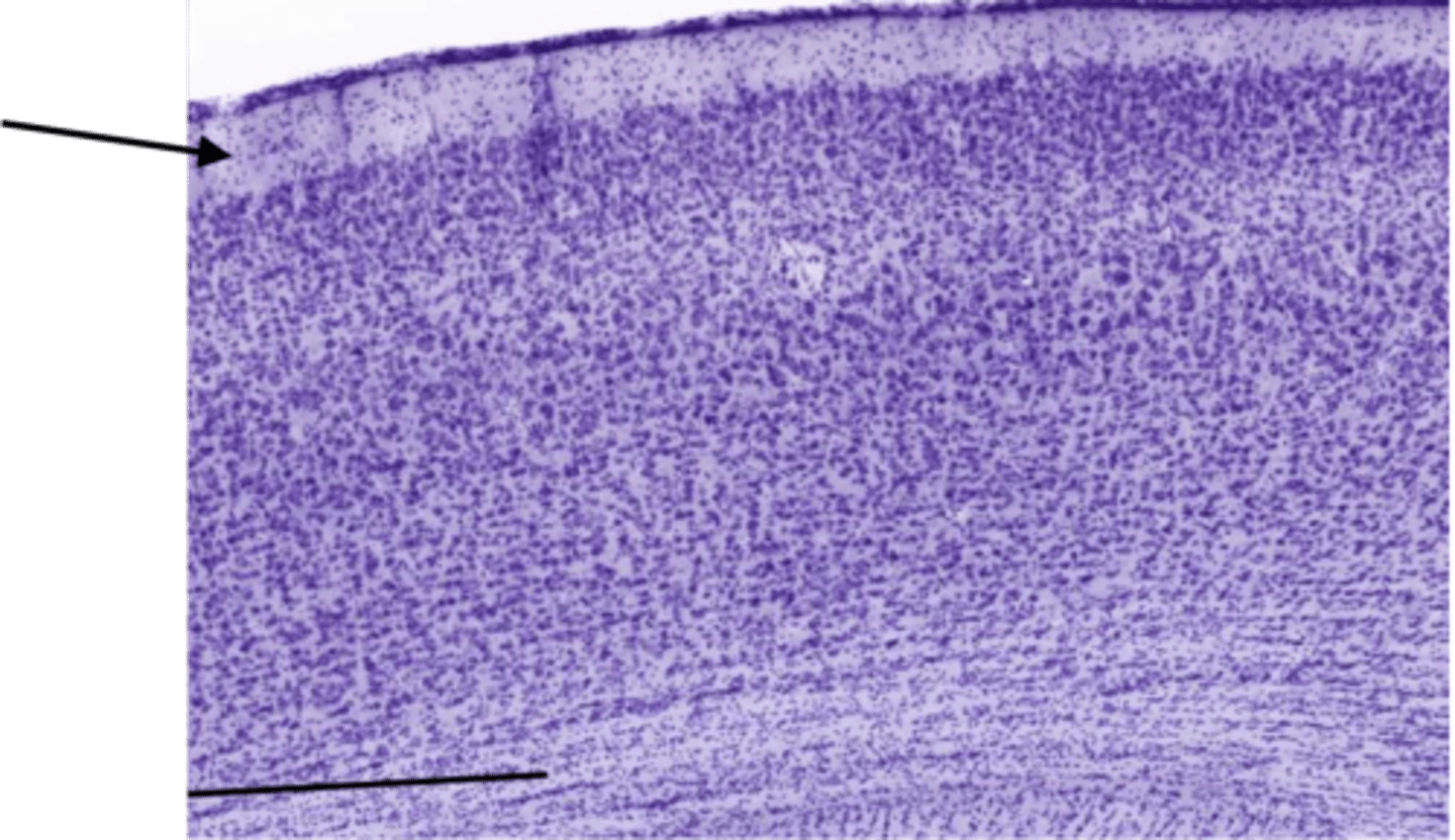
Identify the layer of the cerebrum
molecular layer
Identify the layer of the cerebrum
pia mater
Identify the layer of the cerebrum
arachnoid
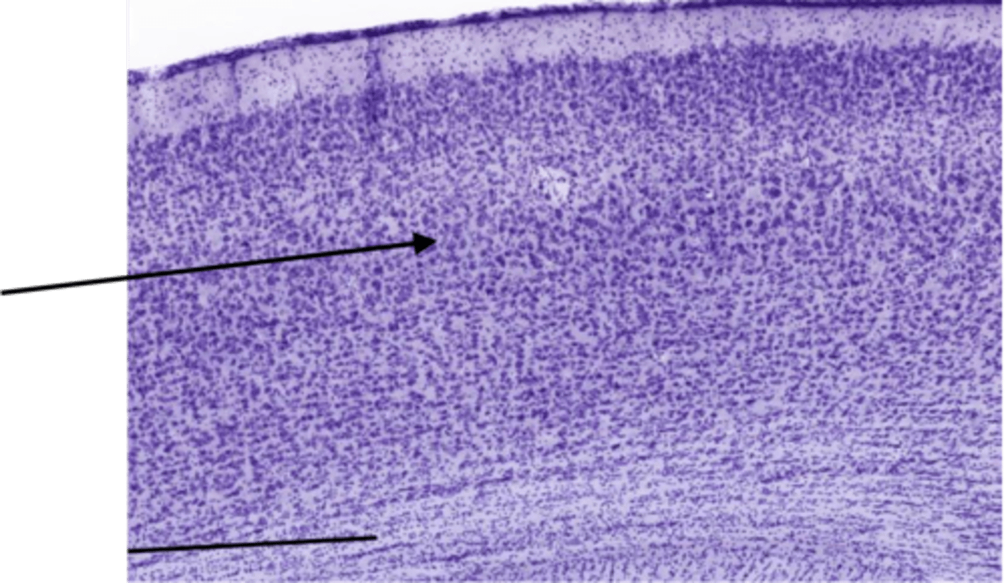
Identify the layer of the cerebrum
pyramidal layer
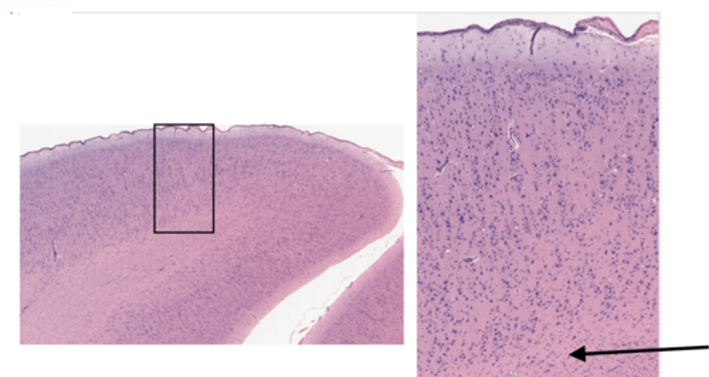
Identify the layer of the cerebrum
white matter
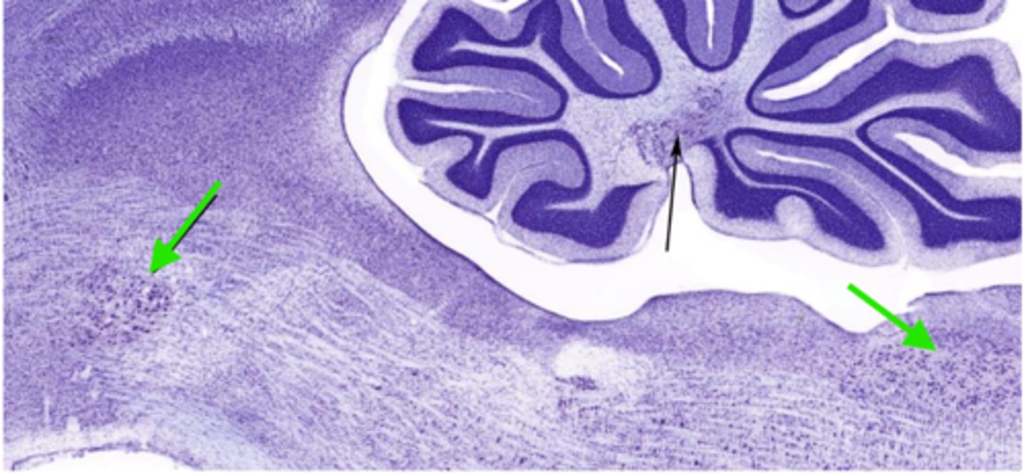
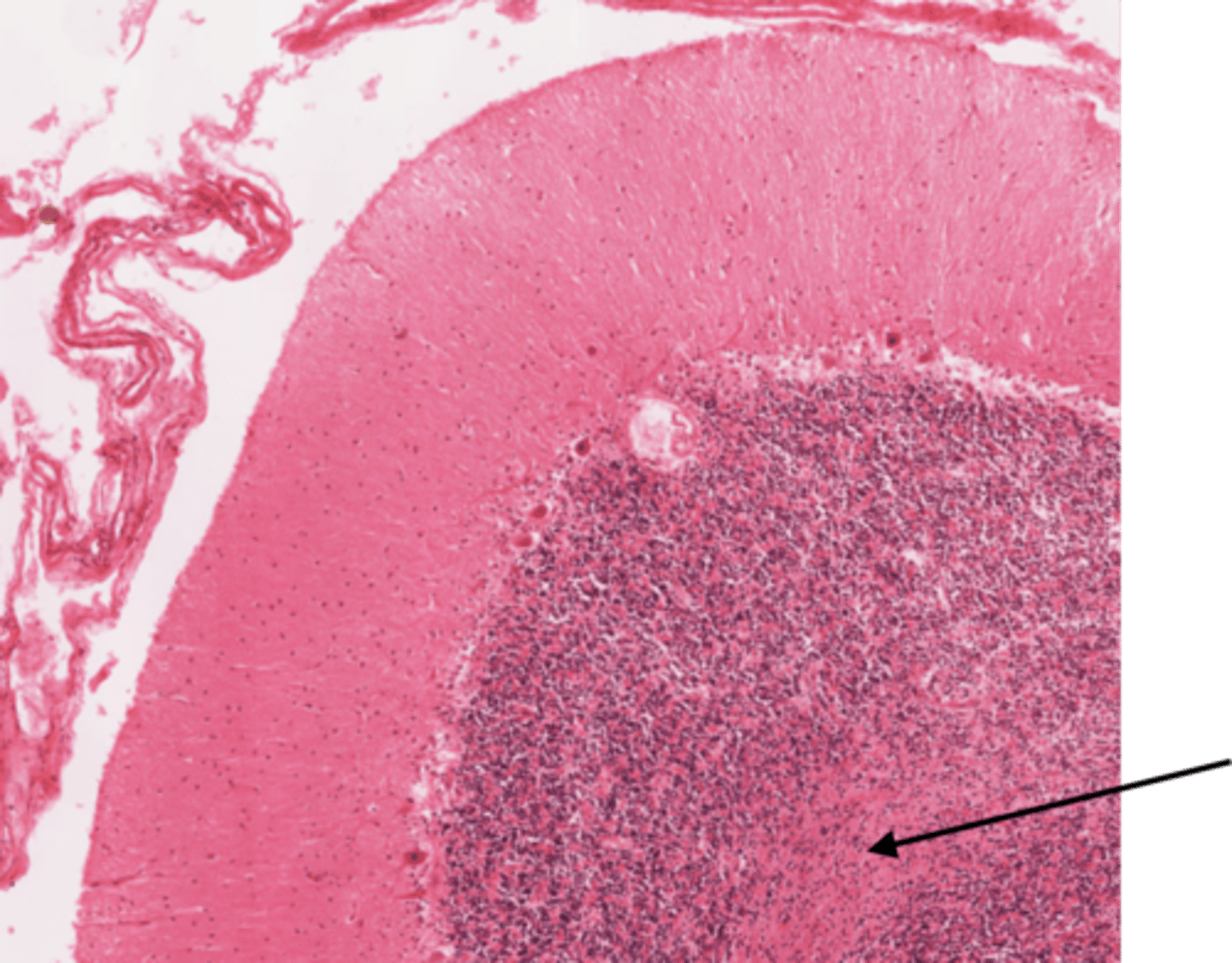
Identify the cluster of neurons at the green arrows
nucleus of the brainstem
Identify the cluster of neurons circled
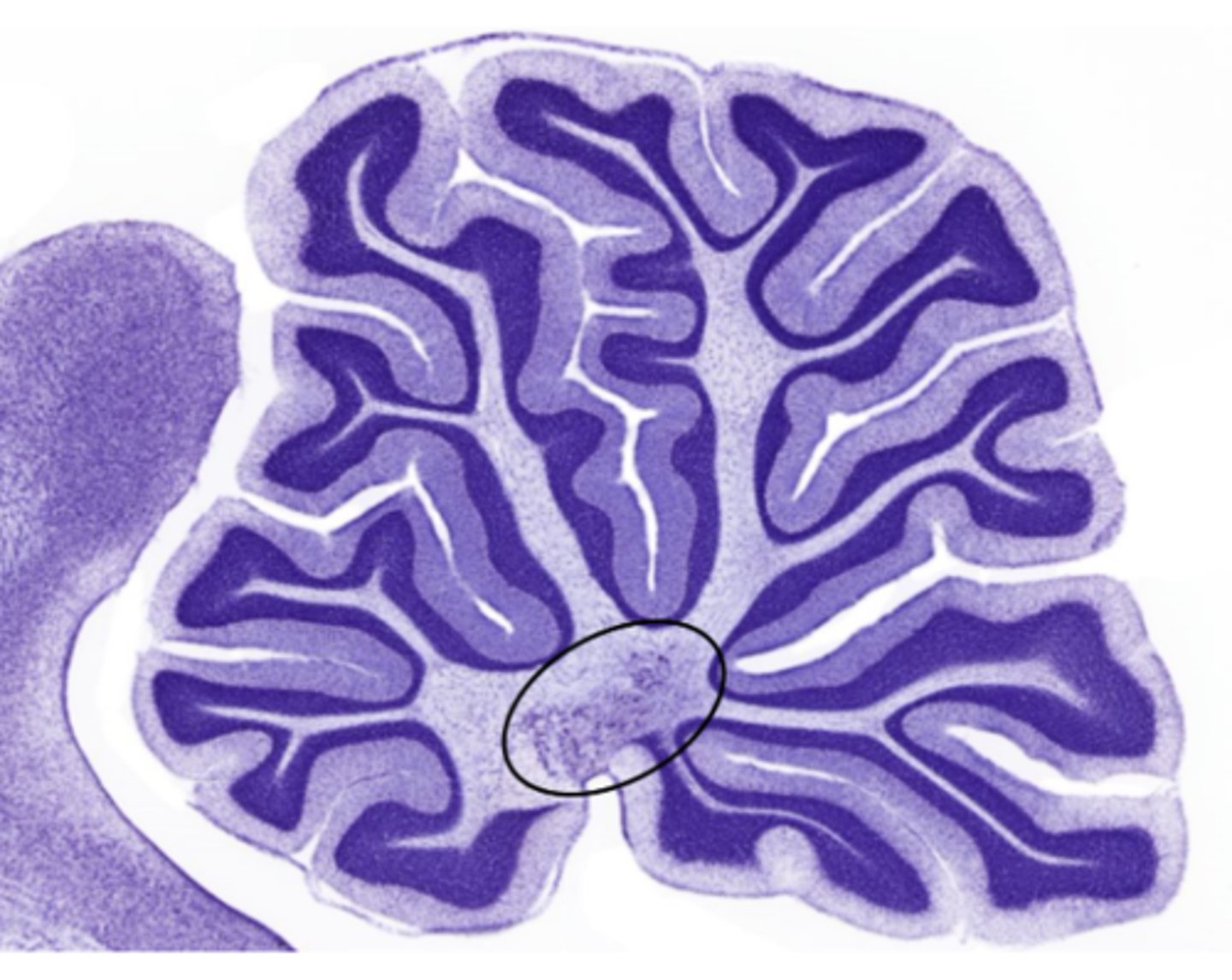
nucleus of the deep cerebellum

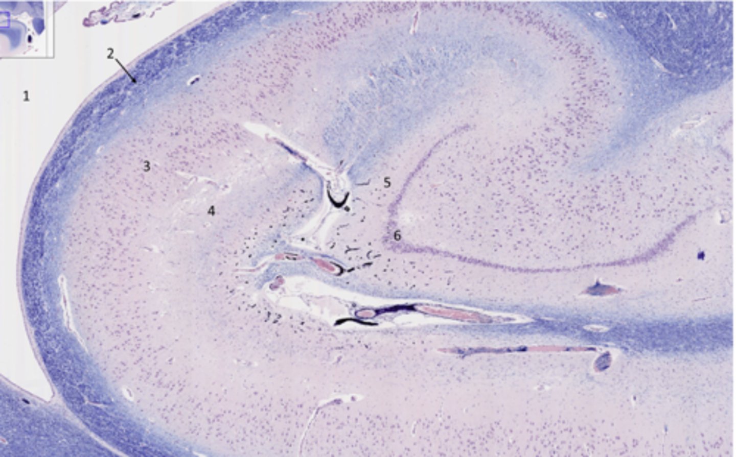
What part of the hippocampus is at 5 and 6
dentate gyrus
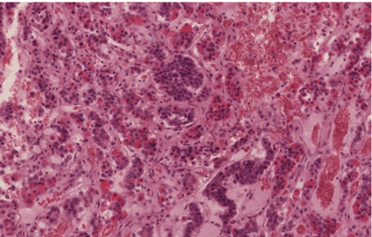
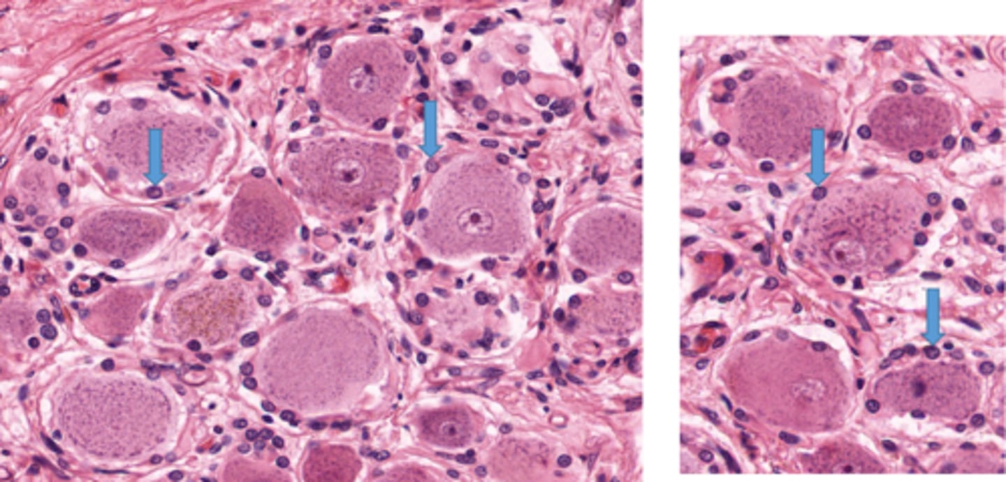
What are the hallmarks of endocrine organs?
cords and clusters of cells and extensive capillary network
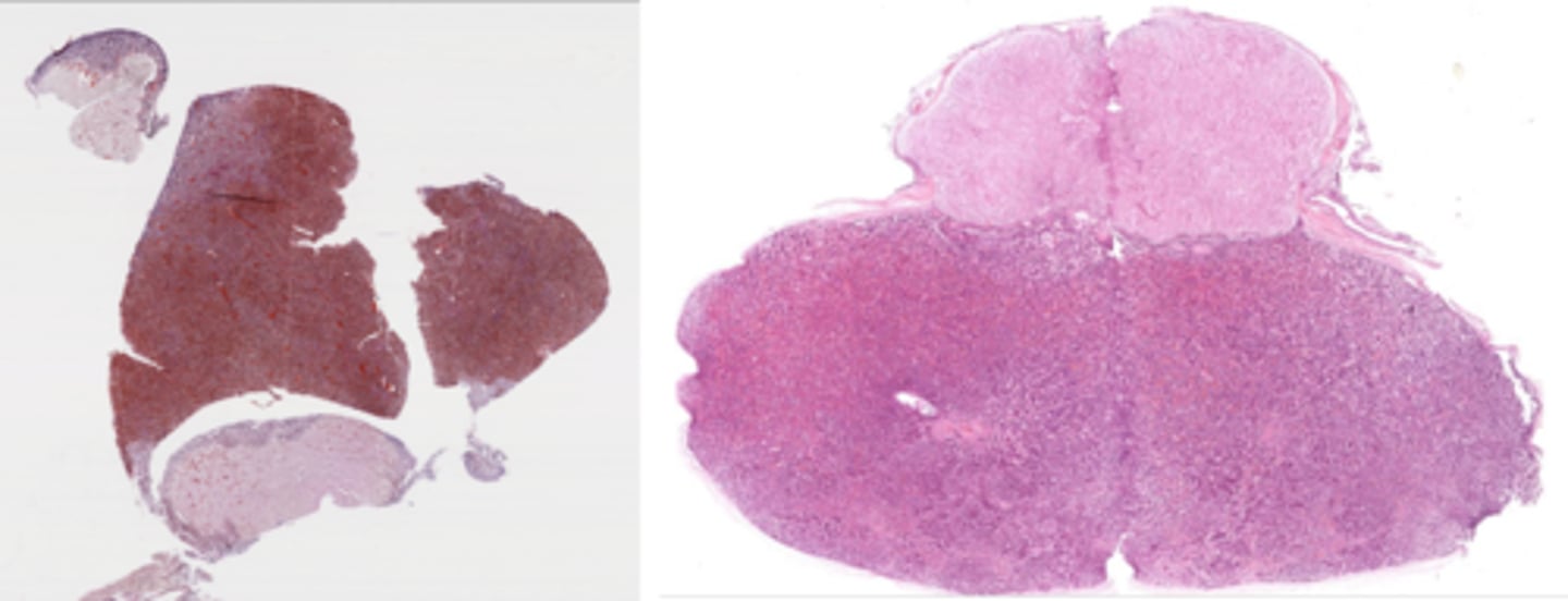
Identify the organ
pituitary
Identify the organ
adrenal gland
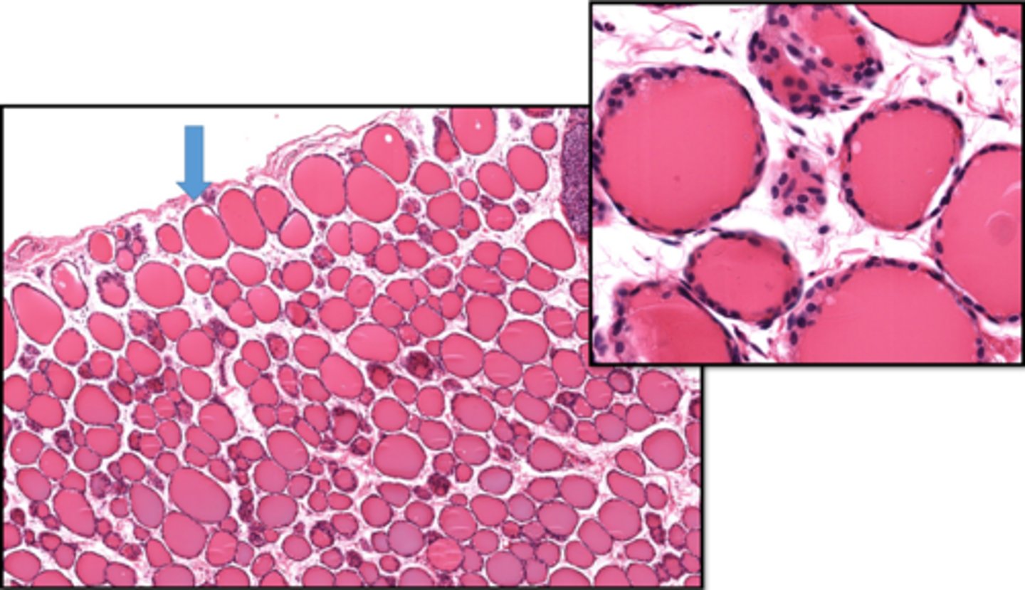
Identify the organ
thyroid