Hock and Distal Limb Muscles (Week 3, Mod 7)
1/19
There's no tags or description
Looks like no tags are added yet.
Name | Mastery | Learn | Test | Matching | Spaced |
|---|
No study sessions yet.
20 Terms
How many rows of bone are in the tarsus? List the names of the bones in each row, from LATERAL to MEDIAL
There are THREE rows of bone (not 2 like the carpus)
Proximal row:
Talus
Calcaneus - PALPABLE
Middle row:
Central + 4th tarsal bone
Distal row:
1st, 2nd, 3rd, and 4th tarsal bone
4th TB bridges the middle and distal rows
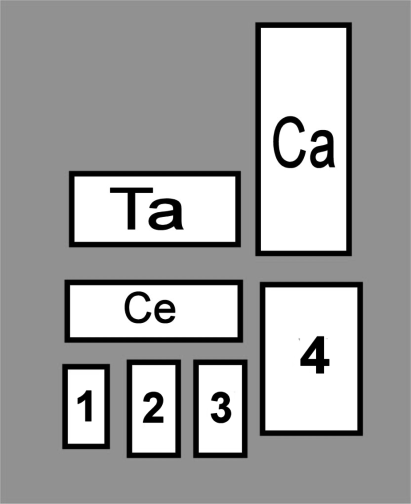
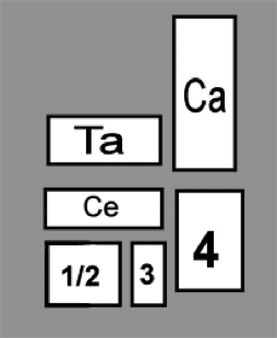
How is the horse tarsus different from the canine tarsus? Think of 2 differences
1) 1st and 2nd TB fused
2) 3rd TB is very large
Articulates with the 3rd metatarsal, which is weight bearing in this species
How many joints are within the hock? Name them (proximal to distal), and list which bones are involved in each.
1) Tibio-tarsal joint (TT joint):
Talus + tibia & fibula
LARGE range of movement here (flexion and extension)
Includes the calcaneus to some degree, but is not articular here
Main function is a lever, not for movement within the joint
Trochlea of the talus are here
2) Proximal Intertarsal joint (PIT joint):
Talus & calcaneus + central + 4th TBs
3) Distal Intertarsal joint (DIT joint):
Central + 1, 2, and 3rd TBs
4) Tarso-Metatarsal joint (TMT joint):
1, 2, 3, and 4, TBs + the metatarsal bones
*** REMEMBER: between rows, its a fibrous joint, so not much movement
Describe the range of motion within the hock… How well can it flex and extend? What is unique about the horse hock that may increase its range of motion?
Has a large range of flexion at the tibio-tarsal joint… little movement at all of the other joints.
In the horse, the trochlea of the talus are NOT VERTICAL… more diagonal, allowing for rotation
Essentially, this means that when the hock is flexed, instead of the distal limb folding in line with the rest of the limb, it will flare out laterally
Prevents “over-reach” injuries, meaning that the horse doesn’t kick its front limbs on accident while sprinting
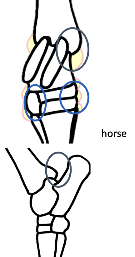
What kind of joint is the hock? What are 2 key features of this joint? (are similar to the stifle…)
The hock is a typical synovial joint…
Has and EXTENSIVE joint capsule
Poor communication between compartments
What areas of the horse hock are palpable? Think of 2 main regions, and where on those regions they can be palpated. Why is this important clinically?
Tibio-tarsal joint
Is separate from the rest of the hock, so has different points that are palpable:
Dorso-medial aspect
Plantaro-lateral aspect
Plantaro-medial aspect (CALCANEUS BONE)
Distal regions of the joint:
Dorsal-medial aspect
Dorso-lateral aspect
Is helpful clinically in diagnosing where effusion may be coming from
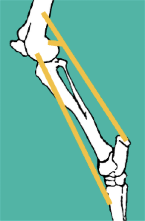
What 4 features contribute to the stability of the hock?
1) Collateral ligaments
Long ligaments: Tibia → metatarsal bones
Short ligaments: bridges bone → bone (in yellow)
2) Fibrocartilagenous plate reinforcement of joint capsule on the PLANTAR aspect
Comes with an accessory check ligament
3) Retinaculum
4) Plantar ligament
Attaches from tip of calcaneus bone to the metatarsals, much like the palmar ligament of the carpus
What is the clinical significance of damage at the tarsus joint in dressage horses?
Since they are encouraged to bear more weight on their hindlimbs, rather than their forelimbs, this can cause damage at the tarsus joint
How many centers of ossification to the tarsal bones have? Who is the exception?
All have a SINGLE ossification center
EXCEPT for the calcaneus bone → has 2
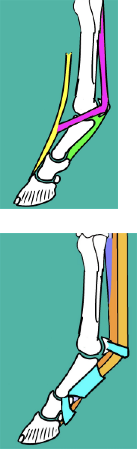
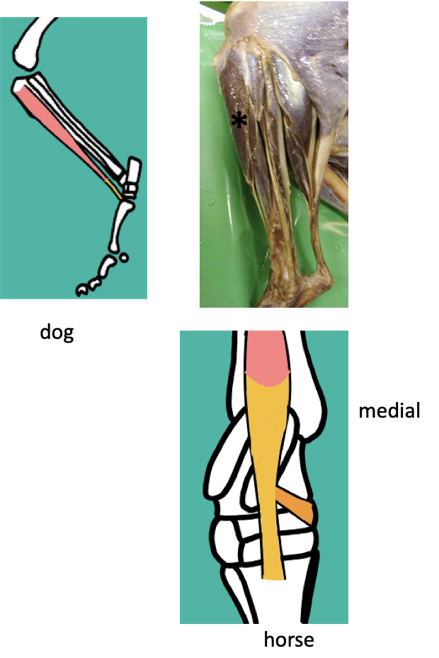
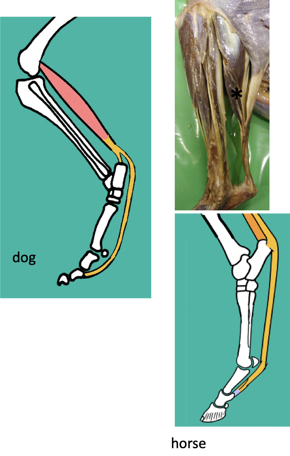
There are 3 muscles associated with the cranial region of the distal limb… what are their names?
1) Cranial tibial muscle
2) Peroneus muscle
Dog and cat → peroneus longus (long fibular muscle)
Horse → peroneus tertius (third fibular muscle)
3) Long digital extensor muscle
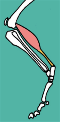
Describe the cranial tibial muscle… origin, insertions, function, and nerve supply.
Origin - proximal tibia
Insertions - has TWO possible insertions… depend on species
Metatarsal bones (all species)
Medial aspect of hock (via cunean tendon) in the HORSE, as well as inserting at the metatarsal bones
Function - hock FLEXION
Nerve supply - Peroneal / fibular nerve (branch of SIATIC)
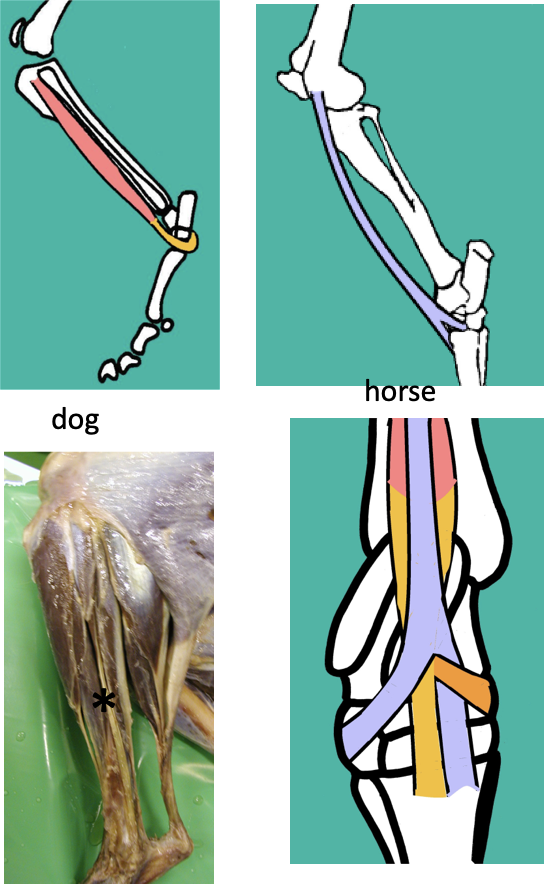
Describe the peroneus muscle… what are the two different kinds? Know what species they belong to, the origins, insertions, function, and nerve supply.
a) Peroneus longus
DOG AND CAT
Origin - lateral tibia & fibula
Insertion - Plantar aspect of tarsus; wraps around from the front to back
b) Peroneus tertius
HORSE ONLY
Origin - Lateral femoral condyle
Insertion - Has TWO
3rd metatarsal (WITH cranial tibial muscle)
LATERAL aspect of the tarsus
Essentially inserts the opposite way from the cranial tibial muscle, forming a sort of girdle around the hock
Is more of a fibrous cord than an actual muscle belly
FUNCTION - hock FLEXION
Nerve supply - Peroneal / fibular nerve (branch of SIATIC)
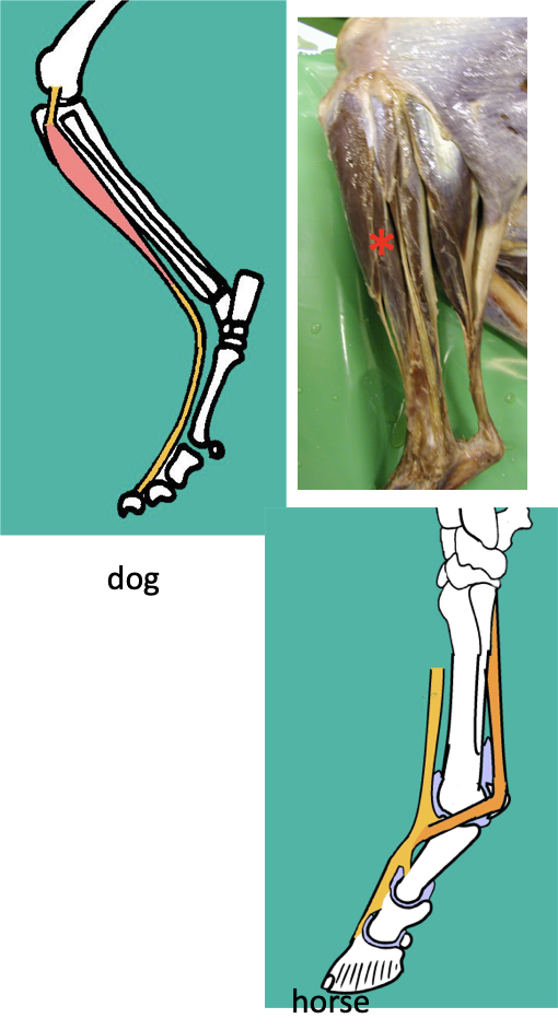
Describe the long digital extensor muscle… origin, insertion, function, and nerve supply.
Origin - extensor fossa (in the FEMUR)
Tendon of origin is incorporated into the stifle joint capsule
Provides LATERAL collateral support to stifle
Insertion - ALL digits (the distal phalanx precisely)
in the horse, the joints form little bursas as the tendon runs dorsally to the digit; done for cushioning and protection, same as the forelimb
Function - hock FLEXION, and digit EXTENSION
Nerve supply - Peroneal nerve
CAUDAL ASPECT OF LIMB
What exactly is the common calcanean tendon?? What 5 muscles is it made up of? Where does it insert, what is its function, and what nerve supplies it?
The calcanean tendon IS THE ACHILLES TENDON
Composed of:
Biceps femoris
Semitendinosis
Gracilis
Gastrocnemius
Superficial Digital Flexor
Insertion - Calcaneus bone
AKA calcanean tuberosity
Acts as a lever for the distal limb
Plantar ligament ALSO attaches here to provide support
Function - hock EXTENSION
Nerve supply - All nerves of the contributing muscles
Describe the Gastrocnemius muscle… origin, insertion, function, and nerve supply.
Origin - Femur
Has 2 tendons of origin
Fabellae (sesamoids of the distal portion of the femur
Insertion - calcaneus bone
is the MOST SIGNIFICANT contributor to the calcanean tendon
Function - hock EXTENSOR, AND acts as a stifle FLEXOR (also crosses behind stifle)
Nerve supply - Tibial nerve (branch of SIATIC)
** Would be comparable to the calf muscle in humans
Describe the Superficial digital flexor muscle… origin, insertions, function, and nerve supply.
Origin - Distal femur (WITH gastrocnemius muscle)
Insertion - has MULTIPLE
Calcaneus bone (part of calcanean tendon)
Branches to ALL DIGITS (middle phalanx precisely)
Function - hock EXTENSION w/ digital FLEXION
Also provides support of the distal limb joints in extension
No accessory check ligament necessary (especially because of calcanean tendon)
Nerve supply - Tibial nerve
Describe the Deep digital flexor muscle… origin, insertion, function, and nerve supply
Origin - tibia
Runs over tarsus to insert at the digits
Is NOT a part of the calcanean tendon
Insertion - ALL digits (distal phalanx)
Function - hock EXTENSOR and a digital FLEXOR
Also functions to support the distal limb joints in extension (has weak accessory check ligaments)
Nerve supply - Tibial nerve
Describe the concept of the hindlimb stay apparatus… where in the hindlimb do horses have “stay apparatuses” in place?
Have them at the level of the stifle (patellar locking mechanism) and the hock (reciprocal apparatus)
Essentially keeps the limbs in the natural weight bearing position of extension
Is able to keep one hindlimb in extension while the other rests, due to hindlimbs not being the weight bearing limbs
What is the reciprocal apparatus composed of? How does it work to keep the limb in extension?
Reciprocal apparatus -> peroneus tertius at cranial limb, superficial digital flexor at caudal limb
Acts as a pulley system
Connects the stifle directly to the tarsus… if the stifle is extended, tarsus must also be extended; if its flexed, tarsus also must be flexed
CAN’T move independently
If the reciprocal apparatus takes care of the proximal limb, then what stay mechanisms are at the distal aspect of the limb? What is their function?
Overall function: prevention of collapse into hyperextension
MTP joint:
Suspensory apparatus:
Suspensory ligament
Proximal sesamoids
Distal sesamoidean ligaments
Long digital extensor
Purpose of suspensory apparatus; same as forelimb
Also has a suspensory ligament that runs down the plantar aspect of the THIRD metatarsal, then wraps around dorsally to attach to long digital extensor muscle (like the common digital extensor of the forelimb)
At the proximal and distal interphalangeal joints:
The SDFT and DDFT provide enough support; these are paired with annular ligaments (in blue) which gives extra stability to the tendons