L8 - How are Proteins Studied?
1/18
There's no tags or description
Looks like no tags are added yet.
Name | Mastery | Learn | Test | Matching | Spaced | Call with Kai |
|---|
No analytics yet
Send a link to your students to track their progress
19 Terms
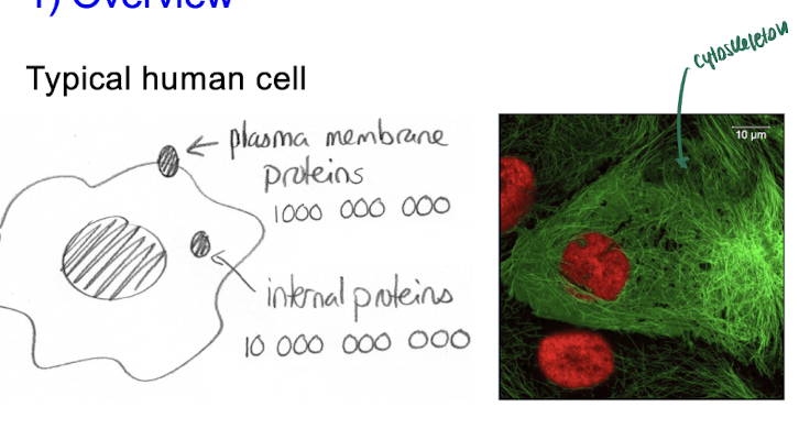
Typical human cell
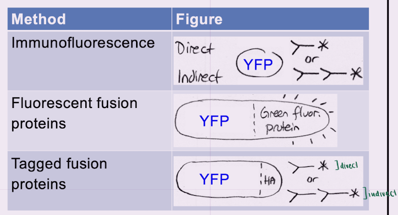
3 Methods of studying Proteins
immunofluorescence (direct/indirect)
= antibodies covalently attached to fl. dye
fluorescent fusion proteins
= gene + fl. protein
tagged fusion proteins (direct/indirect)
= gene + HA-tag + antibodies for tag
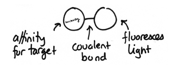
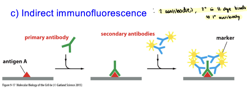
Method 1 : Immunofluorescence
uses a fluorescent probe = antibodies covalently attached to fl. dye
→ antibodies = small proteins made by B-cells
types: IgG (monomer), IgA (dimer) , IgM (pentamer)
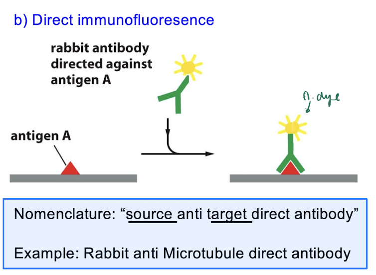
Method 1 : Immunofluorescence - Types
Direct immunofluorescence = probe directly attached to antigen
nomenclature: “source anti-target antibody”
ex. “rabbit anti-microtubule direct antibody”
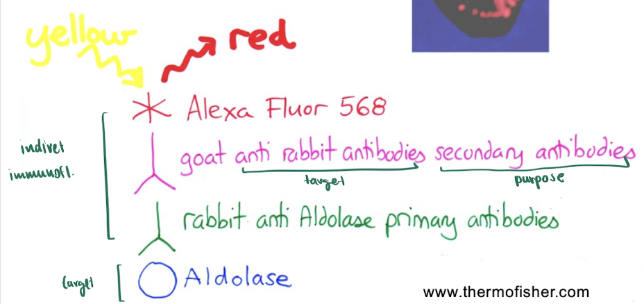
Indirect immunofluorescence = 2 antibodies, secondary w fl. dye binds to primary antibody
more steps, BUT more fluorescence
if it says “source anti-animal ….” : a secondary antibody
if it says “source anti-protein/organelle …” : a primary antibody
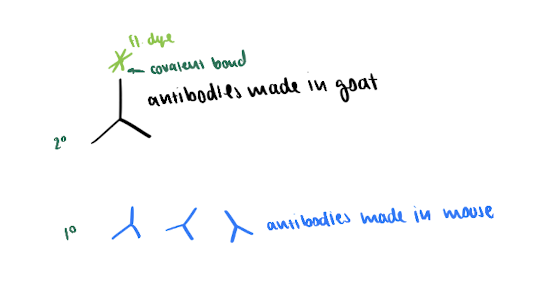
Goat anti-mouse antibody, Alexa Fluor 488 conjugate
these are ___ antibodies made in a __?
secondary antibodies made in a goat
bc it says “anti-animal”, NOT “anti-protein/organelle”
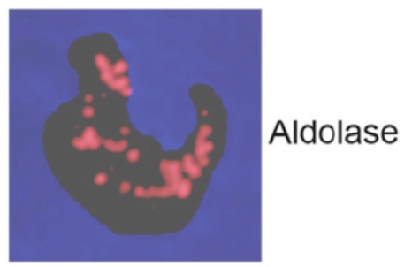
What is red?
Red is adolase, one of the glycolysis enzymes
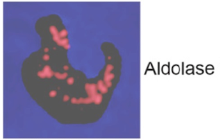
How did they make adolase red?
via indirect immunofluorescence
(answer in image)
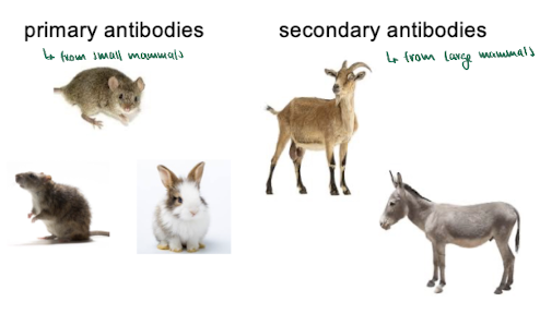
obtaining antibodies from mammals
primary antibodies : from small mammals
secondary antibodies : from larger mammals
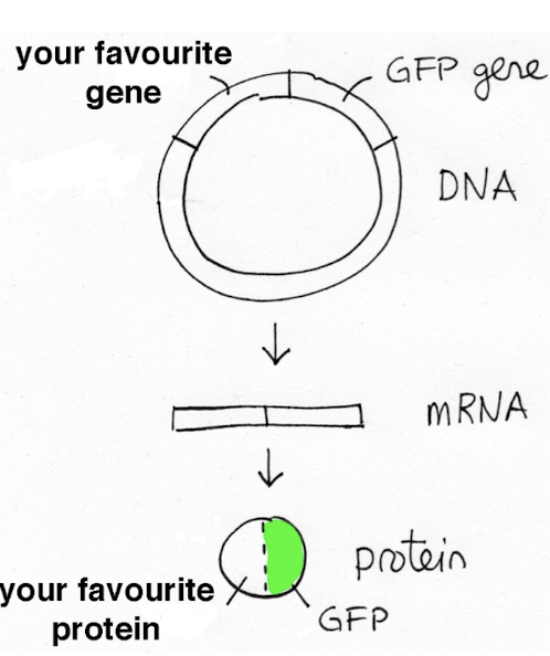
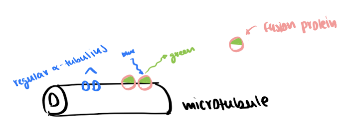
Method 2 : Fluorescent Fusion Proteins
= gene + fl. protein
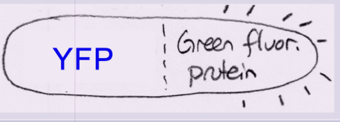
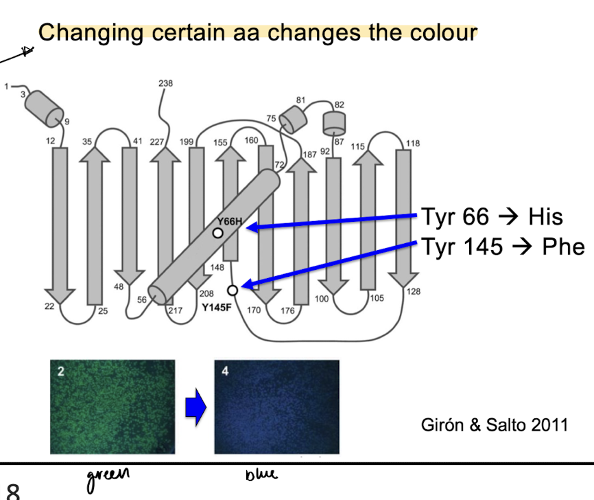
fl. protein is GFP from jellyfish → can change proteins colour with a change in AA
easiest way to detect a new protein
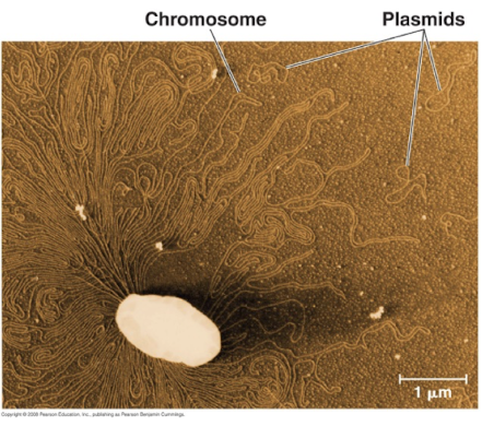
Method 2 : Fluorescent Fusion Proteins - Plasmids
natural plasmids = circles of DNA found in bacteria
recombinant plasmids = made by scientists and put into animal cells via temporary transfection
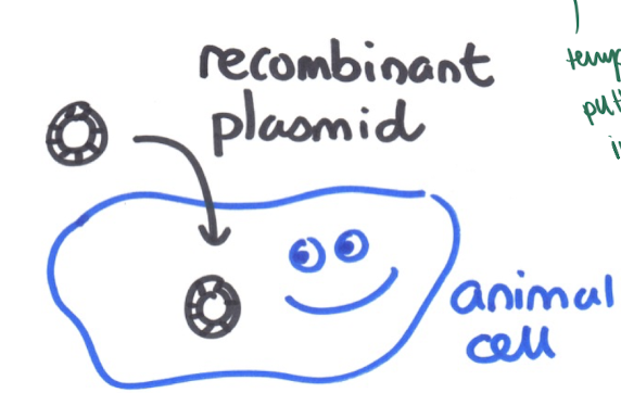
Method 2 : Fluorescent Fusion Proteins - making fusion proteins
make recombinant plasmid
transfect the cells w plasmid
proteins are synthesized inside the cells
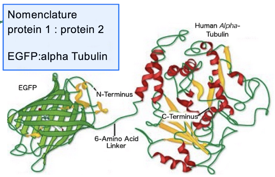
nomenclature: protein 1 : protein 2
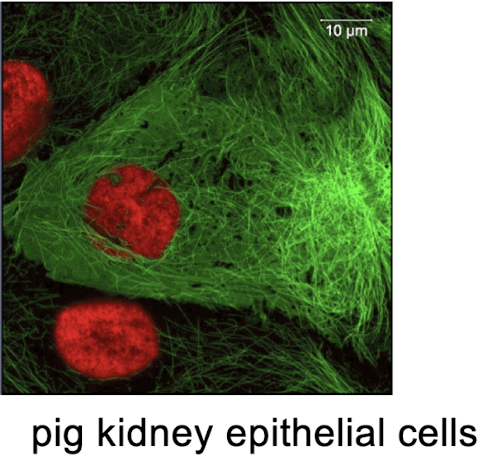
What microscope was used? what is green? what is red?
microscope: confocal fluorescence
green : microtubules
red : chromosomes
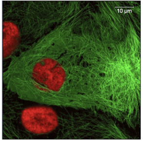
How did they make the microtubules green?
using a fluorescent fusion protein ( EGFP : alpha tubulin )
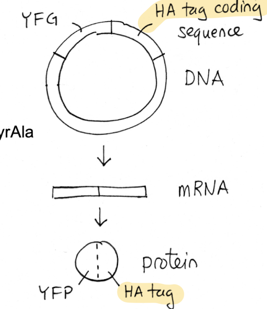
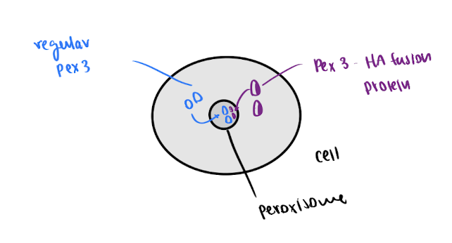
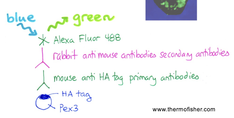
Method 3 : Tagged Fusion Proteins
= gene + HA-tag + antibodies for tag
w/o antibodies for target protein: add HA-tag to plasmid → use anti-HA antibodies
tag adds to either end of protein ( N or C )
protein tags : FLAG, HA, His, Myc
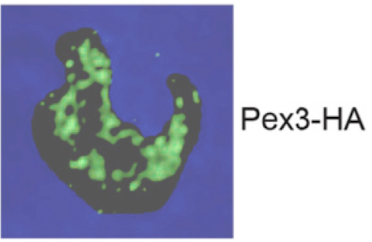
What is green?
green is Pex3, a peroxisome protein
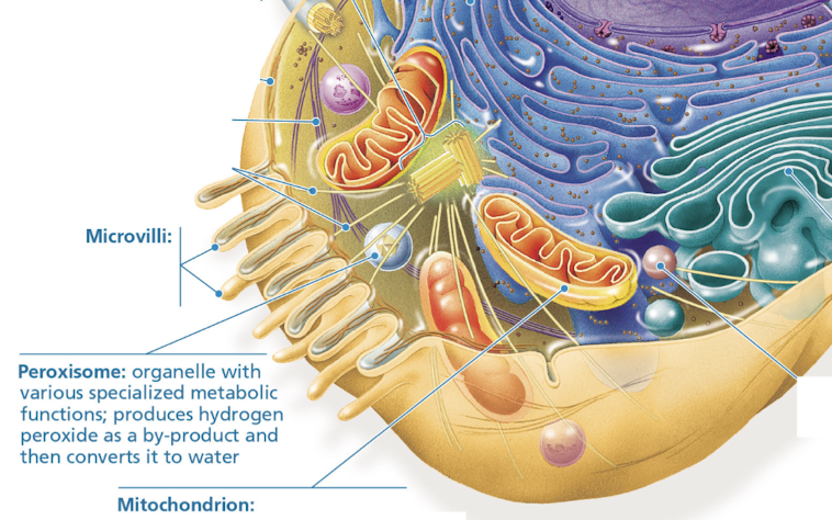
peroxisomes
= small round organelles that import protein from cytosol
use tagged fusion proteins to study
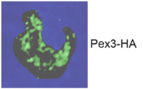
How did they make the peroxisome protein green?
using tagged fusion proteins and Alexa Fluor 488 as fl. dye
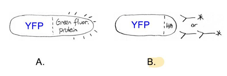
which of these fusion proteins is most likely to go to its proper cellular location?
B, the tagged-fusion protein
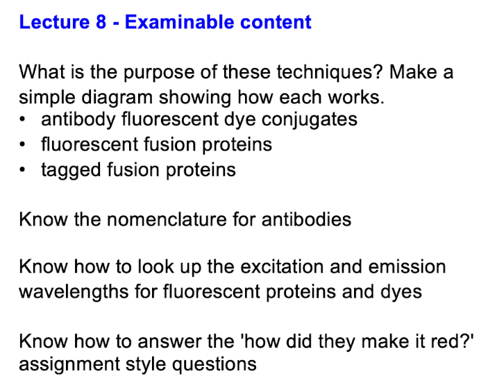
Examinable content