(7VC) regulation of bp by other systems
1/57
There's no tags or description
Looks like no tags are added yet.
Name | Mastery | Learn | Test | Matching | Spaced | Call with Kai |
|---|
No analytics yet
Send a link to your students to track their progress
58 Terms
Describe the Renin-angiotensin system for bp regulation (make sure to note where enzymes come from and what they do)
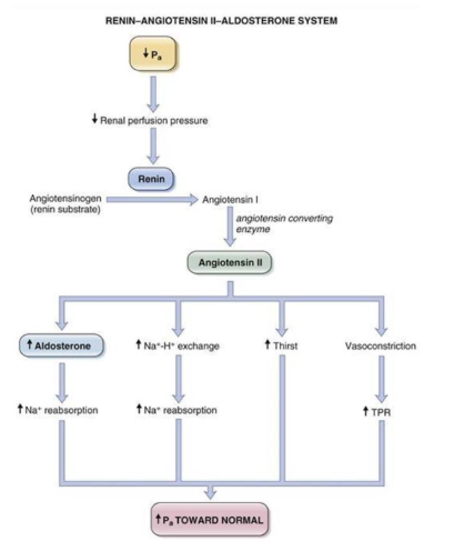
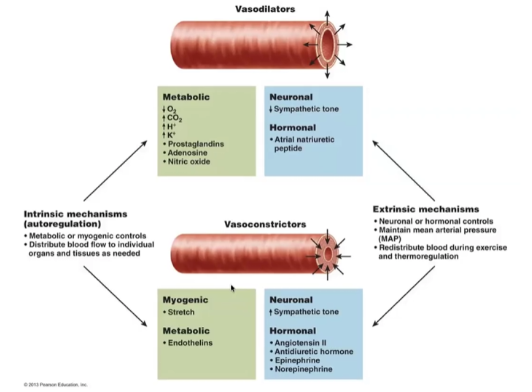
longer-term system that regulates bp via blood volume at kidney level. Fall in bp causes decrease in renal perfusion pressure which triggers release of Renin from juxtaglomerular cells. Renin cleaves angiotensin (from liver) into angiotensin 1(AT1). AT1 is cleaved into AT2 via angiotensin converting enzyme (ACE) in the lungs. AT2 stimulates adrenal gland to secrete the steroid aldosterone and act as a potent vasoconstrictor to increase Pa. aldosterone increases Na+ reabsorption in the kidney and thus restore the volume of the ECF.
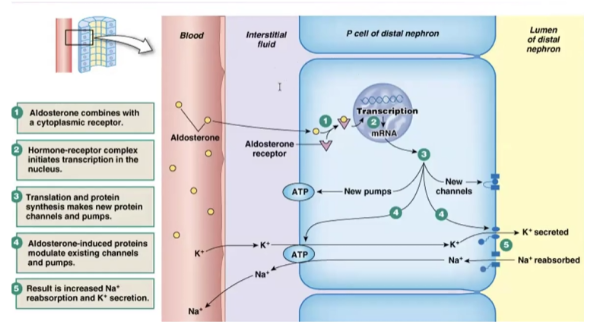
Show how aldosterone acts on cells
Chemoreceptors
located in carotid and aortic bodies, found near the bifurcation of the carotid and the aortic arch. Contains specialized nerve cells (type I glomus cells) that act as sensors for O2, CO2, and pH and will regulate bp accordingly via the ANS.
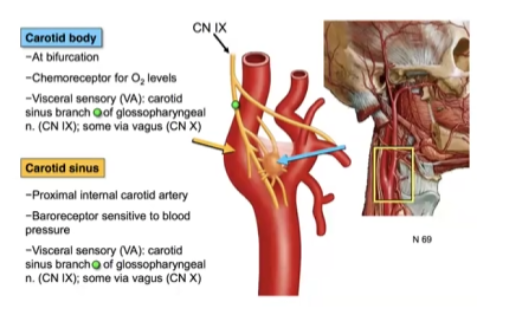
Carotid body
a small cluster of specialized chemoreceptor cells located at the fork (bifurcation) of the common carotid artery in the neck. Its primary function is to continuously monitor arterial blood for oxygen levels, carbon dioxide levels, and pH
Sinusoid
“leaky” capillaries where chemoreceptors are present
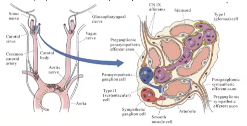
A decrease in arterial PO2 causes what reaction from chemoreceptors
increases firing of sympathetic nerves which increases arterial vasoconstriction in muscle, kidney and gut
Central chemoreceptors are located in the — and are more sensitive to changes in — and —. What do they do if brain becomes ischemic?
medulla; PCo2 and pH; increase TPR and redirect blood away from periphery and towards the brain (brain is intolerant of decreased blood flow)
Low pressure baroreceptors
located atria (right atrium), veins, and pulmonary arteries. Detects changes in atrial stretch which means they detected heightened blood volume. Triggers mechanisms to reduce blood volume which include release of atrial natriuretic peptide, decreased secretion of pituitary hormone vasopressin (ADH), and renal vasodilation to enhance natriuresis. They will also increase HR. If a decrease in blood volume is detected, the response is an increase in pre-load.
How does increase in pressure at the carotid sinus affect HR? How does increase in pressure in the atria affect HR? (explain why for both)
increase in pressure at the carotid sinus lowers HR as baroreceptors are triggered which cause parasympathetic response; increase in pressure in the atria trigger low-pressure baroreceptors which increase HR so as to increase Cardiac output and increase renal perfusion to decrease blood volume
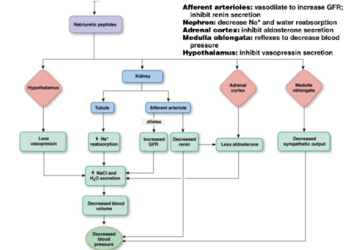
How does Atrial Natriuretic Peptide (ANP)decrease blood pressure? What causes ANP release?
it increases salt and water excretion (thus decreasing blood volume) it does this through many mechanisms shown in the image. ANP is released in response to increased atrial stretch detected by low pressure baroreceptors
Orthostatic hypotension
sudden drop in blood pressure that occurs when standing up after sitting or lying down. Blood pools in legs on standing up due to gravity causing instant drop in VR so there is less blood in ventricles at next contraction. CO falls from 5 L/min to ~3 L/min and BP drops and triggers baroreceptor reflex. This increases CO and RPE so MAP increases and BP returns to normal
BP decrease from orthostatic hypotension is corrected within how many heart beats?
1-2
What happens if corrective mechanisms for orthostatic hypotension do not work?
blood flow to brain is compromised and symptoms such as light-headness, dizziness and weakness are the result
How are bp regulation mechanisms affected by prolonged space flight
blood is evenly distributed in the body instead of pooling to extremities. This causes the kidneys to reduce blood blood volume by ~12%. On return to earth, there is a perceived sudden loss of blood due to blood pooling in extremities again.
Autoregulation
ability of some tissue/capillary beds to regulate blood flow and thereby maintain constant pressure despite changes in flow. Depends on the local environment and release of local factors. Can occur independently or together with systemic pressure evoked by baroreceptors.
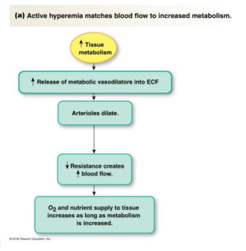
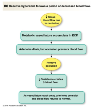
Hyperemia
relaxation of arterioles (vasodilation) in order to allow greater blood flow so as to match metabolic need or in response to lowered blood flow
What are the tissues that autoregulate
renal, cerebral, coronary, skeletal muscle and pulmonary tissue
Active hyperemia
Reactive hyperemia
Lowered [O2] and heightened [CO2] [H+] and [K+]will trigger vasodilation or vasoconstriction?
vasodilation
Serotonin and endothelin
local vasoconstrictor. Useful in repair of blood vessels which have breaks and prevent further hemorrhaging
Extrinsic vs intrinsic mechanisms for vasoconstriction vs vasodilation
Organ with highest O2 consumption
heart
Coronary vasodilation correlates with — and — levels
decreased oxygen and increased CO2 levels
Most important local vasodilator for the coronary circulation
CO2 ( or pH)
When coronary vasodilation is insufficient to meet demand what happens?
ischemic cardiac tissue releases large amounts of adenosine that causes local hyperemia
t/f vasoactive substances in the systemic circulation have little or no effect on cerebral circulation as they are excluded by the blood-brain barrier
true
Why is nitroglycerin given for angina?
it delivers Nitric oxide which is vasodilator that can help coronary circulation deliver more oxygen to the heart
Neural factors for cardiovascular adjustment to exercise
central command, reflexes originating in exercising muscle and baroreceptor reflex
During exercise there is redirection of blood from —- to —-
gut to skeletal muscle
3 sensors that can affect CV system during exercise
proprioreceptors, chemoreceptors, and baroreceptors.
proprioceptors
These pick-up movements in joints and muscles. An increase in intensity of exercise generally means increased amounts of movement. The proprioceptors pick up this change and relay the information to the CCC (cardiac control center), which increases HR and stroke volume (SV) to meet demand.
Show the different type of proprioceptors and what they detect
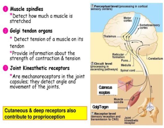
What do chemoreceptors do during exercise?
These pick up chemical changes such as a lowering of blood pH. The increase in acidity (lowering of pH) occurs because as exercise intensity increases, there is an increase in carbon dioxide, which is carried in the blood as carbonic acid. Also, there is an increase in lactic acid production, so combined with the carbonic acid, the blood pH lowers. Such changes are picked up by the chemoreceptors and information is relayed to the CCC(Cardiac Control Center), which increases sympathetic activity which increases HR and SV.
t/f chemoreceptors play a large role in CV control during moderate or mild exercise
false, pH, PCO2 and PO2 do not change much in mild to moderate exercise
What do baroreceptors do during exercise?
(found in aorta and carotid artery) – These pick-up changes in blood pressure as the result of increased exercise intensity.
t/f the CV command center increases sympathetic outflow prior to the onset of exercise
true (remember feed forward mechanism)
Activation of — receptors cause — in gut and kidneys during exercise
alpha1; vasoconstriction
t/f due sympathetic output during exercise, skeletal muscle blood vessels undergo vasoconstriction
false, sympathetic output is overridden by local vasodilators in the muscle
There is (increased or decreased) venoconstriction in muscles during exercise. Why?
increased; party due direct effects of the sympathetic nervous system on vasculature and partly due to skeletal muscle pump
Skeletal muscle pump
pump that forces blood from lower extremities towards the heart as it squeezes large veins when it contracts
t/f respiratory movements help force blood towards the heart
true
Biphasic response to exercise in skin
initial vasoconstriction followed by vasodilation to dissipate heat generated by exercise
There is an overall (increase or decrease) in TPR during exercise. why?
decrease; vasodilation of arterioles in skeletal muscle which outweighs vasoconstriction
What 4 main arteries feed blood to the brain?
two vertebral arteries and two internal carotid arteries
Normal blood flow through the brain
about 50 to 65 ml per 200 grams of brain tissue per minute (750 to 900 ml/min for entire brain)
–% of resting CO is dedicated to the brain
15
When does ischemia occur in the brain? When does tissue death occur?
ischemia results from blood flow dropping below 18-20 ml/100g/min; tissue death occurs if flow drops below 8-10 ml/100g/min
The brain accounts for –% of body weight and —% of resting oxygen consumption. Why is this?
2; 20; high metabolic rate in the brain
3 factors that regulate cerebral blood flow (CBF)
myogenic, metabolic and neurogenic autoregulation
Myogenic autoregulation in the brain
arteries constrict or dilate in response to changes in BP or ICP (intra- cranial pressure)
Metabolic autoregulation in the brain
response to increased CO2, increased [H+] or decreased O2 .
A 70% increase in arterial CO2 will have what effect on CBF?why?
will double CBF. metabolic autoregulation
Decreased CO2 as occurs in hyperventilation, causes cerebral —-, decreased —- and cerebral —-. Increased blood H+ will cause —-- and thus (increase or decrease) CBF.
vasoconstriction, decreased CBF and cerebral hypoxia; vasodilation and thus increase CBF
Neurogenic autoregulation in the brain
SNS will constrict large and intermediate diameter arteries to prevent high pressure reaching smaller vessels where it may cause hemorrhage or stroke. However, under normal conditions this has a minor role in regulation of CBF
Blood brain barrier
tight layer of capillary endothelium that protects the brain from blood contents
Functions of blood brain barrier
maintains constancy of environment of the neurons in the CNS, protects brain from endogenous and exogenous toxins, and prevents escape of neurotransmitters into general circulation
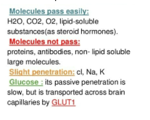
Describe the penetration of substance across the blood-brain barrier