Baylor DPT Anatomy Exam 2
1/273
There's no tags or description
Looks like no tags are added yet.
Name | Mastery | Learn | Test | Matching | Spaced | Call with Kai |
|---|
No analytics yet
Send a link to your students to track their progress
274 Terms
Muscles of the anterior compartment of the thigh
Rectus femoris
Vastus medialis
Vastus intermedius
Vastus lateralis
Sartorious
Pectineus
Iliopsoas
Rectus femoris OINA
0 - anterior inferior iliac spine and rim of the acetabulum
I - tibial tuberosity via the patellar ligament
N - femoral nerve
A - flex the hip and extend the knee
Vastus intermedius OINA
0 - anterior surface of the femur
I - tibial tuberosity via the patellar ligament
N - femoral nerve
A - extend the knee
Vastus lateralis OINA
0 - lateral surface of the femur
I - tibial tuberosity via the patellar ligament
N - femoral nerve
A - extend the knee
Vastus medialis oblique OINA
0 - medial surface of the femur and tendon of the adductor magnus
I - tibial tuberosity via the patellar ligament
N - femoral nerve
A - extend the knee (control tracking of patella)
Sartorius OINA
0 - anterior superior iliac spine
I - superior end of the medial surface of the tibia
N - femoral nerve
A - flexes and lateral rotates thigh at hip joint, weak abductor of thigh and weak flexorof the leg
Pectineus OINA
0 - pectineal line of the superior ramus of the pubis
I - pectineal line of the femur
N - femoral and obturator nerve
A - flex and adduct thigh
Muscles of the medial compartment of the thigh
Gracilis
Adductor longus
Adductor magnus
Adductor brevis
Gracilis OINA
0 - Inferior ramus of the pubis
I - medial surface of tibia, inferior to the condyle
N - adduct thigh, flex leg
A - obturator nerve
Adductor longus OINA
0 - body of the pubis
I - distal 2/3 of the linea aspera
N - obturator nerve
A - adduct and flex thigh
Adductor magnus OINA
0 - tuberosity and ramus of the ischium
I - linea aspera and adductor tubercle
N - obturator nerve and tibial portion of the sciatic nerve
A - adducts and flexes thigh, extends thigh
Adductor brevis OINA
0 - Inferior ramus of the pubis
I - femur
N - obturator
A - adduct and flex thigh
Gluteus maximus OINA
0 - ilium posterior to posterior gluteal line, sacrum, sacrotuberous ligament
I - gluteal tuberosity and iliotibial band
N - inferior gluteal nerve
A - extends the thigh when running and climbing, sit to stand
Piriformis OINA
0 - pelvic surface of the sacrum
I - greater trochanter
N - nerve to piriformis
A -lateral rotation and extension of thigh
Gluteus medius OINA
0 - ilium between iliac crest ad superior gluteal line
I - greater trochanter
N - superior gluteal nerve
A -abducts and medially rotates thigh
Obturator internus OINA
0 - obturator membrane
I - greater trochanter
N - nerve to the obturator internus
A - laterally rotates and extends thigh
Superior gemellus OINA
0 - ischial spine
I - greater trochanter
N - nerve to obturator internus
A - laterally rotates and extends thigh
Inferior gemellus OINA
0 - ischial tuberosity
I - greater trochanter
N - nerve to the quadratus femoris
A -laterally rotates and extends thigh
Quadratus femoris OINA
0 - ischial tuberosity
I - intertrochanteric crest
N - nerve to the quadratus femoris
A - laterally rotates and extends thigh
Gluteus minimus OINA
0 - ilium between superior and inferior gluteal lines
I - greater trochanter
N - superior gluteal nerve
A - abduct and medially rotate the thigh
Obturator externus OINA
0 - obturator membrane
I - trochanteric fossa
N - obturator nerve
A - adduct and laterally rotate the thigh
Tensor fascia lata OINA
0 -iliac crest
I - iliotibial band
N - superior gluteal nerve
A - abducts and flexes thigh
Muscles of the posterior compartment of the thigh
Biceps femoris
Semitendinosus
Semimembranosus
Biceps femoris OINA
Long head
O - ischial tuberosity
Short head
O -linea aspera
I - head of the fibula
N -long head by the tibial portion of the sciatic, short head by the common peroneal portion of the sciatic
A - extend thigh and flex leg (knee)
Semitendinosus OINA
0 - ischial tuberosity
I - medial surface of tibia inferior to the condyle
N - tibial portion of sciatic
A - extend thigh, flex leg and medially rotate leg
Anterior compartment of the leg
Tibialis anterior
Extensor digitorum longus
Extensor hallucis longus
Peroneus tertius
Tibialis anterior OINA
0 - superior 2/3 lateral surface of tibia
I - medial cuneiform and base of first metatarsal
N - deep peroneal
A - dorsiflexion and inversion of foot
Extensor digitorum longus OINA
0 - superior 2/3 of the fibula
I - middle and distal phalanges of the lateral four toes
N - deep peroneal
A - dorsiflexion of the foot and extension of the toes
Extensor Hallucis longus OINA
0 - middle 1/3 fibula
I - base of distal phalanx of hallux
N - deep peroneal
A - dorsiflexion of the foot and extension of the hallux
Peroneus tertius OINA
0 - distal end of the fibula
I - base of the fifth metatarsal
N - deep peroneal
A - dorsiflexion of the foot and eversion of the foot
Muscles of the dorsum of the foot
Extensor digitorum brevis
Extensor hallucis brevis
Extensor digitorum brevis OINA
0 - calcaneus
I - long extensor tendons of digits 2-4
N - deep peroneal
A - extension of digits 2-4
Extensor Hallucis brevis OINA
0 - calcaneus
I - base of proximal phalanx of hallux
N - deep peroneal
A - extension of the big toe
Muscles of the lateral compartment of the leg
Peroneus longus
Peroneus brevis
Peroneus longus
0 - superior 2/3 of the fibula
I - base of the 1 st metatarsal and medial cuneiform
N - superficial peroneal
A - eversion and plantarflexion of the foot
Peroneus brevis
0 - distal end of the fibula
I - base of the 5th metatarsal
N - superficial peroneal
A - eversion and plantarflexion of the foot
Muscles of the posterior compartment of the leg
Gastrocnemius
Soleus
Plantaris
Flexor digitorum longus
Flexor hallucis longus
Tibialis posterior
Popliteus
Gastrocnemius OINA
0 -lateral head from lateral condyle of femur, medial head from medial condyle of femur
I - calcaneus
N - tibial nerve
A - plantarflexion of foot, flexion of leg (knee)
Soleus OINA
0 - posterior surface of fibula and tibia
I - calcaneus
N - tibial nerve
A - plantarflexion of the foot
Plantaris OINA
0 - lateral condyle of femur
I - calcaneus
N - tibial nerve
A - plantarflexion of the foot, flexion of leg
Flexor digitorum longus OINA
0 - tibia
I - distal phalanx of lateral four digits
N - tibial nerve
A - flexion of digits 2-5 and plantarflexion of the foot
Flexor Hallucis longus OINA
0 - fibula
I - distal phalanx of big toe
N - tibial nerve
A - flexion of hallux and plantarflexion of the foot
Tibialis posterior OINA
0 - tibia, fibula and interosseous membrane
I - navicular, cuneiform, cuboid and base of the 2nd, 3rd, and 4th metatarsal
N - tibial nerve
A -inversion and plantarflexion of the foot
Popliteus OINA
0 - upper end of tibia
I - lateral condyle of femur
N - tibial
A - rotation of knee joint, knee flexion
Muscles of the first layer of the sole of the foot
Abductor hallucis
Flexor digitorum brevis
Abductor digiti quinti
Abductor hallucis OINA
0 - calcaneus
I - proximal phalanx
N - medial plantar nerve
A - abduct and flex hallux
Flexor digitorum brevis OINA
0 - calcaneus
I - middle phalanges of lateral four toes
N - medial plantar nerve A - flexes toes
Abductor digiti quinti OINA
0 - calcaneus
I - proximal phalanx
N - Lateral plantar nerve A - abduct and flex little toe
Muscles/tendons of the second layer of the sole of the foot
Flexor digitorum longus tendon
Flexor hallucis longus tendon
Quadratrus plantae
Lumbricals
Flexor digitorum longus tendon OINA
0 - tibia
I - distal phalanx of lateral four digits
N - tibial nerve
A - flexion of digits 2-5
Flexor hallucis longus tendon OINA
0 - fibula
I - distal phalanx of big toe
N - tibial nerve
A -flexion of hallux
Quadratus plantae OINA
0 - calcaneus
I - tendon of the flexor digitorum longus
N -lateral plantar nerve
A - redirect the line of pull of the tendons of the flexor digitorum longus
Lumbricals OINA
0 - tendons of the flexor digitorum longus
I - dorsal digital expansions
N - medial one by the medial plantar nerve, lateral three by the lateral plantar nerve
A - flexes the MPJ and extends the proximal IPJ of the lateral four toes
Muscles of the third layer of the sole of the foot
Flexor hallucis brevis
Flexor digiti quinti
Adductor hallucis
Flexor hallucis brevis OINA
0 - cuboid and lateral cuneiform
I - Proximal phalanx of the big toe
N - medial plantar nerve A -flexes the great toe
Flexor digiti quinti OINA
0 - base of the 5th metatarsal
I - proximal phalanx of the 5th toe
N -lateral plantar nerve
A -flex the little toe
Adductor hallucis OINA
0 - oblique head: base of the 2nd, 3rd, and 4th metatarsals Transverse head: plantar ligaments of the lateral four MPJ
I - proximal phalanx
N -lateral plantar nerve
A - adducts the great toe, assists in maintaining the transverse arch of the foot
Muscles of the fourth layer of the sole of the foot
Plantar interossei
Dorsal interossei
Plantar interossei OINA
0 - medial sides of the 3rd, 4th, and 5th metatarsals
I - medial sides of the proximal phalanges of the 3rd, 4th, and 5th digits and extensor expansions
N - lateral plantar nerve
A -adduct to the line of the second toe
Dorsal interossei
0 - adjacent sides of the two metatarsals
I - #1 to medial side of the 2nd digit #2 to lateral side of the 2nd digit base of the #3 to lateral side of the 3rd digit proximal phalanx #4 to lateral side of the 4th digit
N -lateral plantar nerve
A - abducts from line of the 2nd toe
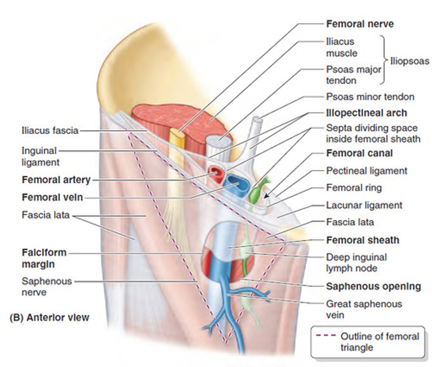
Contents of Femoral Triangle
Lateral femoral cutaneous nerve
Femoral nerve
Femoral artery and vein in femoral sheath
Profunda Femoris
Profunda Femoris artery
main supplier of blood to the thigh
originates from femoral artery
gives rise to the medial and lateral femoral circumflex arteries
Also called the deep femoral artery
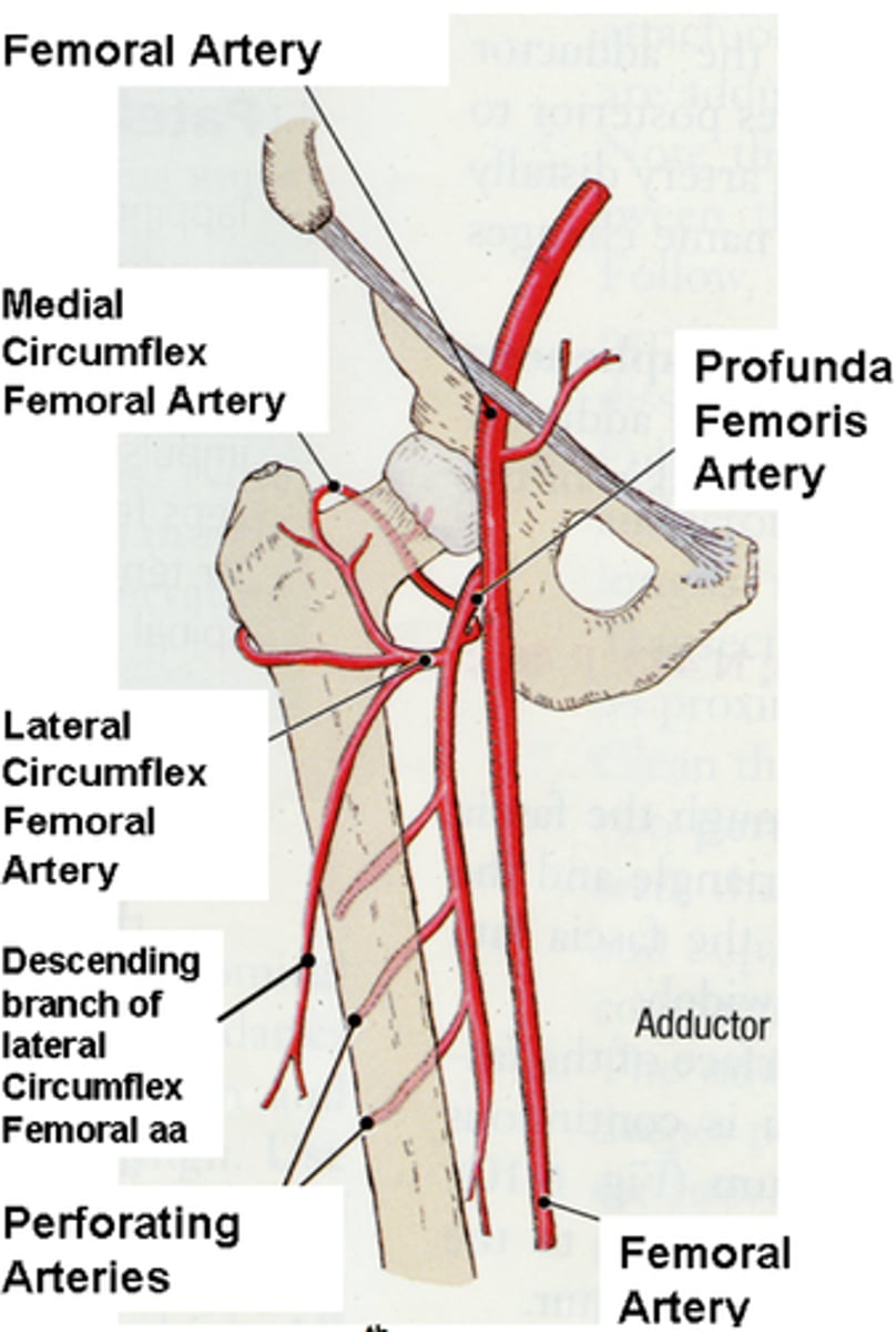
Medial and lateral circumflex arteries
main suppliers to the head and neck of the femur

Dorasal venous arch
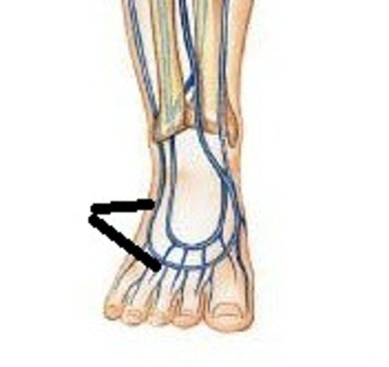
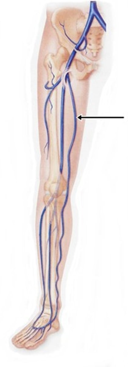
great saphenous vein
Perforating branches of veins
pump blood horizontally in venous system
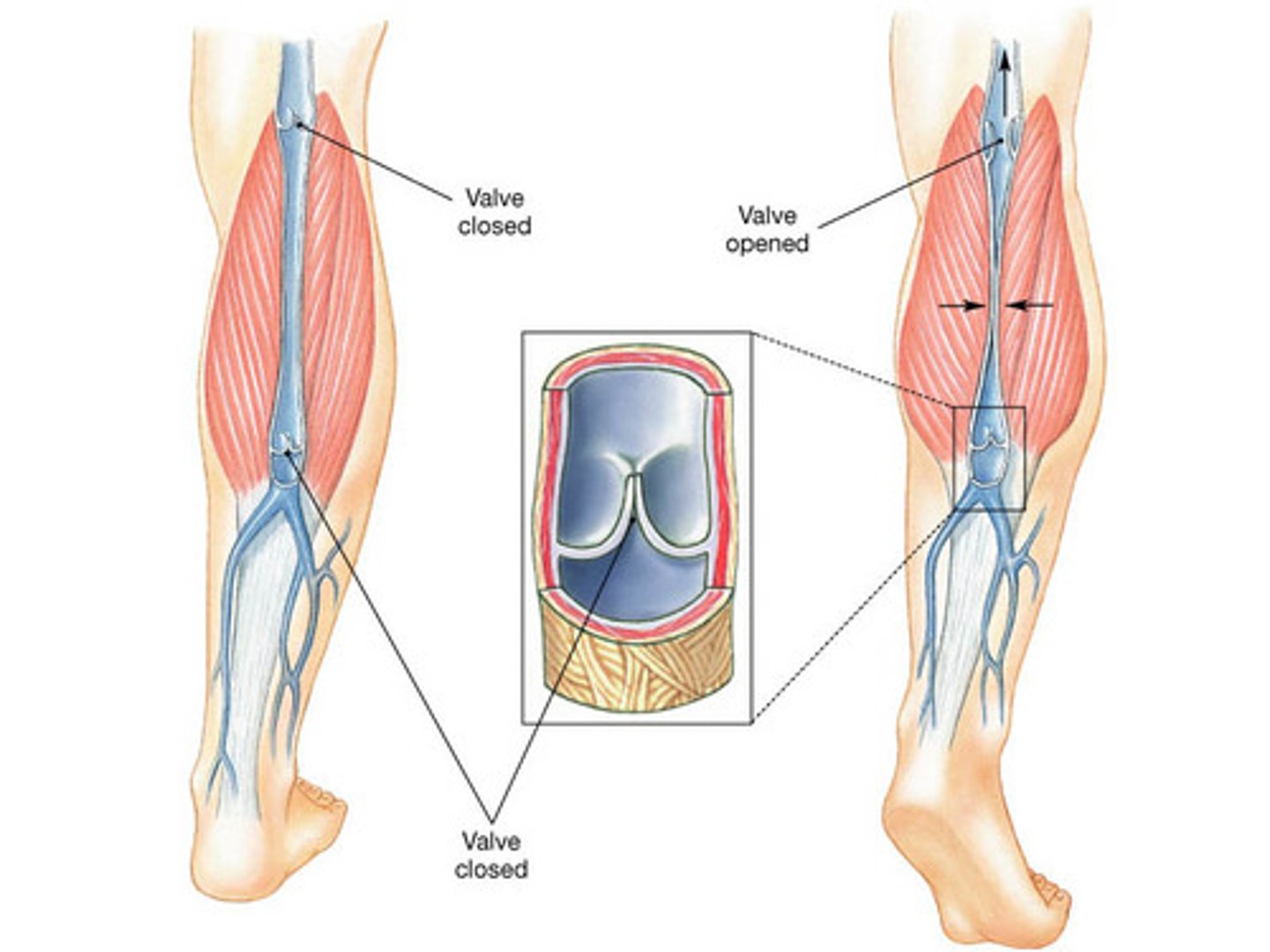
Venous valves
Cup like endothelium that fill from above
Prevent reflux of blood distally
valve mechanism allows blood to overcome force of gravity
Valve mechanism allows blood to overcome force of gravity
Adductor canal location
The canal begins at the point where the sartorius muscle passes over the adductor longus muscle
It ends at the adductor hiatus in the tendon of the adductor magnus muscle
Adductor canal contents
Femoral artery and vein and saphenous nerve
Anterior compartment of the thigh rules
Knee extensors
(exceptions: only the quads extend the knee)
innervated by femoral nerve
(exception: pectineus is also innervated obturator nerve)
Medial compartment of the thigh rules
Hip adductors
innervated by the obturator nerve
(exception: adductor magnus is also innervated by the tibial nerve)
Posterior compartment of the thigh rules
Knee flexors and hip extensors
(exceptions: short head of biceps femoris only flexes the knee)
innervated by the sciatic nerve
(exceptions: all are innervated by the tibial portion of the sciatic nerve except the short head of the biceps femoris which is innervated by the fibular (peroneal) portion)
Thigh cutaneous innervation
Subcostal nerve (light purple)
Iliohypogastric nerve (posterior superior blue)
Ilioinguinal nerve (anterior green)
Genitofemoral nerve (anterior purple)
Lateral femoral cutaneous
Femoral nerve
Anterior femoral cutaneous nerve

Gluteal region muscles
Gluteus maximus
Gluteus medius
Gluteus minimus
Tensor fascia lata
Piriformis
Superior gemellus
Inferior gemellus
Obturator externus
Obturator internus
Quadratus femoris
Gluteal region boundaries
Superior- iliac crest
Medial- sacrum and coccyx
Inferior- sacrotuberous ligament and ischial tuberosity
Lateral- greater trochanter
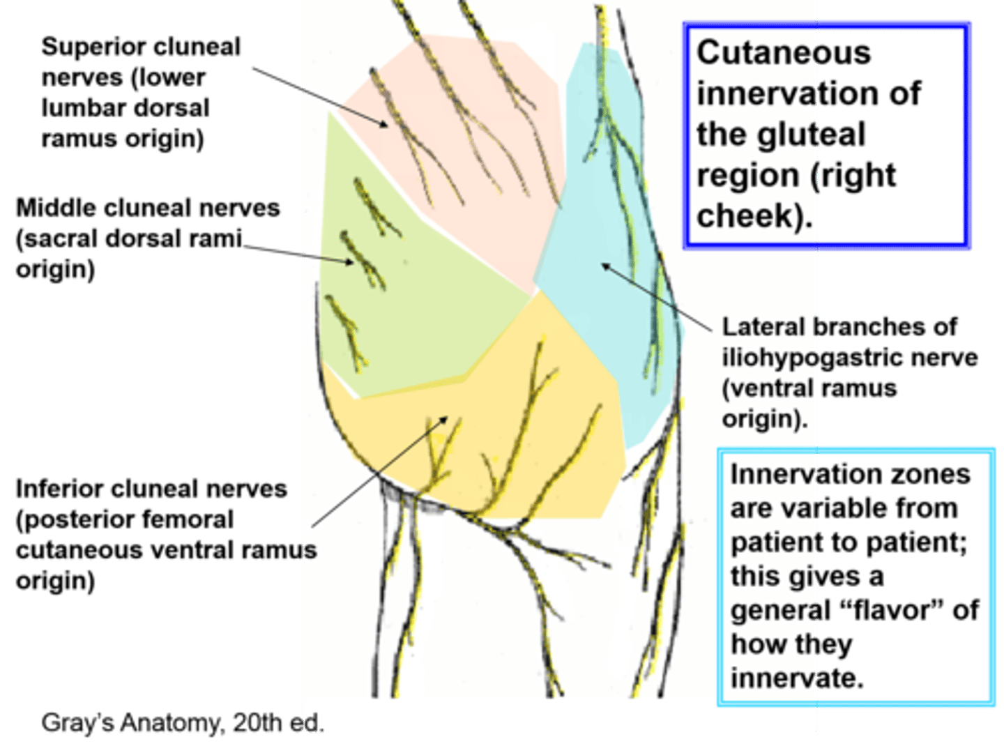
Gluteal region cutaneous innervation
Superior cluneals (from dorsal rami of first 3 lumbar nerves)
Middle cluneals (dorsal rami of first 3 sacral nerves)
Inferior cluneals (posterior femoral cutaneous)
Subcostal, Iliohypogastric, and lateral femoral cutaneous may also reach this area)
Gluteal region rules
Primarily involved in abduction and lateral rotation of the thigh (ALL muscles in some degree)
Innervated by the superior and inferior gluteal nerves
and
smaller branches of the lumbosacral plexus (nerve to piriformis, superior gluteal nerve, nerve to obturator internus, nerve to quadrartus femoris, and obturator nerve)
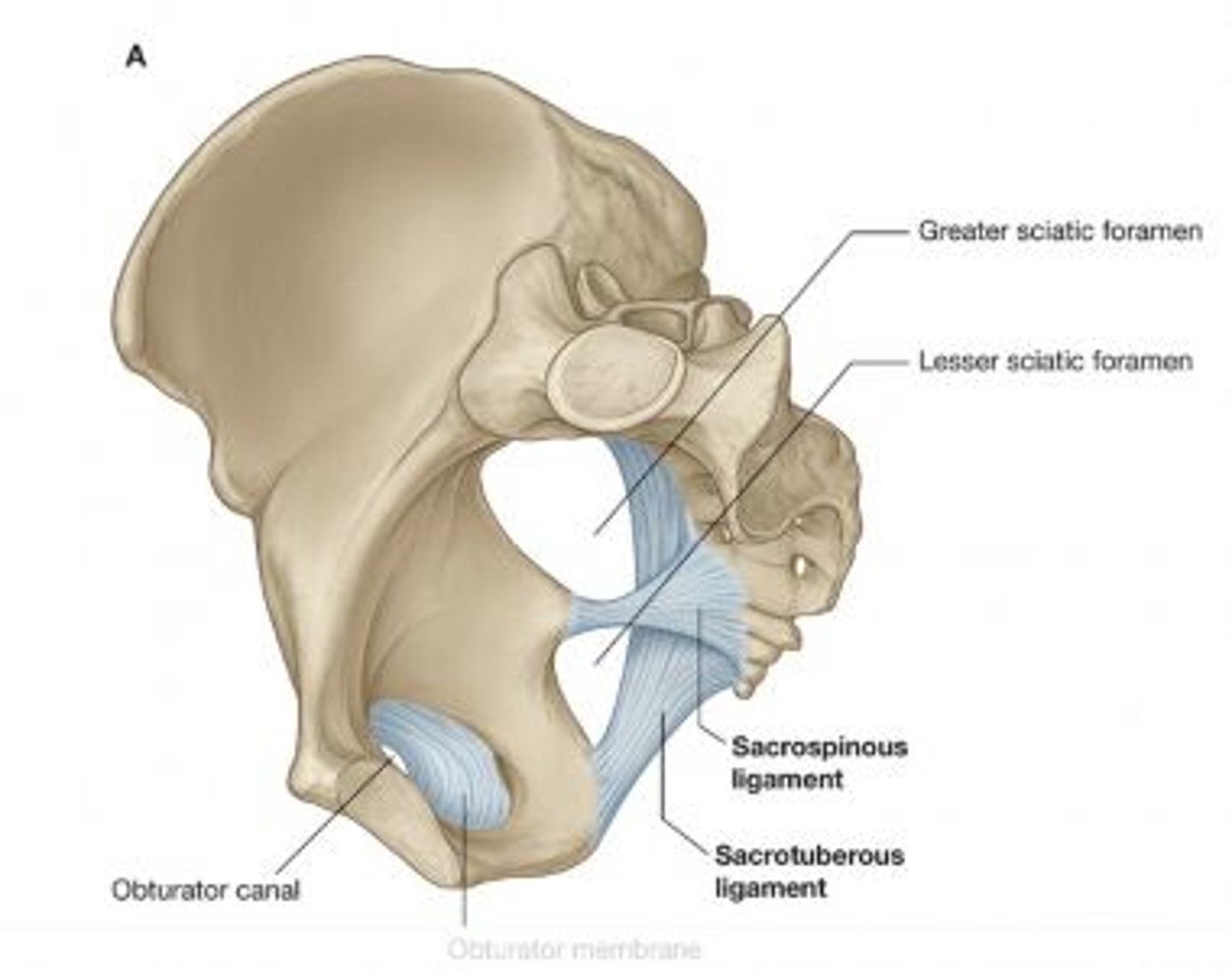
Ligaments of gluteal region
sacrotuberous ligament (posterior)
sacrospinous ligament (anterior)
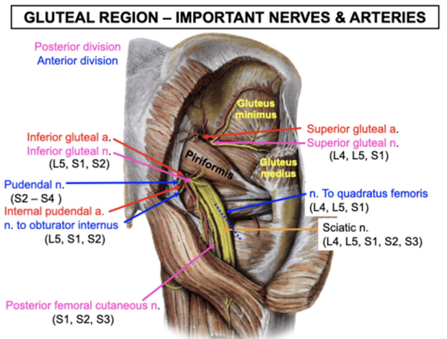
Gluteal arteries and nerves
All of the following can be visualized; read to know what to look for
Superior gluteal nerve and vessels
-Pass through the greater sciatic foramen
-Superior gluteal vessels provide superficial branches to the Gluteus maximus, pass between the Gluteus medius and minimus to reach the TFL
Inferior gluteal nerve and vessels
-Exit the greater sciatic foramen inferior to the piriformis and enter the deep surface of the gluteus maximus
Sciatic nerve and vessels
-Passes through the greater sciatic foramen
-It descends posterior to the obturator internus, gemelli, and quadratus femoris to enter the thigh
Posterior femoral cutaneous
-Runs medial to the sciatic nerve
-It supplies cutaneous information to the posterior thigh
Nerve to the obturator internus
-Exits greater sciatic foramen and passes immediately into the lesser sciatic foramen
-It supplies motor innervation to the obturator internus and superior gemellus muscles
Pudendal nerve and vessels
-Exit through the greater sciatic foramen and passes immediately into the lesser sciatic foramen just medial to the nerve to the obturator internus
Nerve to the quadratus
-Exits the greater sciatic foramen inferior to the piriformis and descends anterior to the obturator internus and gemelli muscles
-It innervates the inferior gemellus and quadratus femoris muscles
Posterior thigh cutaneous innervation
posterior femoral cutaneous nerve

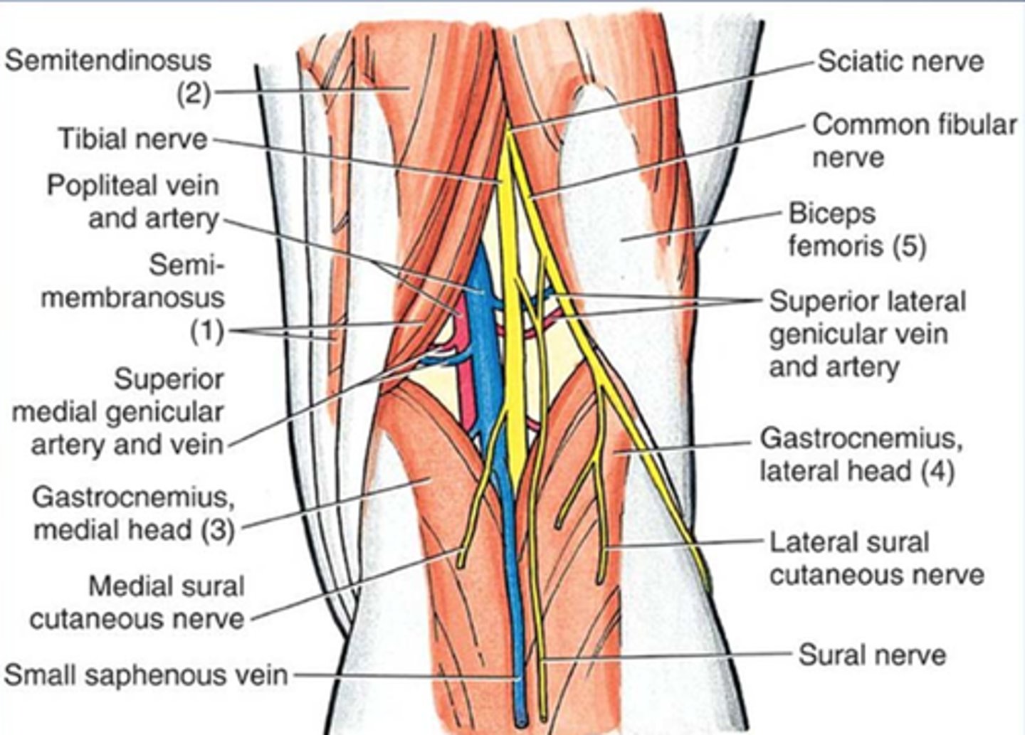
Popliteal fossa boundaries
Superior lateral - biceps femoris
Superior medial - semitendinosus and semimembranosus
Inferior lateral - lateral head of the gastrocnemius
Inferior medial - medial head of the gastrocnemius
Roof - Fascia lata with branches of the posterior femoral cutaneous nerve and the lesser saphenous vein
Floor - Popliteal surface of the femur, Capsule of the knee joint, and Oblique popliteal ligament
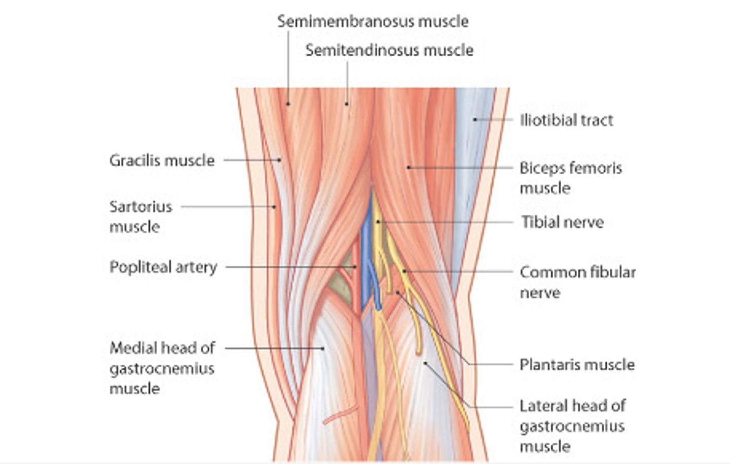
Popliteal fossa contents
-Tibial nerve
-Common peroneal (fibular) nerve
-Popliteal vessels
-Origins of sural nerve
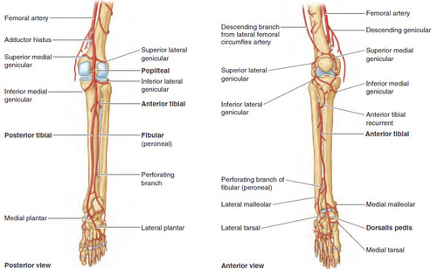
Popliteal artery branches
Anterior tibial artery (supplies anterior compartment of leg)
Posterior tibial artery (supplies posterior compartment of the leg)
Genicular anastomoses (supply the patella, knee capsule and surrounding bone)
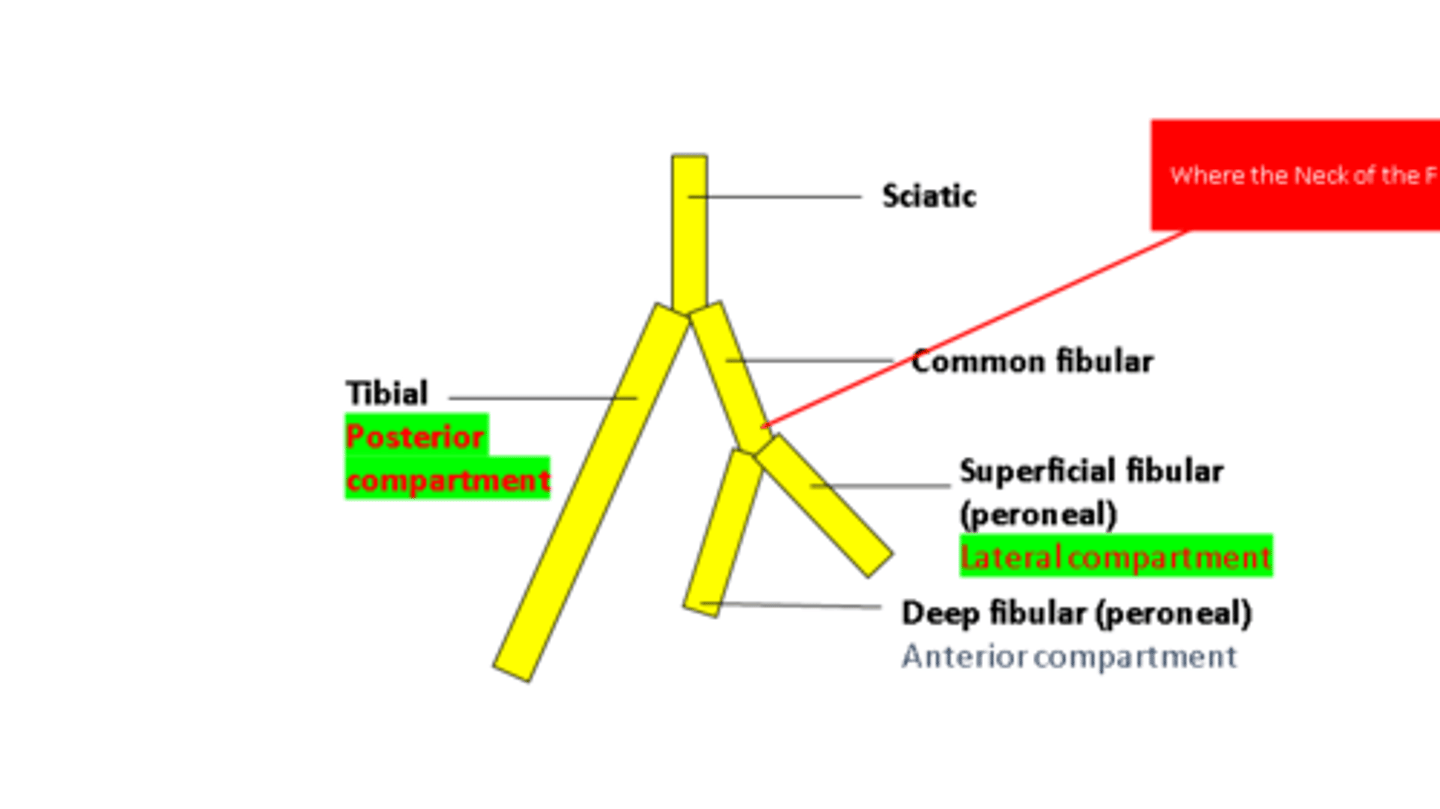
Popliteal nerve branches
Tibial nerve
Common peroneal nerve
Deep peroneal nerve
Superficial peroneal nerve
Leg compartments
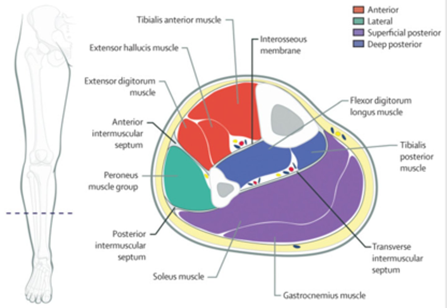
Anterior compartment of the leg rules
supplied by anterior tibial artery
innervated by deep peroneal nerve
foot dorsiflexors
Lateral compartment of the leg rules
supplied by the peroneal artery, (branch from the posterior tibial artery)
innervated by superficial peroneal nerve
evert and plantarflex the foot
Posterior compartment of the leg rules
supplied by the posterior tibial artery
innervated by the tibial nerve
plantarflex the foot
(exception popliteus)
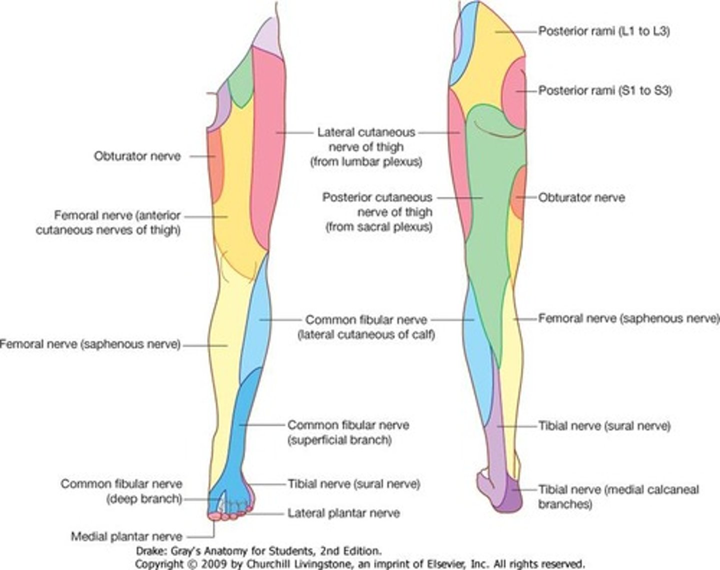
Leg Cutaneous Innervation
Saphenous nerve
Sural nerve
Superficial peroneal nerve
Deep peroneal nerve
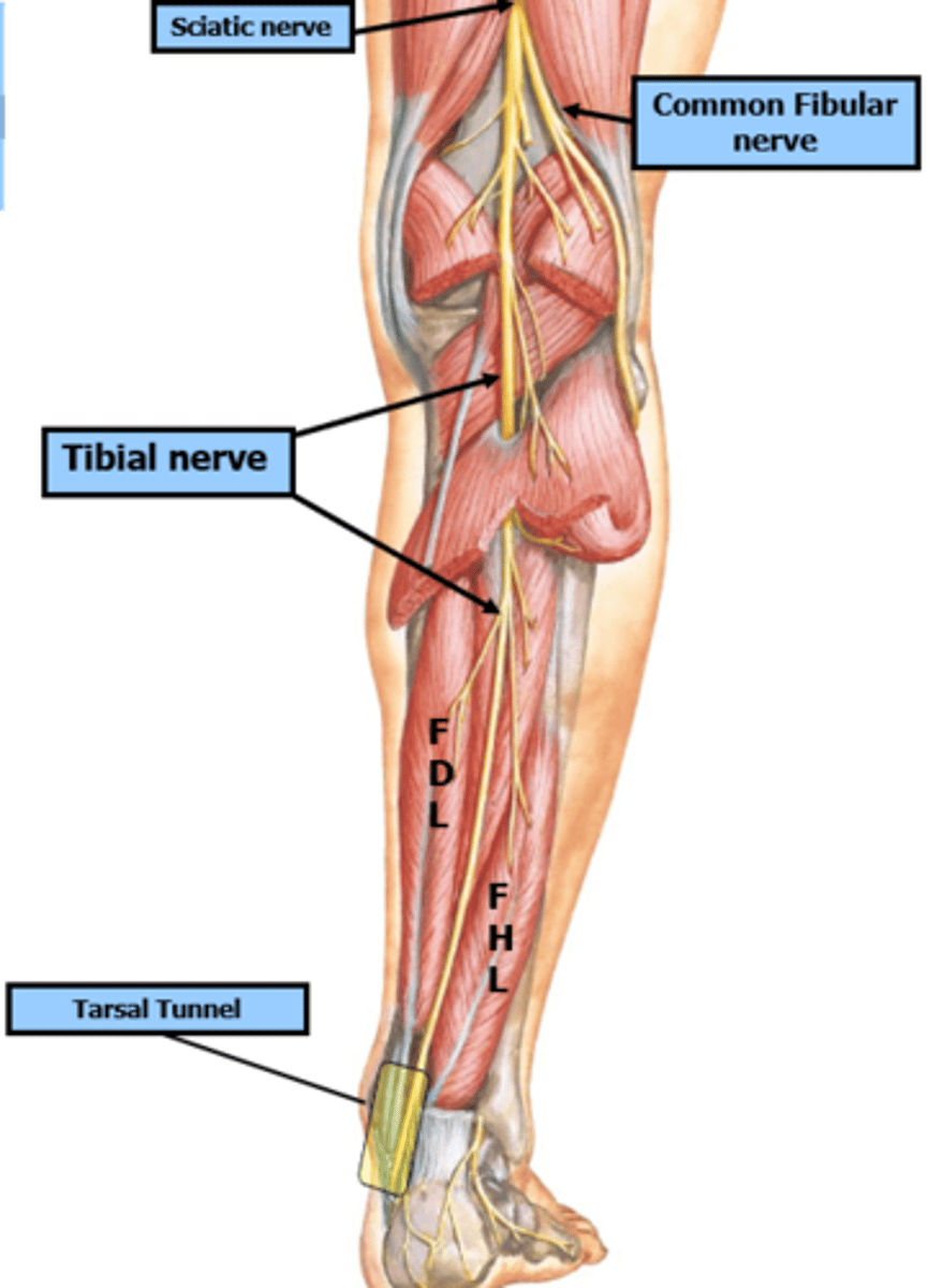
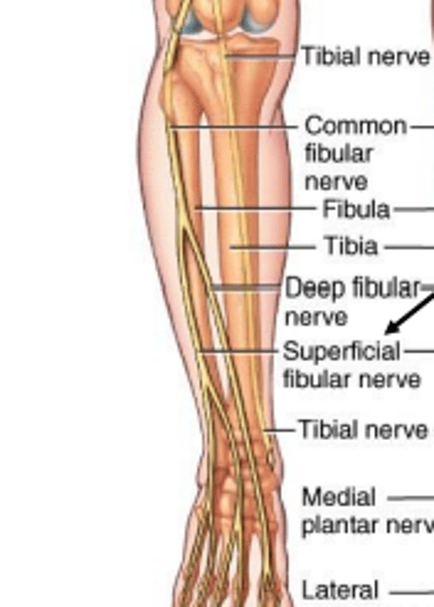
Path of tibial nerve
Tibial nerve travels with the posterior tibial artery down the back of the leg. It passes posterior to the medial malleolus and divides into medial and lateral plantar nerves in the sole of the foot
Path of the deep peroneal nerve
Deep peroneal nerve supplies muscular innervation to the anterior compartment of the leg.It runs with the anterior tibial artery and ends with branches innervating the ankle joint andskin between the first and second digits.
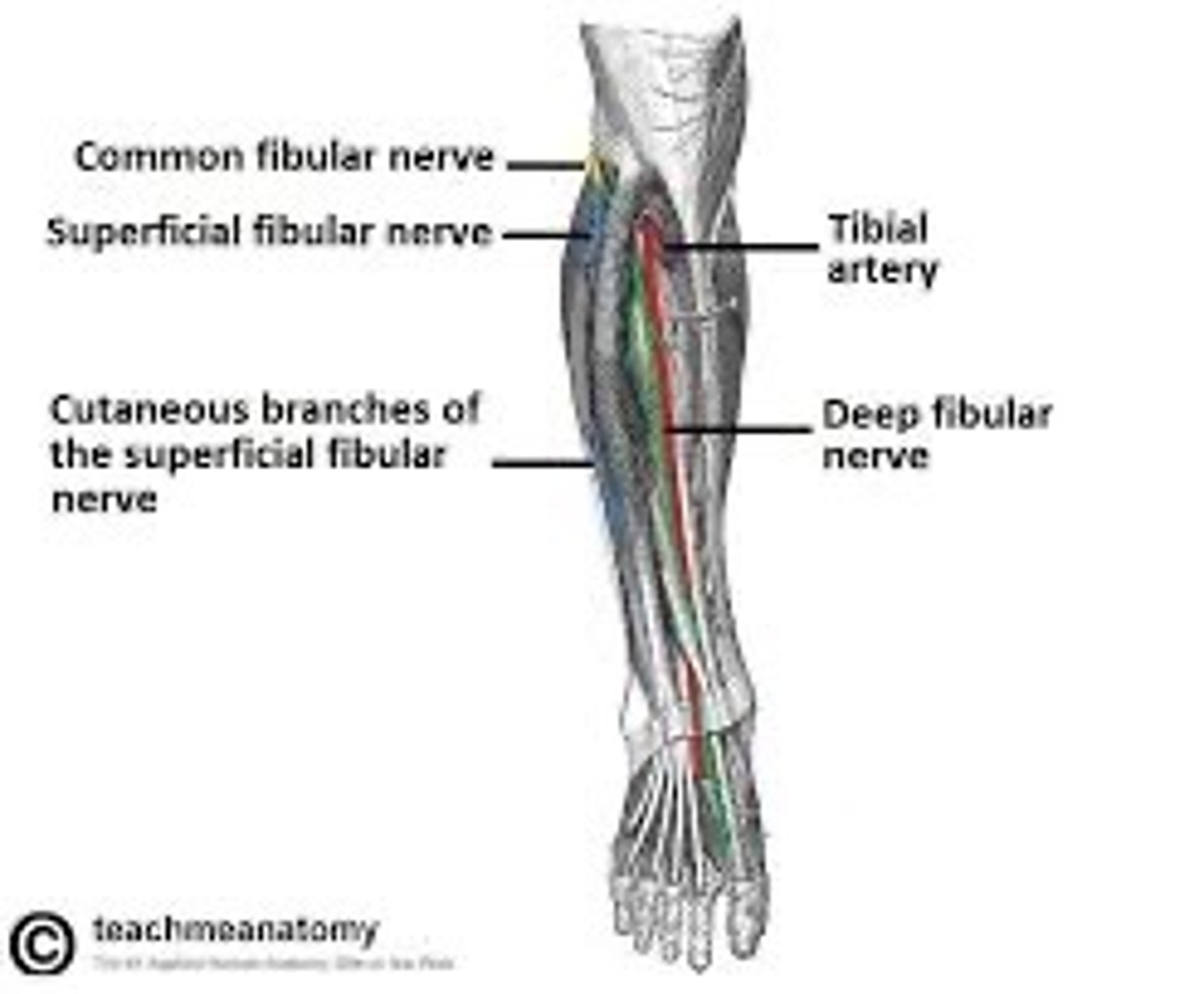
Path of the superficial peroneal nerve
Superficial peroneal supplies innervation to the muscles of the lateral compartment of the leg. It then innervates the skin along the distal aspect of the anterior surface of the leg, dorsum of the foot and digits 3 to 5.
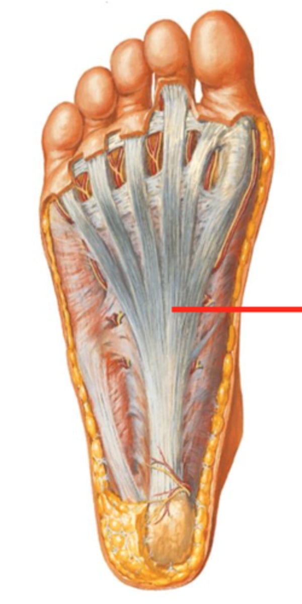
plantar aponeurosis
thickening of the plantar fascia, similar to that found in the palm of the hand
acts
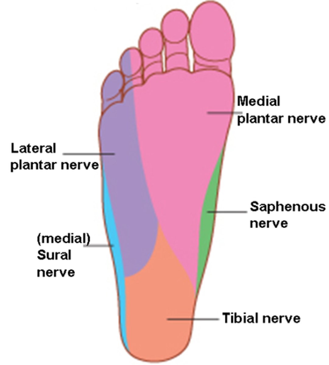
Cutaneous innervation of the sole of the foot
Medial plantar nerve
Lateral plantar nerve
Saphenous nerve
Sural nerve
medial plantar nerve innervates
Crural definition
refers to the leg, specifically the part of the lower limb between the knee and the ankle
What does ASIS stand for
Anterior superior iliac spine
What does AIIS stand for
anterior inferior iliac spine
What does PIIS stand for
posterior inferior iliac spine
what does PSIS stand for
posterior superior iliac spine