Basic Histology - Tissues Practical Notes
1/30
Earn XP
Description and Tags
Comprehensive vocabulary flashcards covering basic histology including epithelial, connective, blood, cartilage, bone, muscular, and nervous tissues.
Name | Mastery | Learn | Test | Matching | Spaced | Call with Kai |
|---|
No analytics yet
Send a link to your students to track their progress
31 Terms
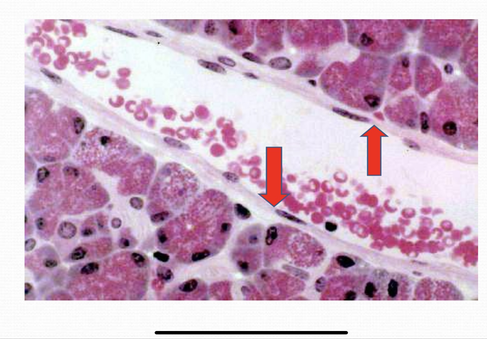
Epithelial Tissue Epithelium) (LM): (Red arrows) pointed on simple squamous epithelium stained with general stains
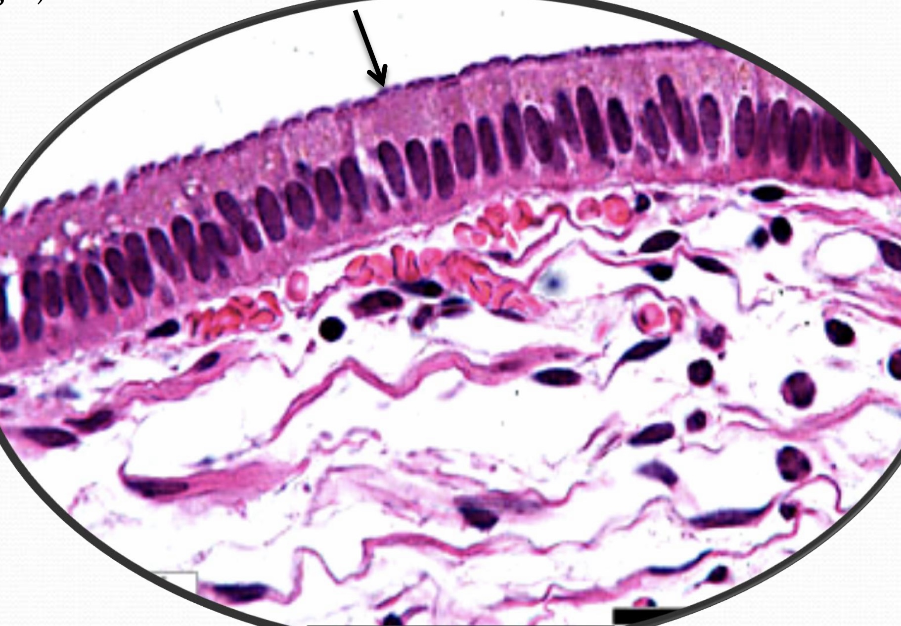
Epithelal Tissue Epithehum (LM): Black arrow pointed on columnar (non ciliated) epithelium stained with general stains
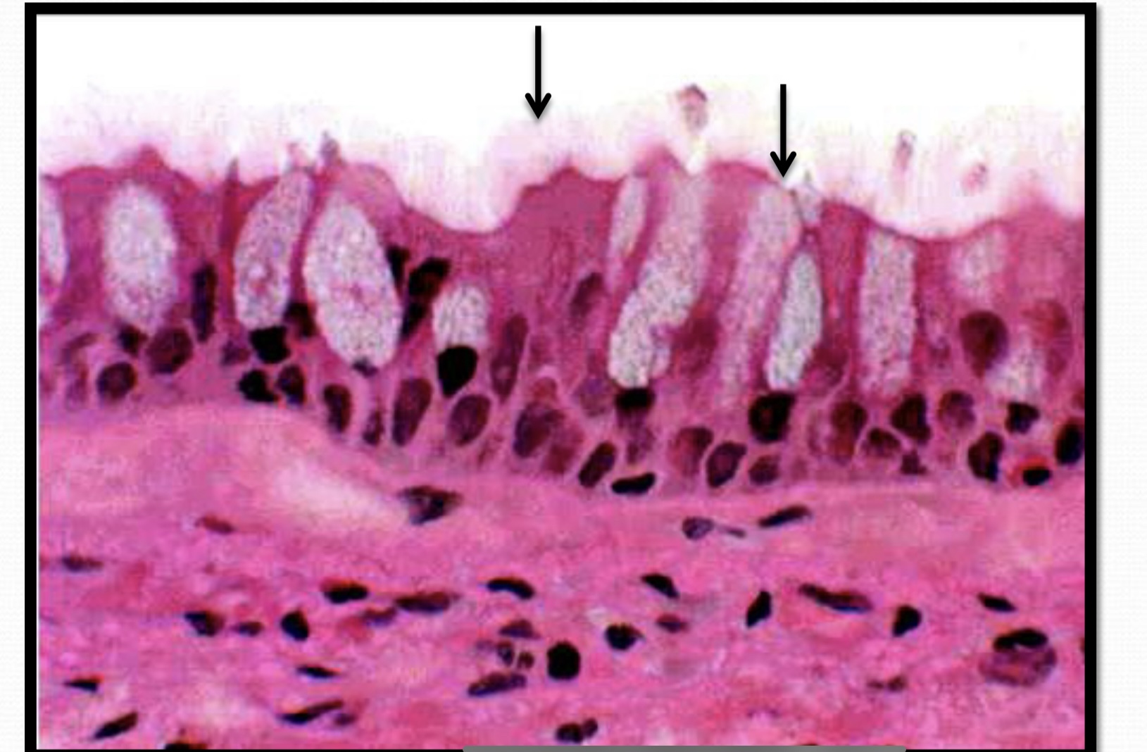
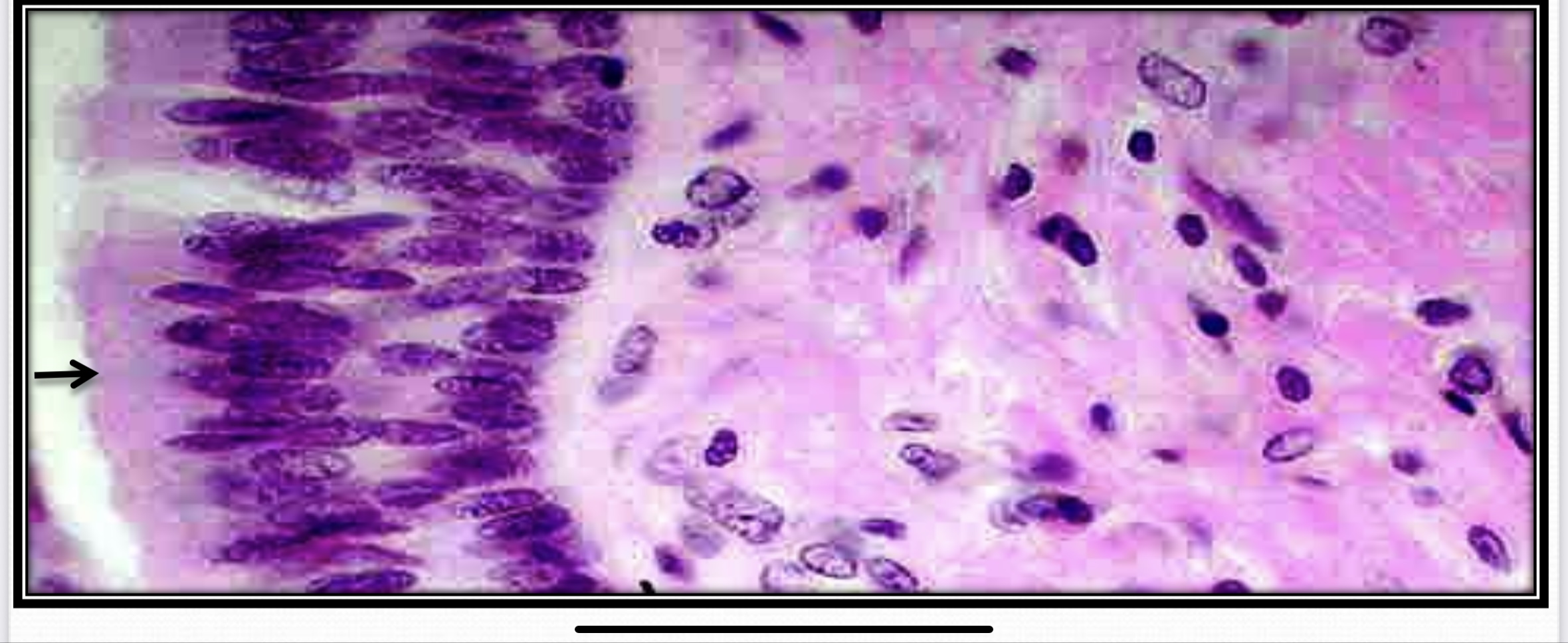
Epithelial Tissue (epithelium). (LM): (Black arrows), pointed on pseudo-stratified columnar ciliated (motile) epithelium with goblet cells stained with general stains
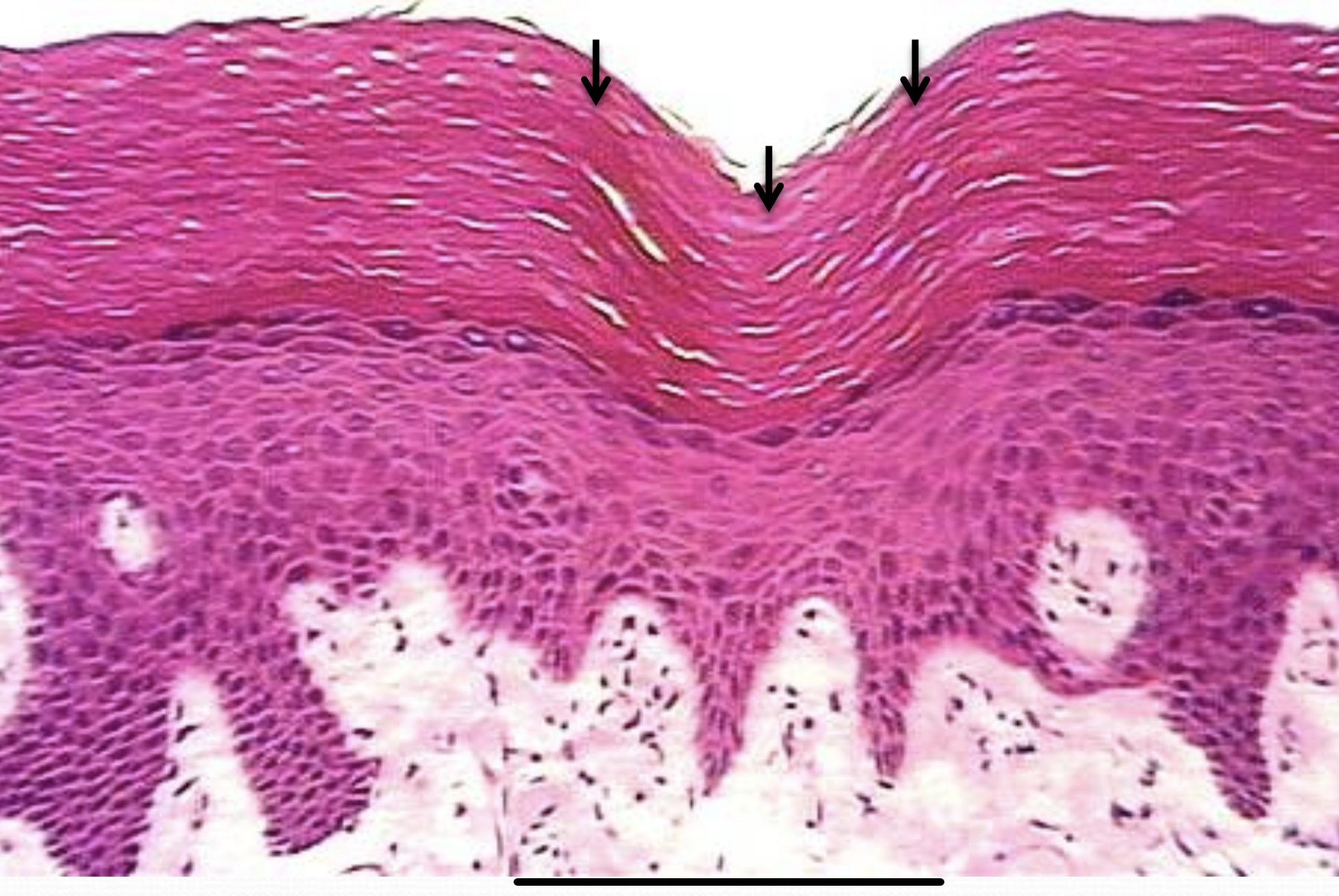
Epithelial Tissue (Epithelium) (LM): (Black arrows) pointed on keratinized stratified squamous epithelium stained with general stains
Keratinized stratified squamous epithelium
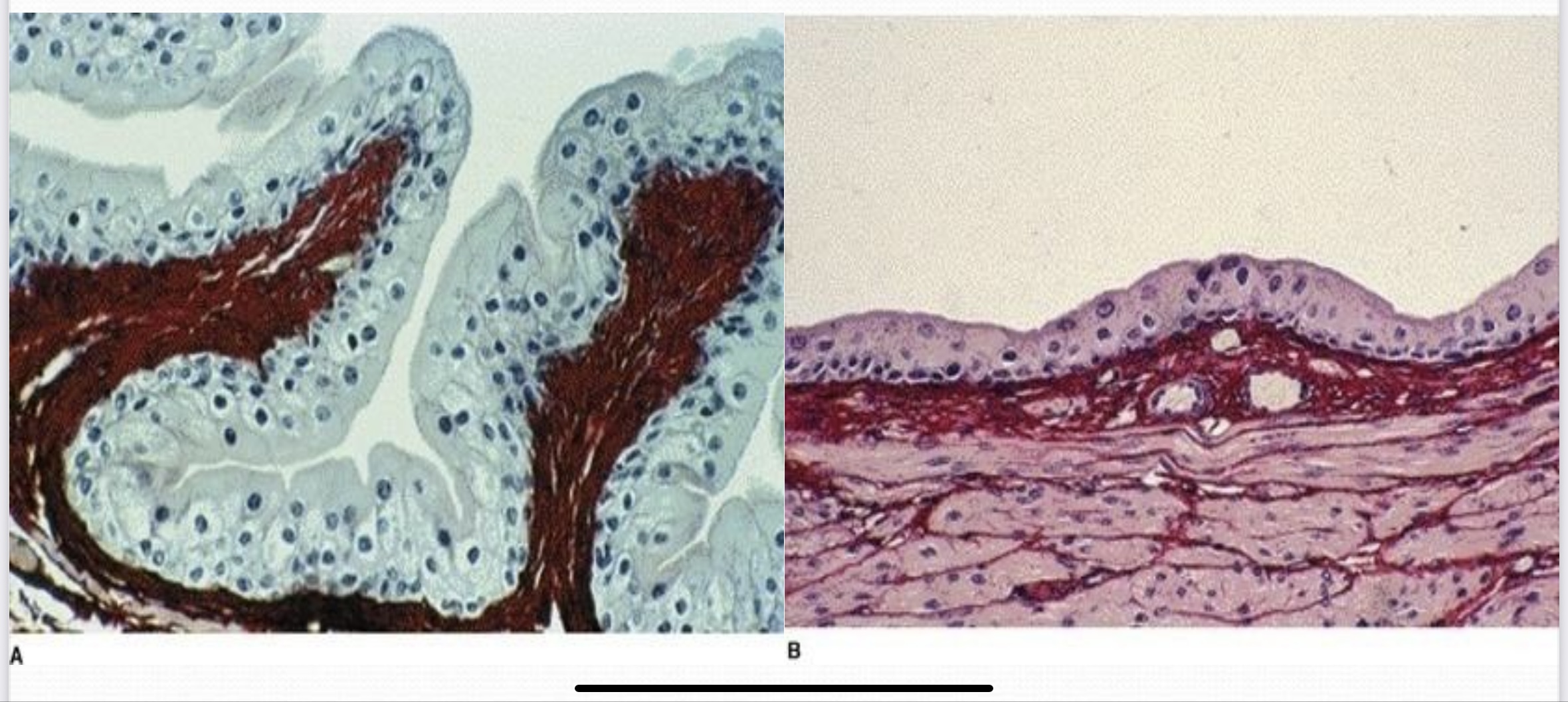
Iransitional Epithelium
Urothelum (LM): (A) pointed on urothelum ir
empty
urinary bladder
B pointed on urothelium in full urinary bladder
stained with general stains
Transitional Epithelium (Urothelium)
Epithelial Tissue (Epithelium) (LM):. (Black arrow).pointed on stratified columnar epithelium
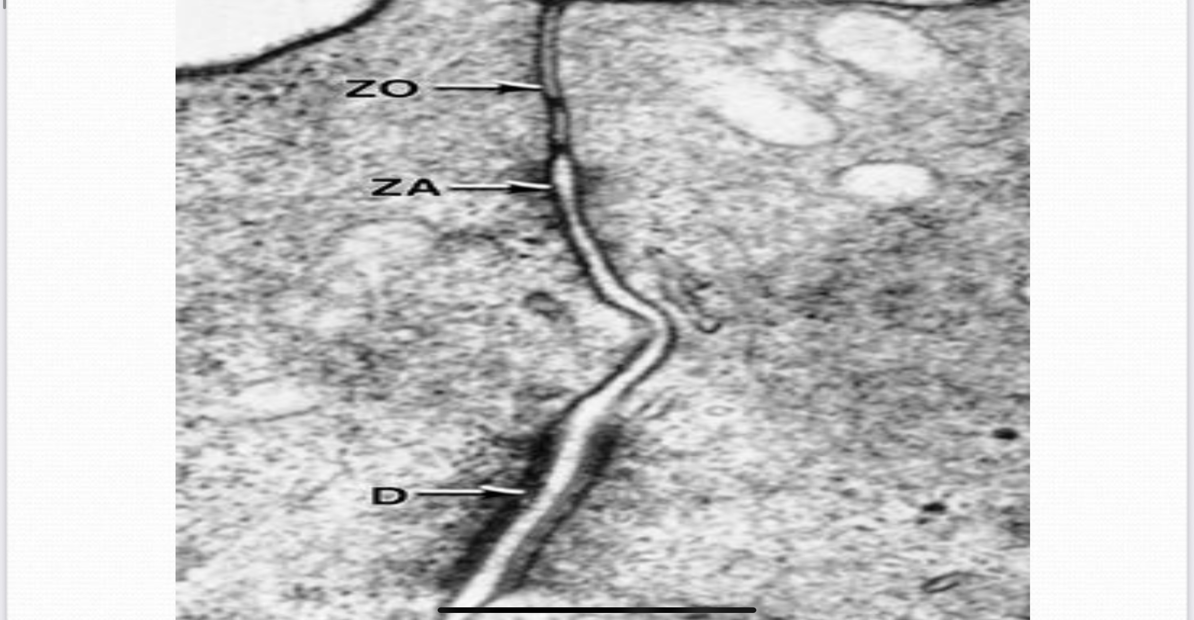
Zonula occludens
uncons on donsome matt adheren achere canes.on zonula occludens(adherence/anchoring junctions de
Zonula adherens
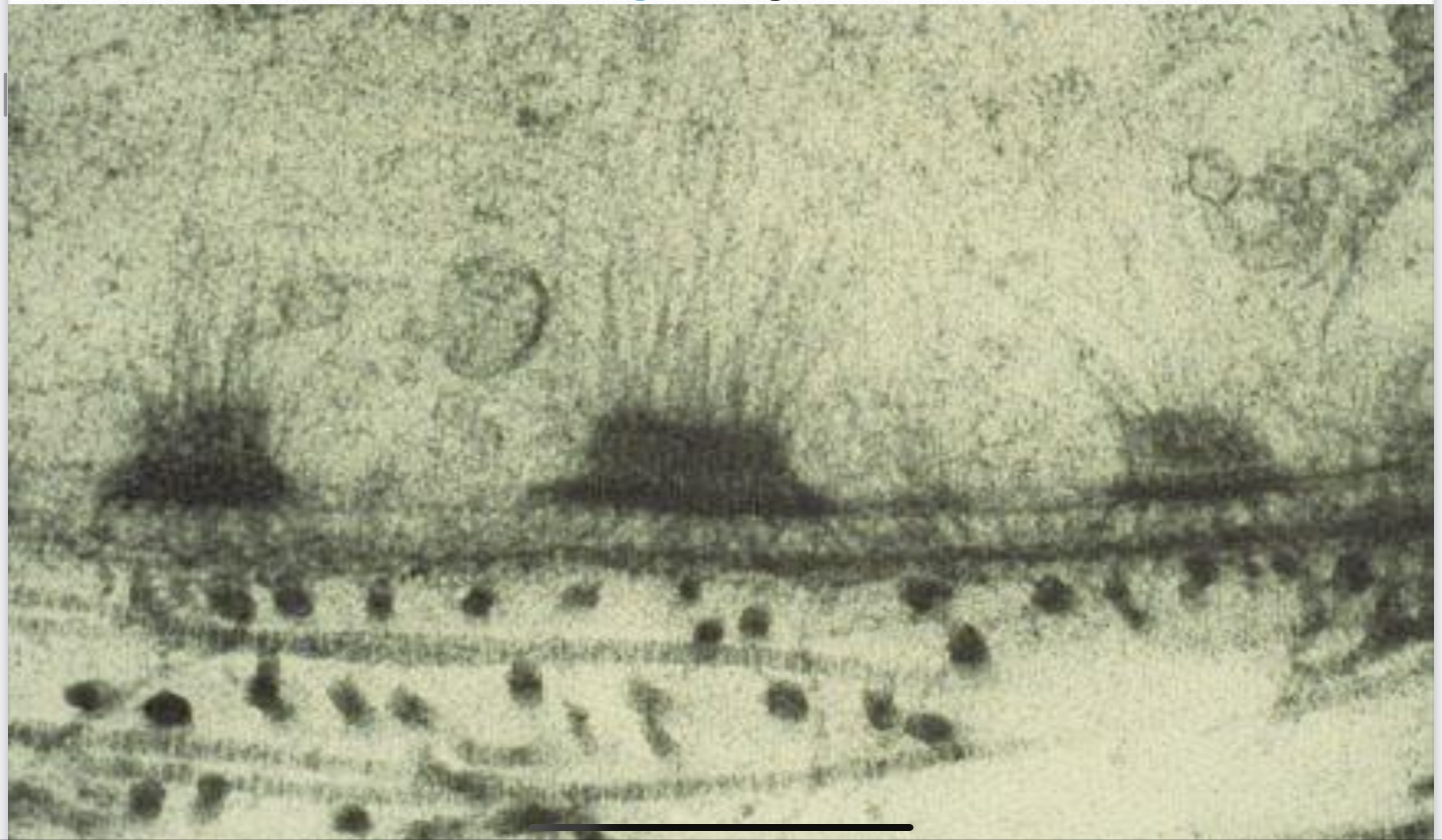
Hemidesmosome (TEM):
Desmosome (macula adheren)
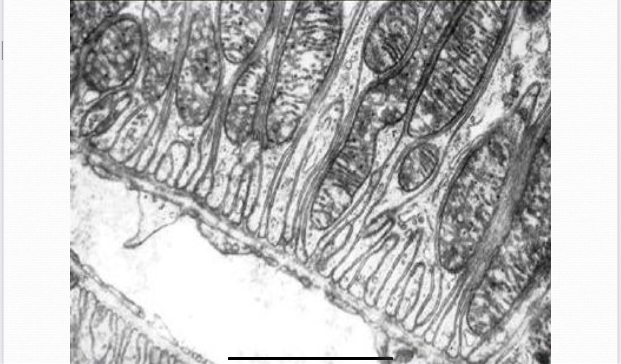
Basal membrane infoldings (TEM):
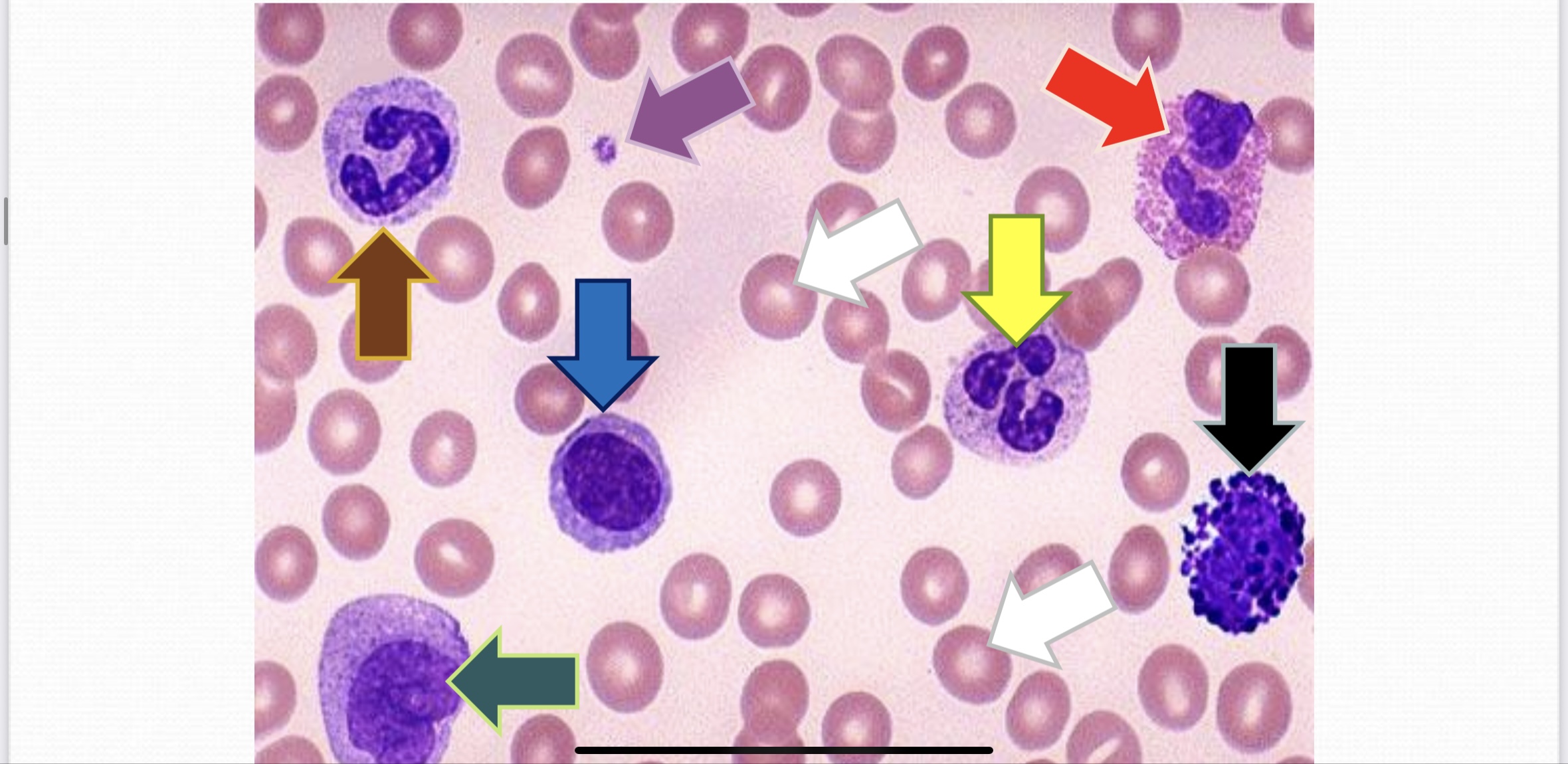
Thin Blood film (LM): (Red arrow) points on esinophil, (white arrow) points on erythrocytes, (blue arrow) points on lymphocyte, (black arrows) points on basophils, (brown arrows) points on banded neutrophils, (yellow arrow) points on neutrophil, (purple arrow) points on platelet, & (green arrow) points on monocyte, slide stained with giemsa stains
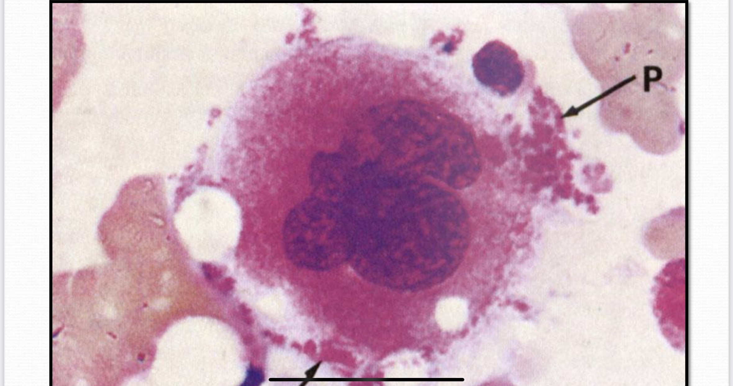
Bone Marrow Smear - Megakaryocyte (LM): (P) points on platelets stained with giemsa stains
Mesenchymal Connective Tissue
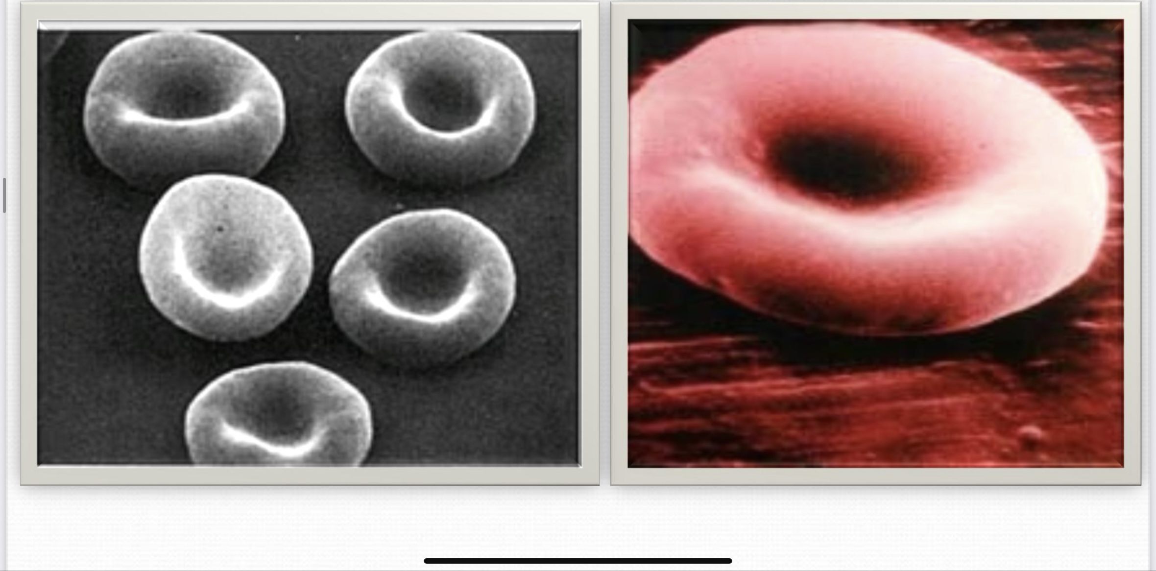
Red Blood Corpuscles (erythrocytes) (SEM):
Reticular Connective Tissue
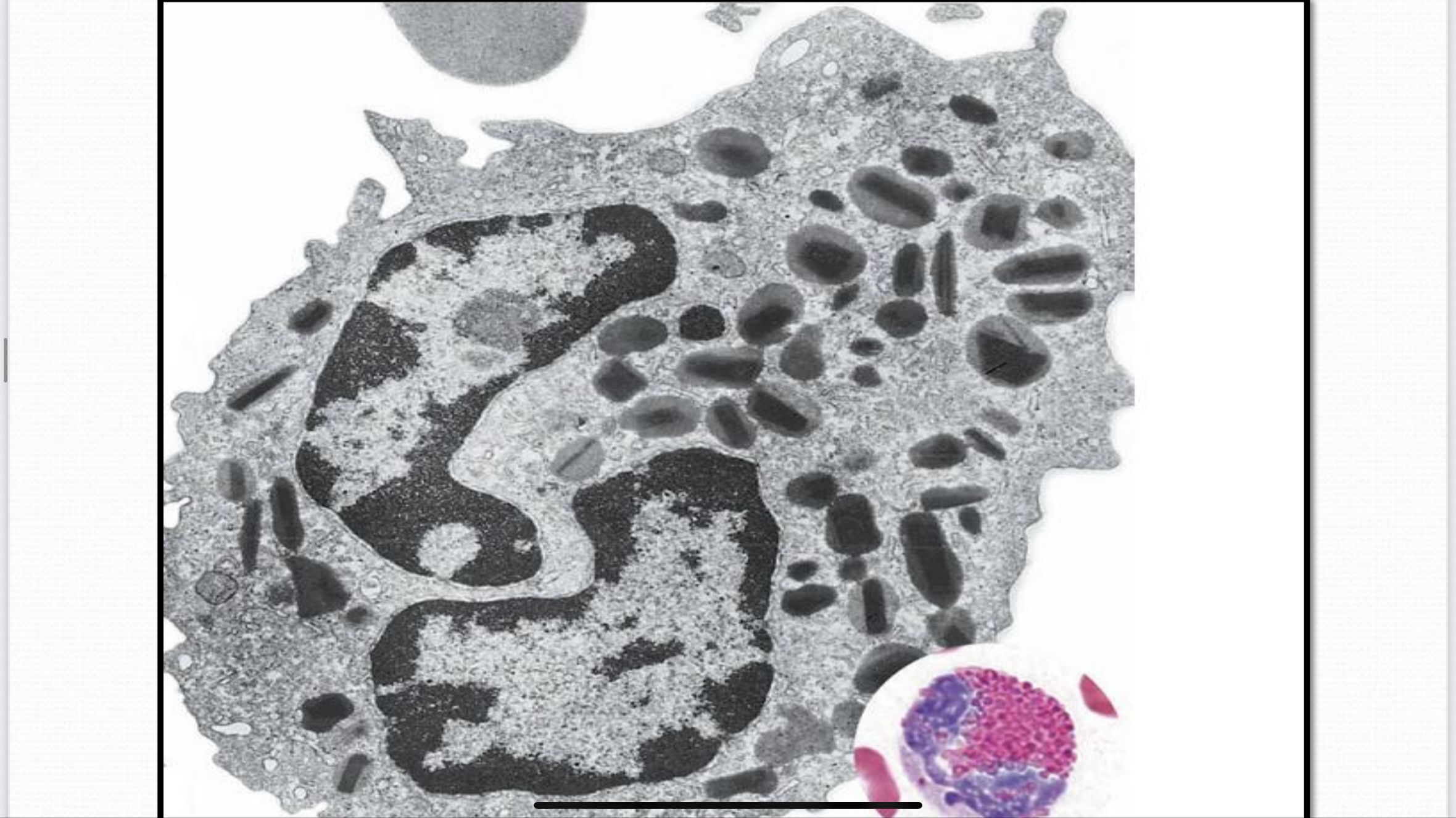
Esinophil (TEM):
White Adipose Connective Tissue
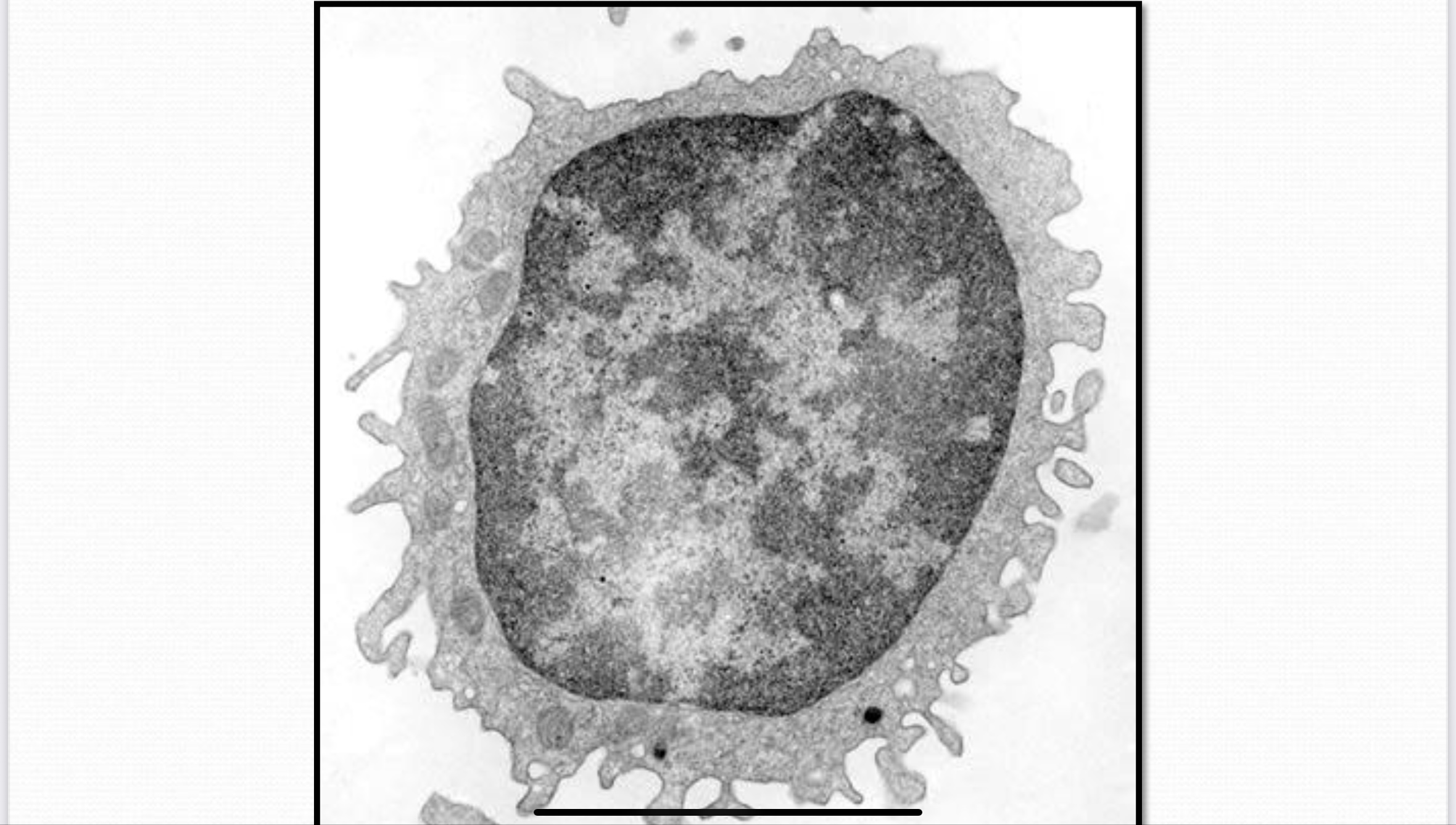
Lymphocyte (TEM):
Brown Adipose Connective Tissue
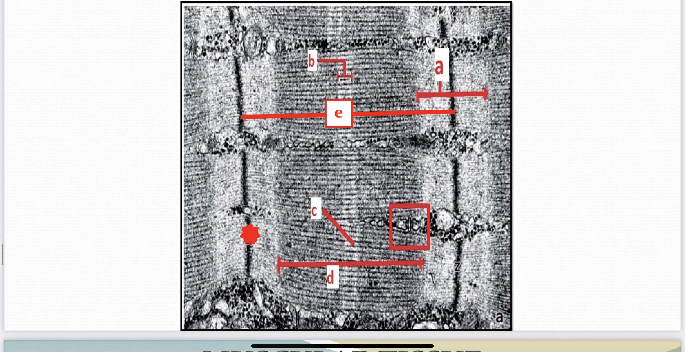
Skeletal Muscle Tissue (TEM): (a marked) points on light I-band, (b marked) points on H-zone, (c marked) points on M-line, (d marked) points on dark A-band, (e marked) points on sarcomere, (star marked) points on Z-disc, & (square marked) points on triad tubular system
Dense Yellow Elastic Connective Tissue
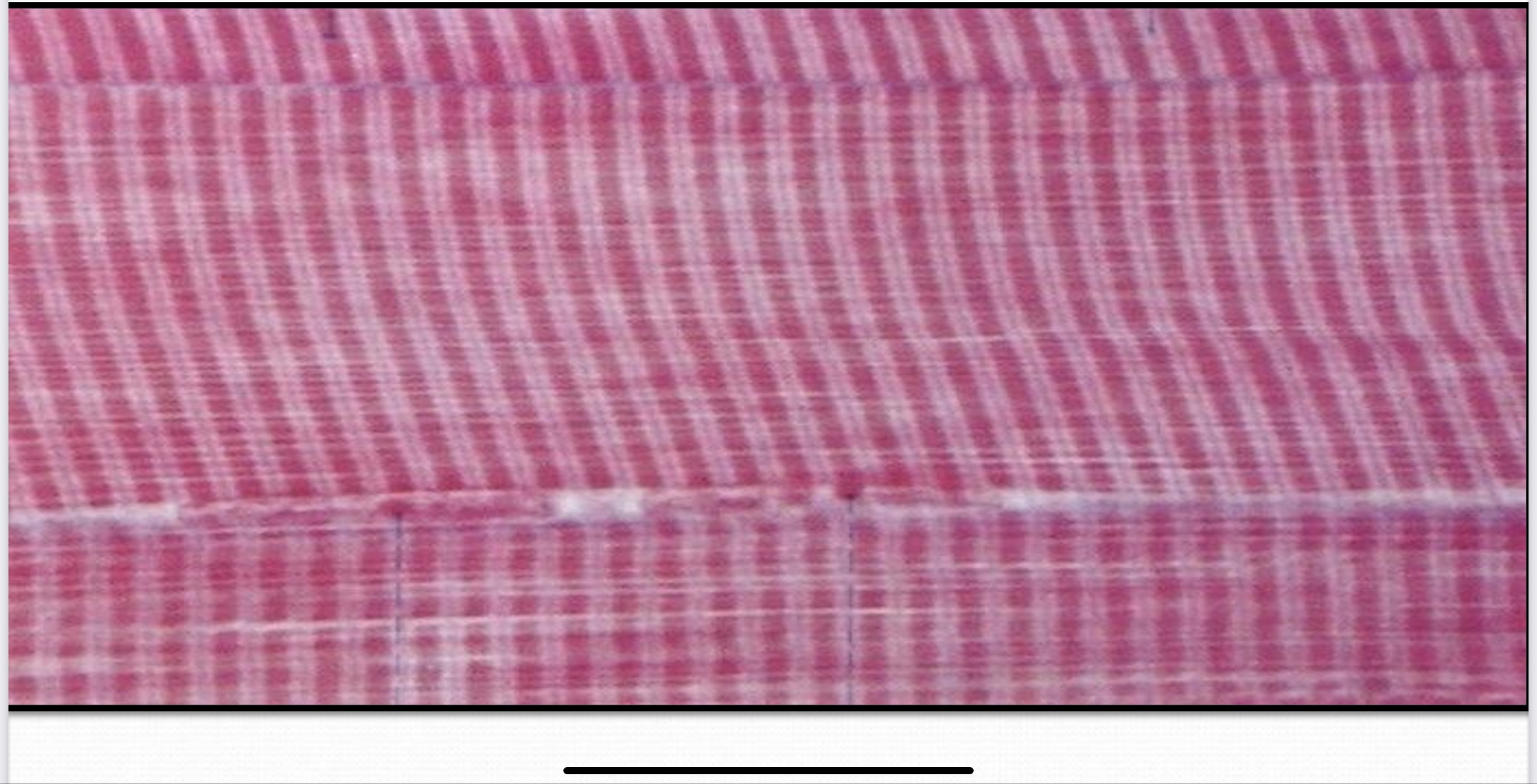
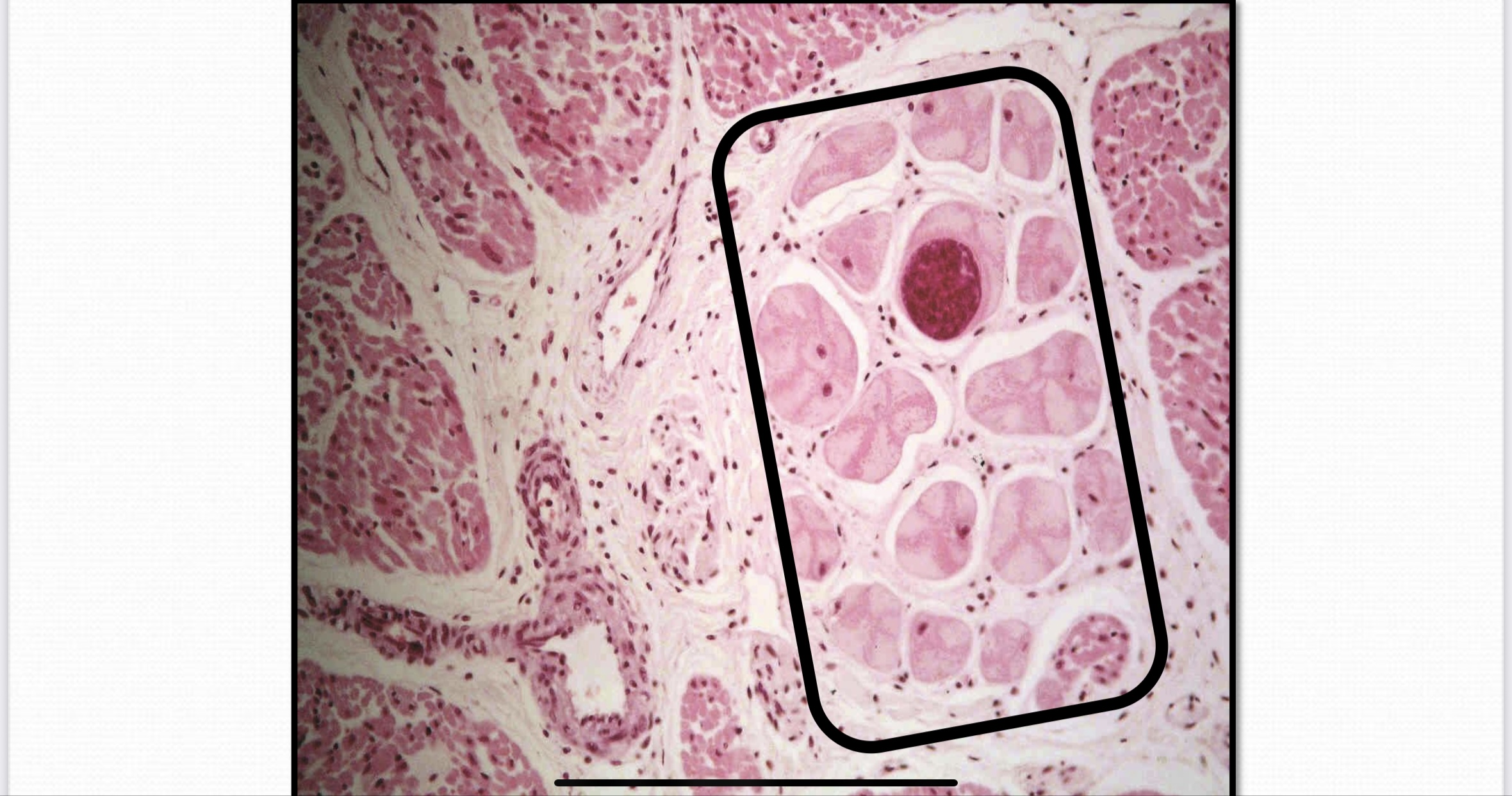
Skeletal Muscle Tissue (L.S) (LM): Stained with general stains
Mast cell
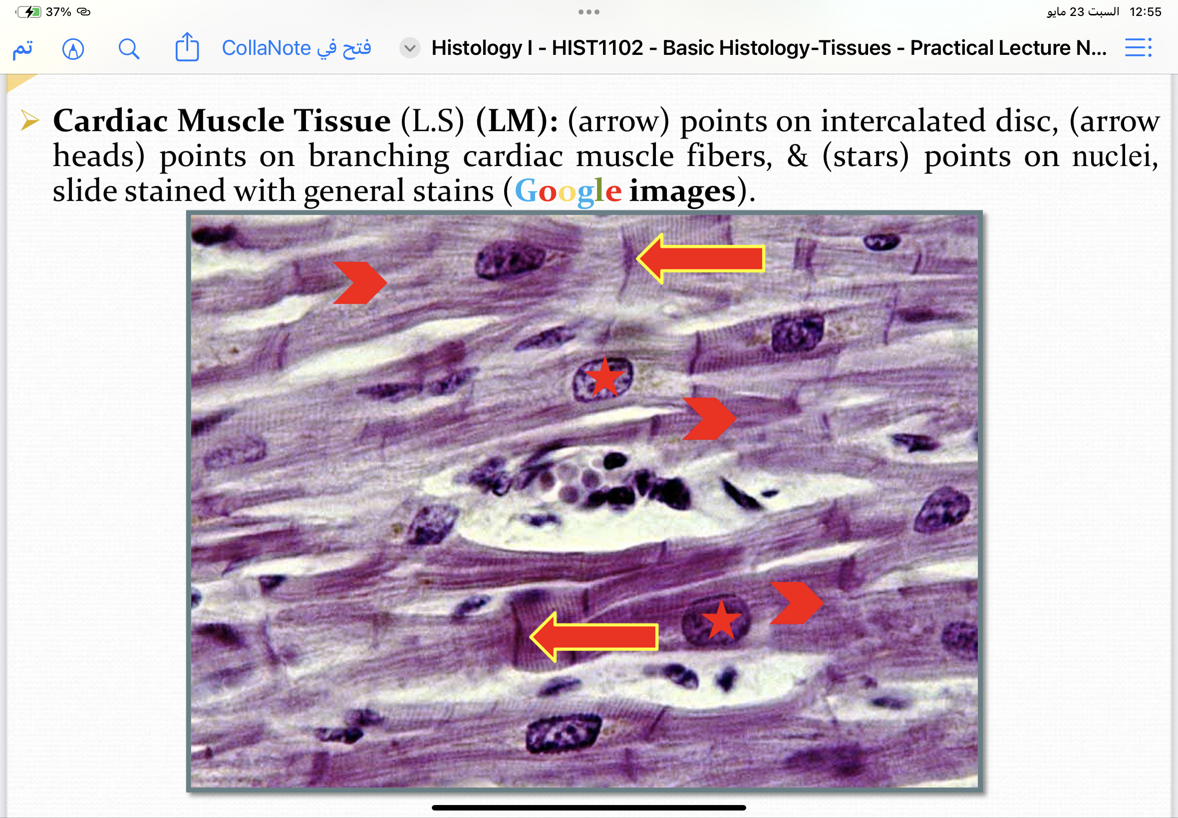
Cardiac Muscle Tissue (L.S) (LM): (arrow) points on intercalated disc, (arrow heads) points on branching cardiac muscle fibers, & (stars) points on nuclei, slide stained with general stains
Cardiac Muscle Tissue (LM): (Square marked) points on purkinje fibers stained with general stains
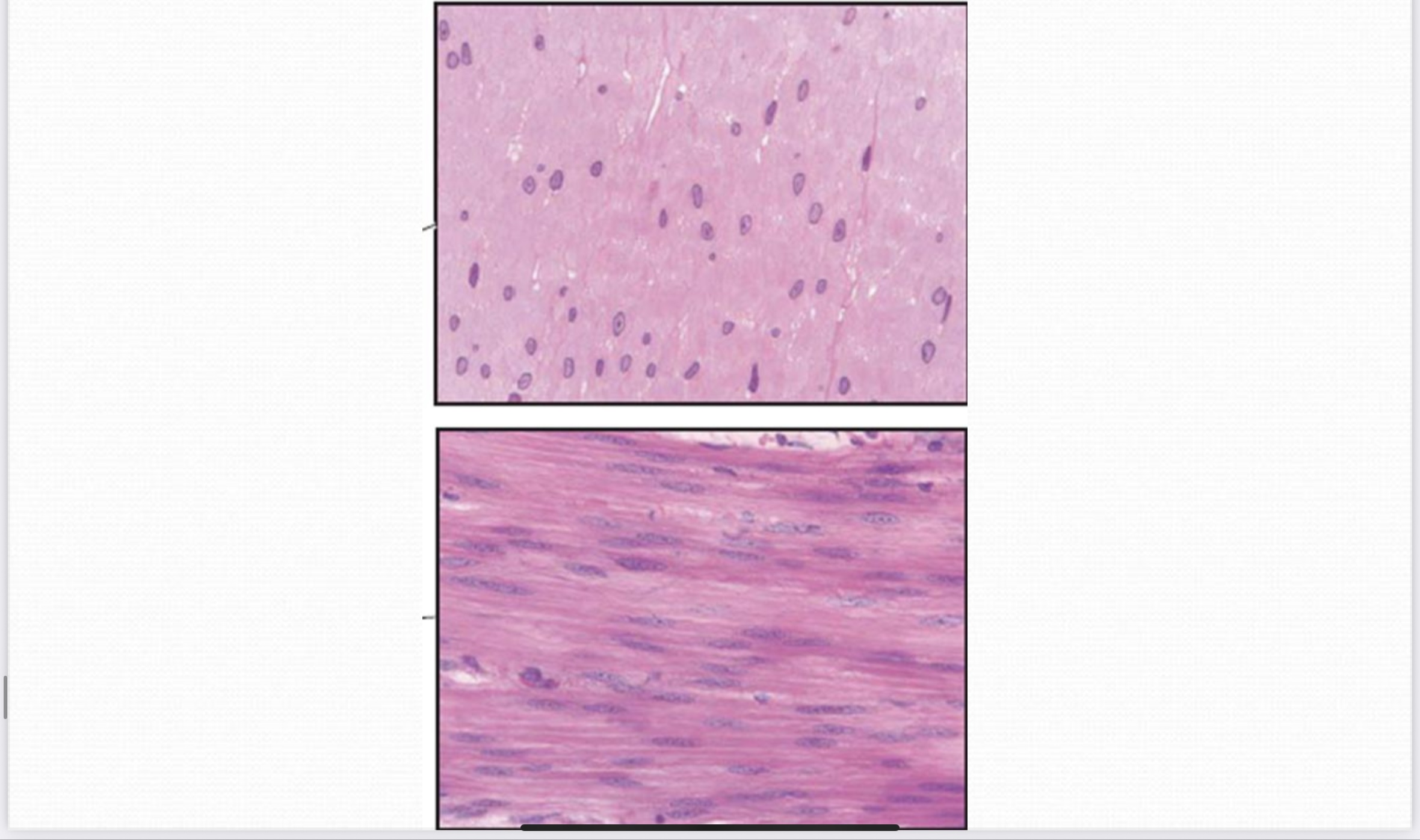
Smooth Muscle Tissue (LM): Stained with general stains
Megakaryocyte
A large bone marrow cell responsible for the production of platelets (P).
Hyaline Cartilage
A type of cartilage with a glass-like appearance, visualized in Light Microscopy.
Chondroblasts
The young chondrocytes responsible for the growth of cartilage.
Chondrocytes
Mature cartilage cells that can be stained with PAS (Periodic acid-Schiff) to detect glycogen and mucous.
Compact Bone
Bone tissue characterized by Haversian canals (HC), Interstitial lamellae (IL), and Lacunae (L).
Osteoclasts
Large bone cells involved in the resorption and removal of bone tissue.
Sarcomere
The functional unit of skeletal muscle, identified in TEM along with structures like the Z-disc, I-band, and H-zone.
Intercalated disc
A specialized connection between cardiac muscle fibers that allows for synchronized contraction.
Purkinje fibers
Specialized cardiac muscle fibers involved in the electrical conduction system of the heart.
Neuroglia
Also known as 'nerve glue', these are the supporting cells of the nervous system.
Nissl's granules
Structures within neurons composed of polyribosomes or polysomes.