perception 12 -physiology
1/28
There's no tags or description
Looks like no tags are added yet.
Name | Mastery | Learn | Test | Matching | Spaced | Call with Kai |
|---|
No analytics yet
Send a link to your students to track their progress
29 Terms
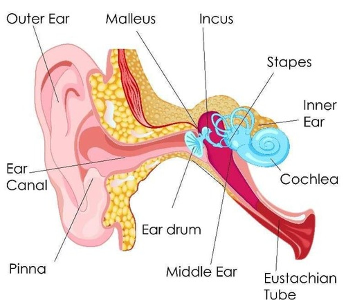
ear divisions
-outer ear with pinnea and external auditory canal
-middle ear has the 3 bones (malleus, inchs, stapes)
-then inner ear with the cohlear
Pinnae
They enhance hearing sensitivity
-need to be smaller than the wave to funnel and control it maybe
- therefore used for high frequencies and uniquelty mammalian
-some animals can direct and control their pinnea to control sound also
Less clear why primates have fixed ears
-may be due to the role in sound localisation (maybe more important than picking u sound ore sensitively from diff locations)
For non-mammal no pinnae
-just ear with hole
-for the frequencies it can hear would need a pinnae bigger than its head
Owl basically only animal with an analogy to a pinnae - its whole face!,
works a bit like a radar dish (effectively enormous pinnea for frequencies it hears)
after pinna go through...
ear canal
-keeps the delicate things further in the ear away from harm -that's his thoughts
-but does also lead to resonance
-doesn't physically amplify but leads to sound being a bit louder
-therefore consequence for hearing
consequences of resonance in ear canal
3 kHz particularly sensitive because of ear canal and the resonance as it resonates at about 3 kHz
after ear canal reach
-eardrum with the ossicles (3 small bones): the malleus (hammer), incus (anvil), and stapes (stirrup). [1, 2]
Their job is to take sound waves in ear and turn them into sound waves in water
-using their mechanical adavantage - sometimes called stiletto effect
why waves need to e transferred to water
-water is a very good reflector of sound (about 99% reflects)
-cochlear is a fluid filled cavity in skull
how wave transferred water to air
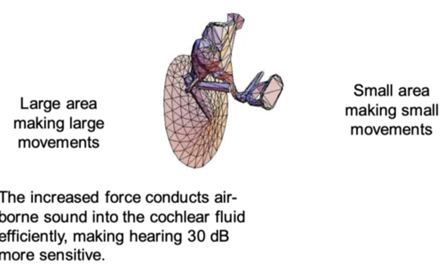
Finite-element model of the human tympanic membrane (before ossicles between them and canal) and ossicles.
Large area making large movements to Small area making small movements
The increased force conducts air- borne sound into the cochlear fluid efficiently, making hearing 30 dB more sensitive. (leverage effect on bones also does this)
-motion of the bones in the smaller area - end bit is stapes footplate
-this neatly fits onto a hole which opens into the cochlear called and oval window
-membrane over the window which it pushes on driving sound into the inner ear
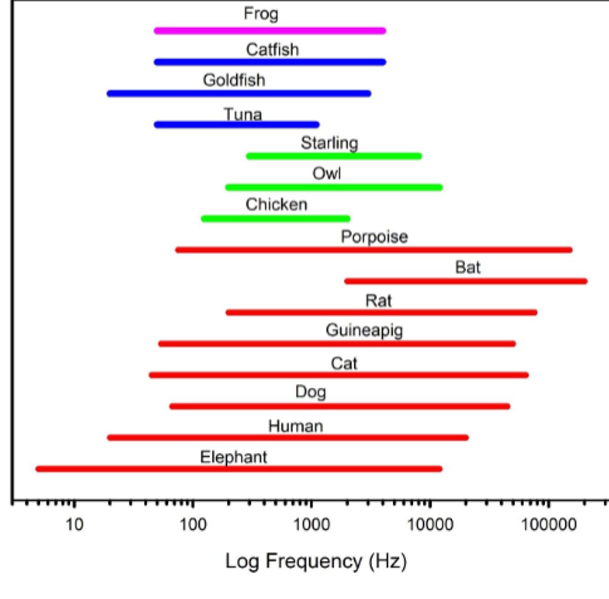
The detectable frequency ranges for different species
Mammals in red have wider range of frequencies
-due to middle ear - mammals have 3 whereas other animals 1 (from the jaw somehow!)
Range of hearing is also generally scaled ith saze (elephants big low frequ, rats small high)
-this is because it scales with sizze of middle ear structures
-bottom end scaled to size of middle ear cavity
(although guinea pig has extra cavity improve it like some others)
--but basically scales with head size
osicles and frequency
-smaller osicles the higher the frequency you can hear
-they are already tiny!
After middle ear get to
The cochlear is where tranduction occurs -wave of sound tuned into nerve impulses
Cochlear cross-section
ADD
cochlear + frequencies
Mammals are curled up - probably as needs to be quite long
-we can hear lots of frequencies and these are arranged along the cochlear
-high frequncies excite the base and low the apex -counter intuitive
how sound travels in the cochlear
Waves of pressure in the fluid sent from the oval plate
-length of cohlear divided into 3 compartments - top scala vestibuli, bottom scala tympani and in middle between 2 membrane is the scala media
-sound comes in on red path in the scala vestibuli
-sound wave travels coiling all the way up to the top then finds a little hole and goes all the way bac down again through the scale tymapni (these meet at the top)
- then has to go somewhere (fluids not compressible)
Goes out of the round window (little whole on left)
membranes in cochlear
The 3 compartments are divvied up by membranes
The most impoortant is the basilar membrane at the bottom (first arrow cross)
-this analyses sounds to diff frequencies
-at bottom tuned to only vibrate to very high frequencies and changes properties as goes up to do the reverse
auditory nerve
Auditory nerve is whole bunch of axons sending if info to nerve
Spiral ganglion is all the cell bodies of all the auditory nerve fibres
basilar membrane
-this analyses sounds to diff frequencies
-at bottom tuned to only vibrate to very high frequencies and changes properties as goes up to do the reverse
-like a harp
Similarly, the basilar membrane is narrow, light and stiff at the bottom and wide, heavy and floppy at the top.
Both with vibrate "in sympathy" with sounds of different frequencies in the air.
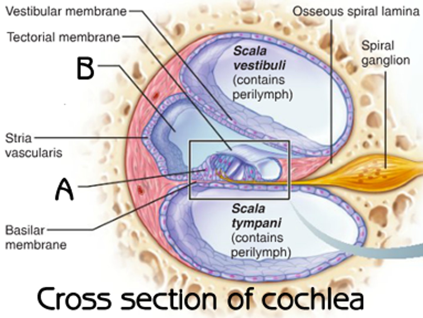
Structure of cochlear zoomed in
Scala vetsibuli + tympani are filled with perilymph whereas sala media is filled with ednolymph (different fluid chemically + electricall)
-the basilar membrane separates scala tympani and scala media
-organ of corti sort of within scala media
-the stria vasculari -provide lots of blood vessels to scala media
Spiral ganglian has dendrite from cell bodies to organ of corti - touching specialised cells
ADD PIC
what potential differences we are interested in
The one btween the scala media and organ of corti
-stria vasculari pumps potassium ions into the scala media increasing the postive charge and creating potential differe
(hence the endolymph)
Organ of Corti structure
-specialised hair cells with stereocillia
-inner vs outer depending where they are
-inner har cells connect to afferent nerves and outer to effecerent
-these sit on the basiliar membrane which can move
-and under the tectorial membrne which can't
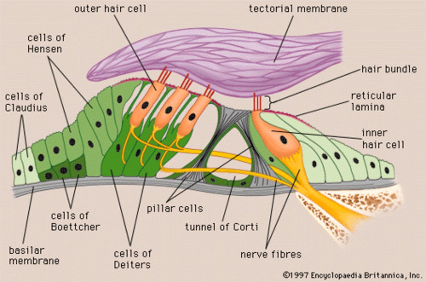
what happens in organ of corti when there is a sound wave
-when a sound wave moves the basiliar membrane up and down the hairs get moved back and forth - especially as the tectorial membrane will brush across the top of the organ corti
Tip links (string like connection) connect each stereocilium in the bundle to the next.
They pull open ion channels (mechanically gated) during deflection of the bundle. (they open and close with the movement)
Ledds to release of voltage in scala media (all those positive ions)
Changes the voltage of the cell leading to realease of nt at base where connected to dendrites in spiral ganglion
(inner hair cells, afferent nerves, trnaduce)
What about outer hair cells
-oddly the outer hair cells only has efferent nerves (from the brain rather than to it)
-suggests under control of brain in some way -we're not sure what
-most likely probability is that rain is trying to tell them to turn the volume down
outer hair cells study set up
Hudspeth, (2008)
-micropipettes can be attached OHCs aand fuse with the membrane
-this means electrical signals can be sent through them and they can affect voltage
See music causes the cell to move and certain things cause it to move more than others (other way round to what we think of)
what ohc studies suggest
This shows that the hair cells themsevles not just the basilar membrane are frequency tuned
We think that the movement of the cells increase the basiliar movement
Positive feedback application
-increases what you can hear - phsycial amplifications
when amplification does and doesnt happen
-these amplificaations can be 60 dB for low level sounds, but reduce with increasing sound explaning slow loudness growth
-could be because they cant amplify it any more or respond differently -consider theyre efferent
studies have stimulate relevant bits of brain and found reduction which could suggest less effort
Providing some kind of controlled amplification
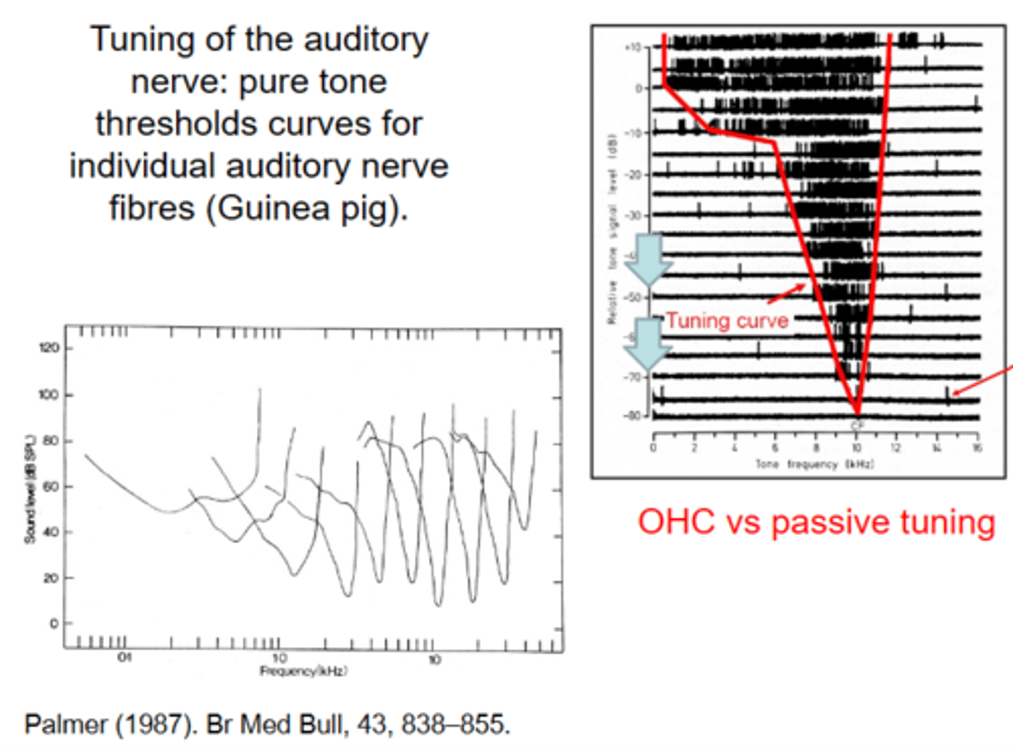
Tuning of the auditory nerve: (study)
pure tone thresholds curves for individual auditory nerve fibres (guinea pig) - Palmer
-put electrode in auditory nerve
-they play a tone sweep
-when it reaches the preferred frequency the cell starts firing (cells come from different points of basiliar membrane tuning)
-very discrete region (frequency) which gets wider and wider with increasing intesnsity (dB)
If you do tis for lots of auditory nerve fibres this creates a spectrum where different frequecnies are picked up by different auditory nerve fibres
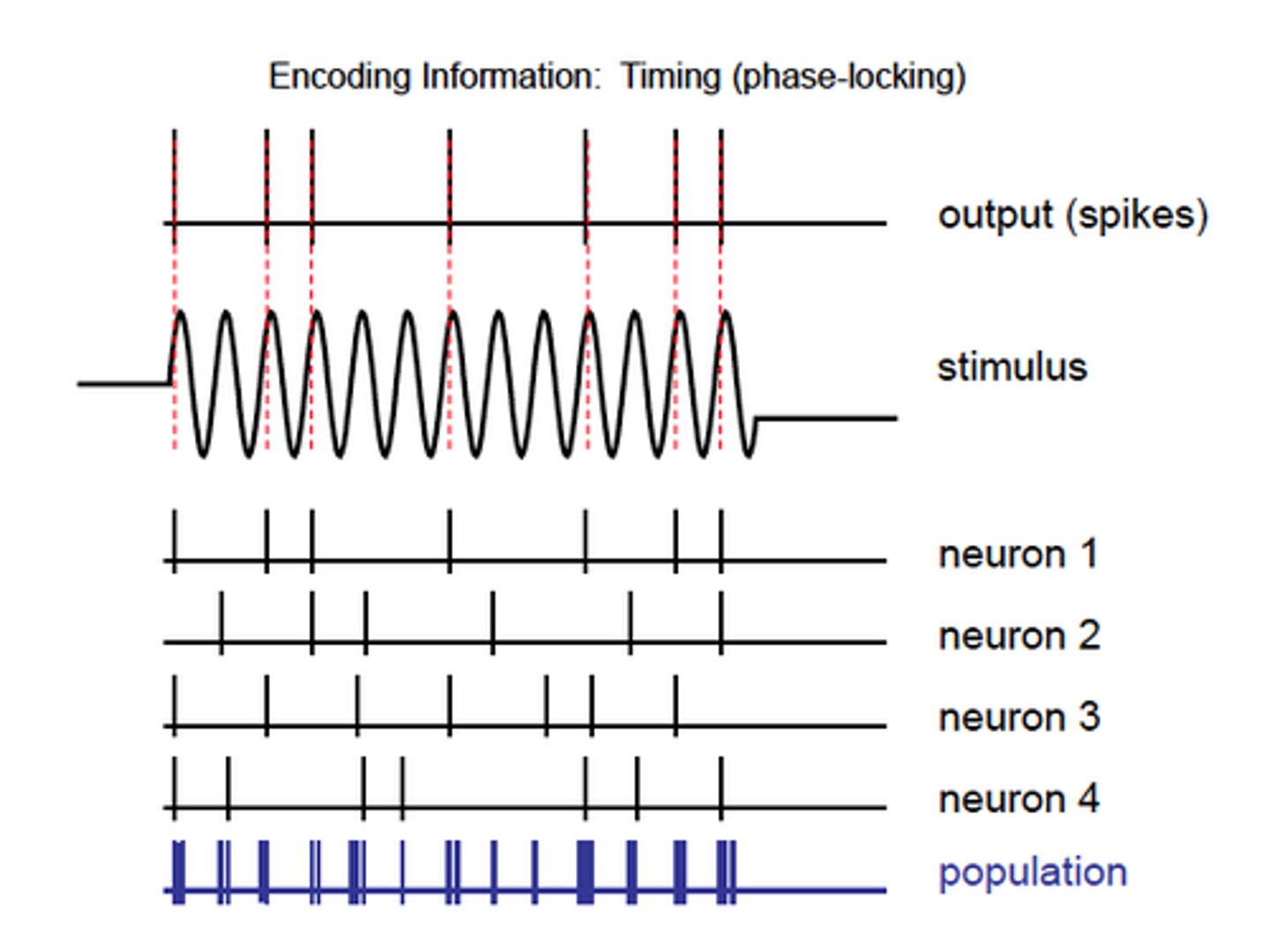
Phase Locking
When stimulated get an action potential but people noticed these are not random
-in most sensory fires its more aps with more intense stimulus but they are occuring randmly in time
Auditory nerve seems to respond to the same part of the cycle
-not responding to the whole thing but trying to convey the waveform
-not just sound at the frequency but sound at this frequency of this shape
Use lots of different waveforms to do this by different ones firing at different points of wave
Volley principle as combine lots of fibres
gross electoride study
Wever and Bray
-gross electrode on whole auditory nerve
-from this got the actual sound - could hear what someone as saying
Relies on having lots of fibres - humans about 30,000 about 10 for each hair cells
Auditory transduction: an overview.
1) Sound waves set the ear drum (tympanic membrane) in motion.
2) The vibrations are passed through the middle-ear bones (the ossicles) to the inner ear.
3) Pressure waves in the inner ear fluid set the basilar membrane in motion, different frequencies in different places.
4) Stereocillia of hair cells on the basilar membrane flex back and forth.
5) Tip-links at the tops of the stereocillia pull open and close ion channels.
6) Current flows into hair cells in time to vibration.
7) Outer hair cells change length amplifying basilar membrane vibration.
8) Inner hair cells stimulate phase-locked action potentials on the auditory nerve.