NEUR200 Exam 1
1/69
There's no tags or description
Looks like no tags are added yet.
Name | Mastery | Learn | Test | Matching | Spaced |
|---|
No study sessions yet.
70 Terms
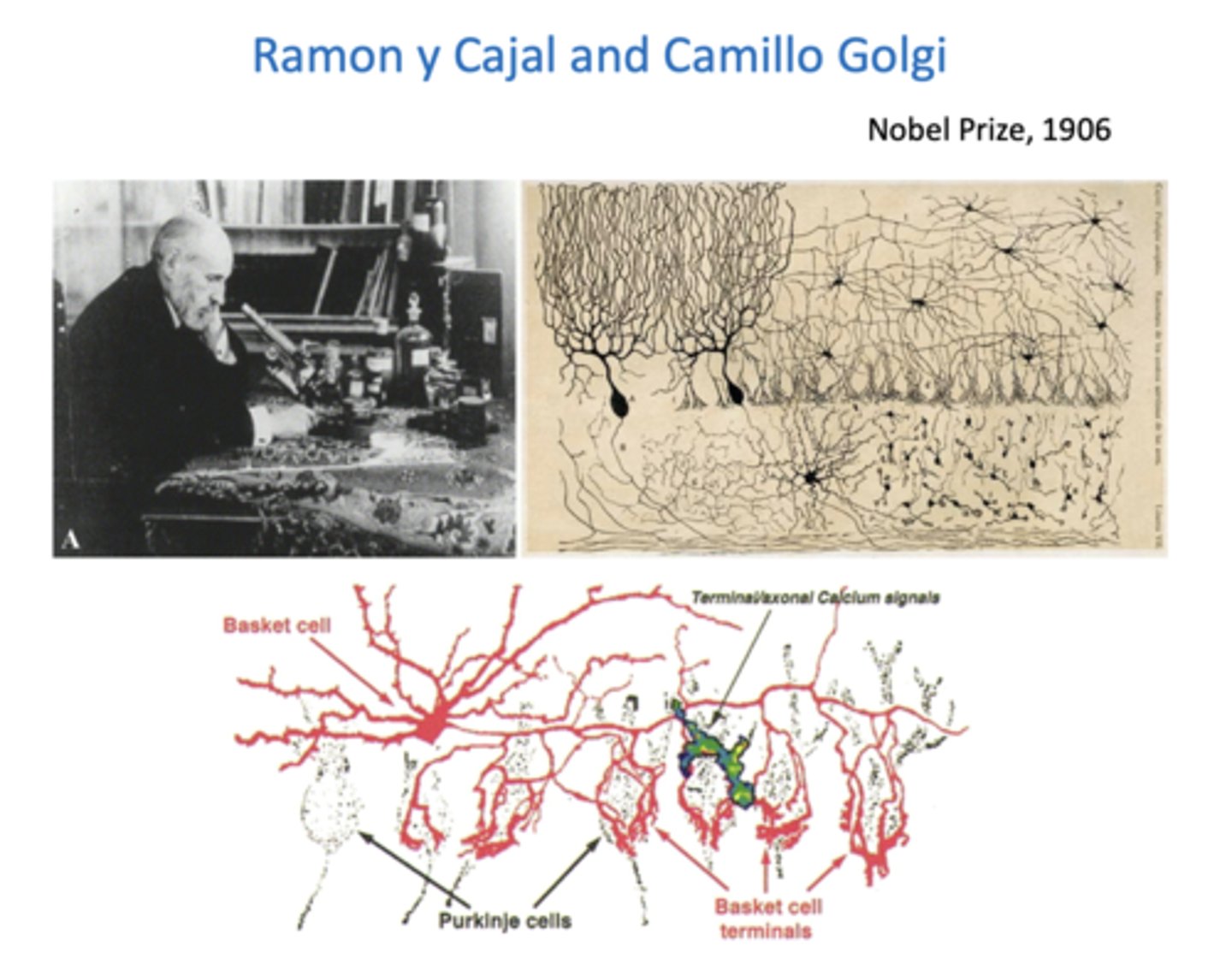
Ramon y Cajal contributions to neuroscience
- determined nerve cells remain separated
- separation between neurons = synapse
- looked at neurons under microscopes and drew what he saw, drawings still used today
- determined structure of neurons
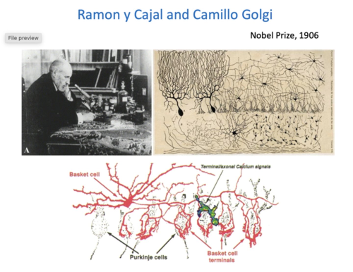
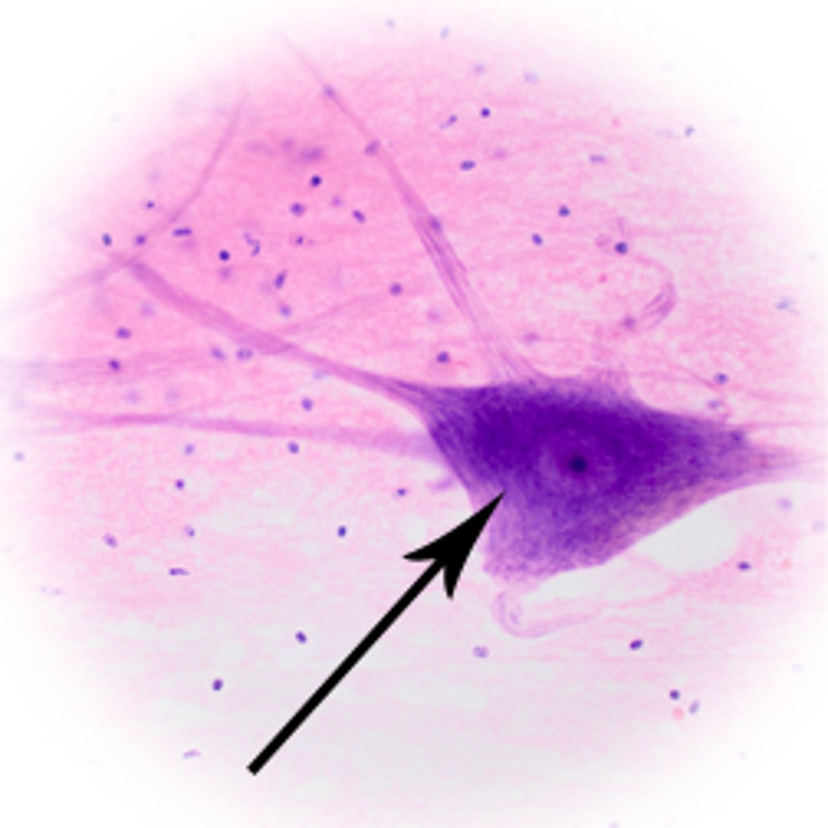
Golgi contributions to neuroscience
- neuroscientist that developed method (silver staining technique) to stain limited number of nerve cells
- revealed shape and organizations of neurons in the brain
- discovered black reaction -> hardens brain tissue and stains sections of it, only some brain cells are clearly stained
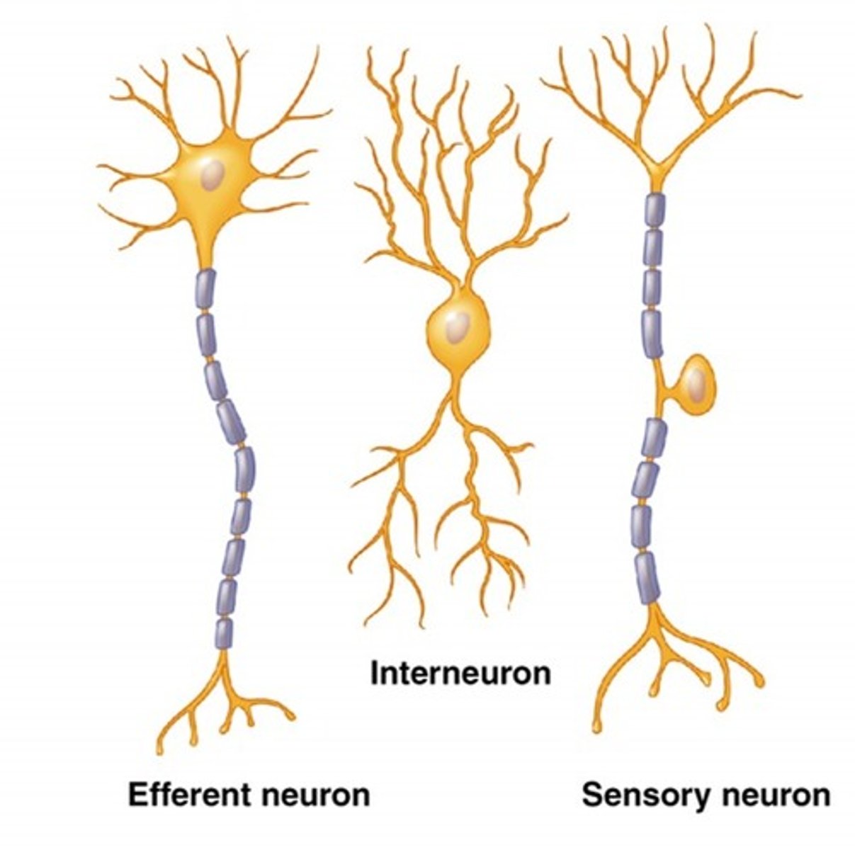
the three functional classifications of neurons
sensory neurons, motor neurons, interneurons
the three structural classifications of neurons
multipolar, bipolar, unipolar
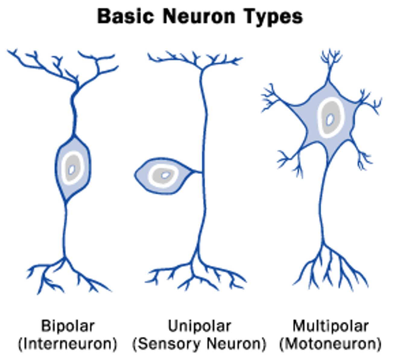
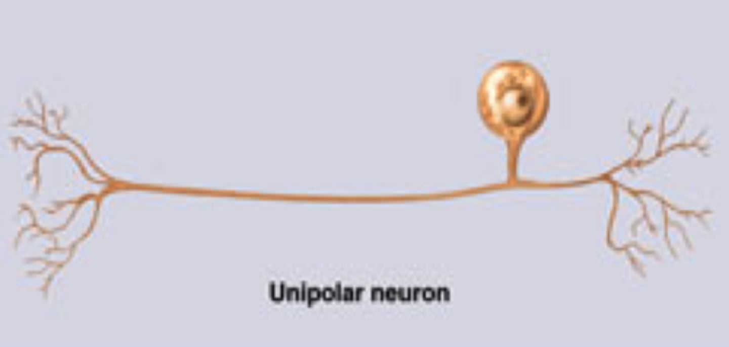
unipolar neurons structure/function
- a single process extending from the cell body
- one axon divides into peripheral and central processes: one serves as an input zone and the other as an output
- dendrites found at the receptor and the axon leads to the spinal cord or brain
- conduct impulses toward the CNS
- most of the body's sensory neurons are unipolar
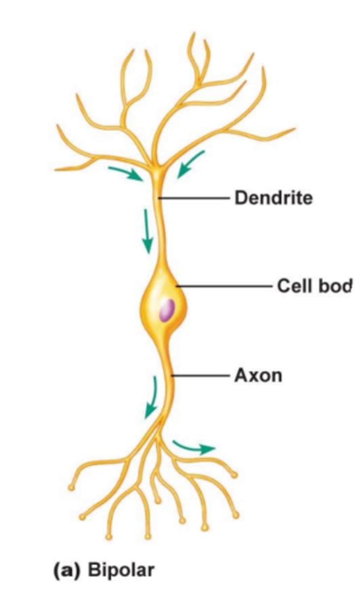
bipolar neuron structure/function
- two processes attached to the cell body: one single dendrite and one single axon
- found only in the eye, ear, and olfactory mucosa
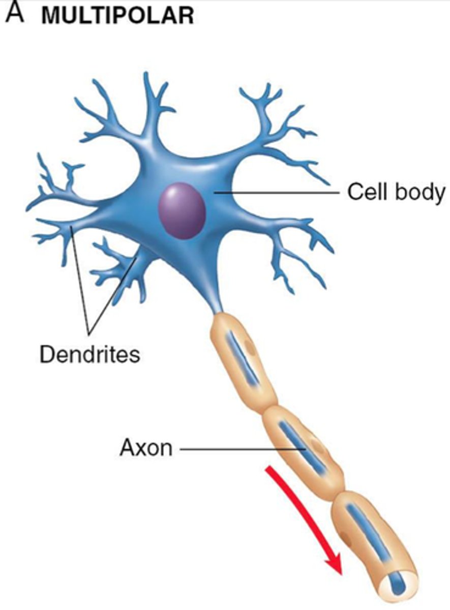
multipolar neuron structure/function
- at least 3 processes attached to the cell body: at least two (often many) dendrites and one axon
- the most abundant type of neuron in the body
- multipolar neurons integrate many pieces of information (from many dendrites) to produce a single response which is transmitted through the axon
sensory (afferent) neurons
afferent -> "arrive"
- carry impulses from sensory receptors in the skin, internal organs, muscles and special sense organs toward the CNS
- light, sound, touch
- the soma of sensory neurons is located on the trunk
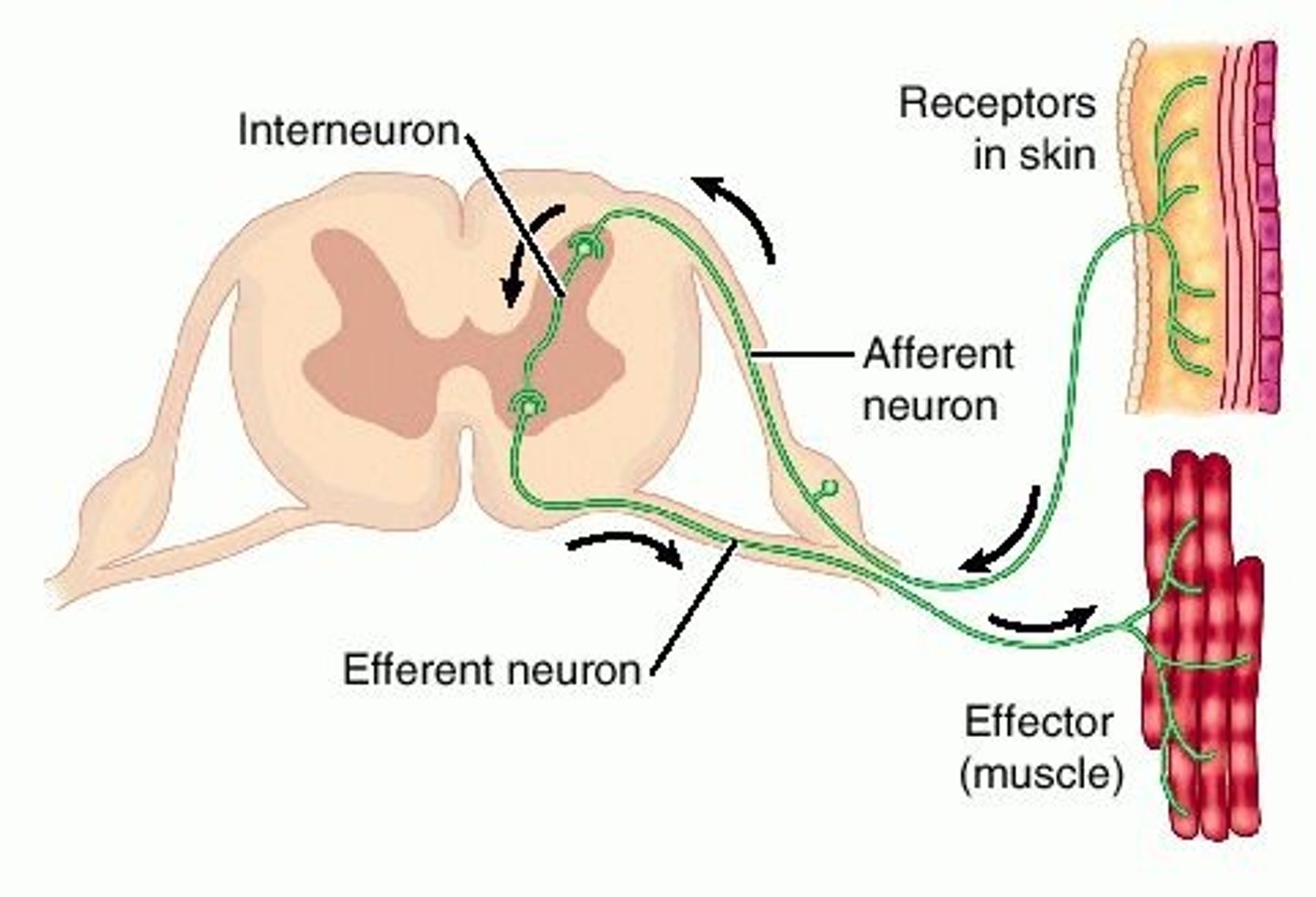
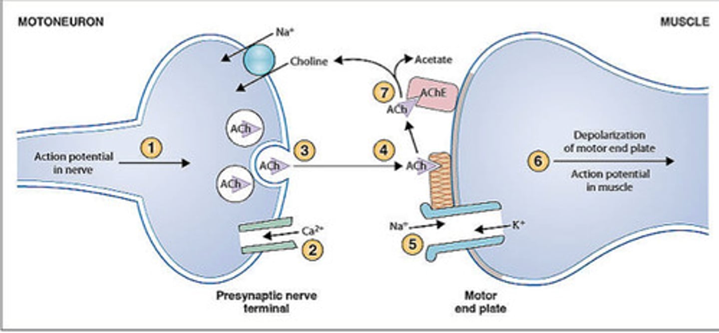
motor (efferent) neurons
efferent -> "exit"
- carry impulses away from CNS to organs, muscles, and glands
- the soma of motor neurons is located in the spinal cord + conducts impulses along its axon to muscle
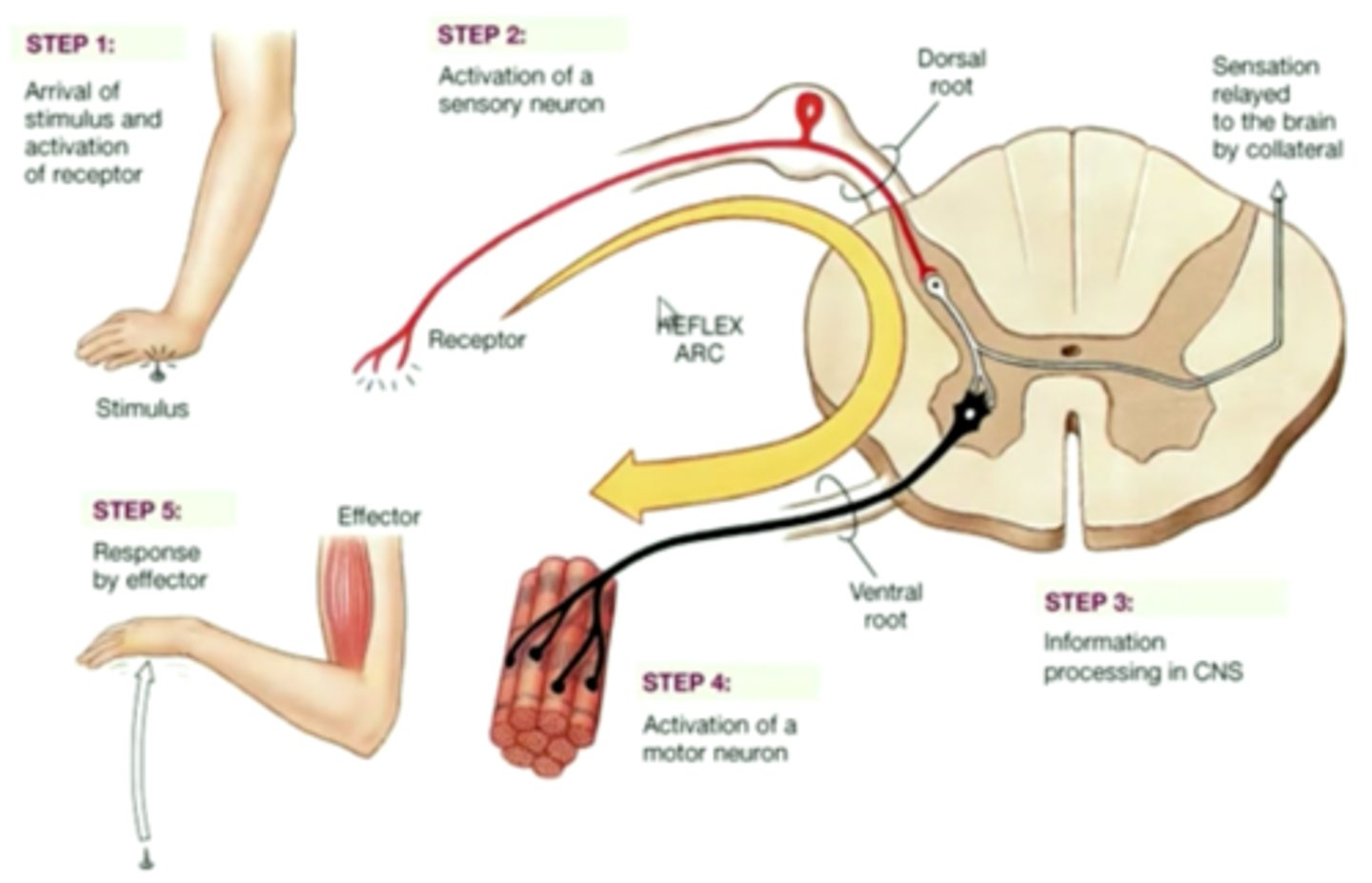
association (intrinsic) neurons
- located in the CNS between sensory and motor neurons
- conduct impulses within the CNS

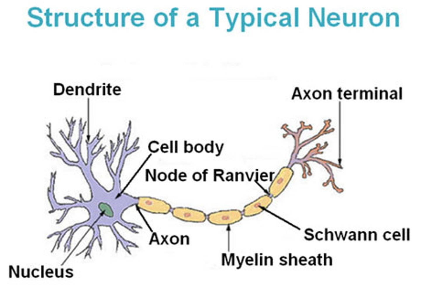
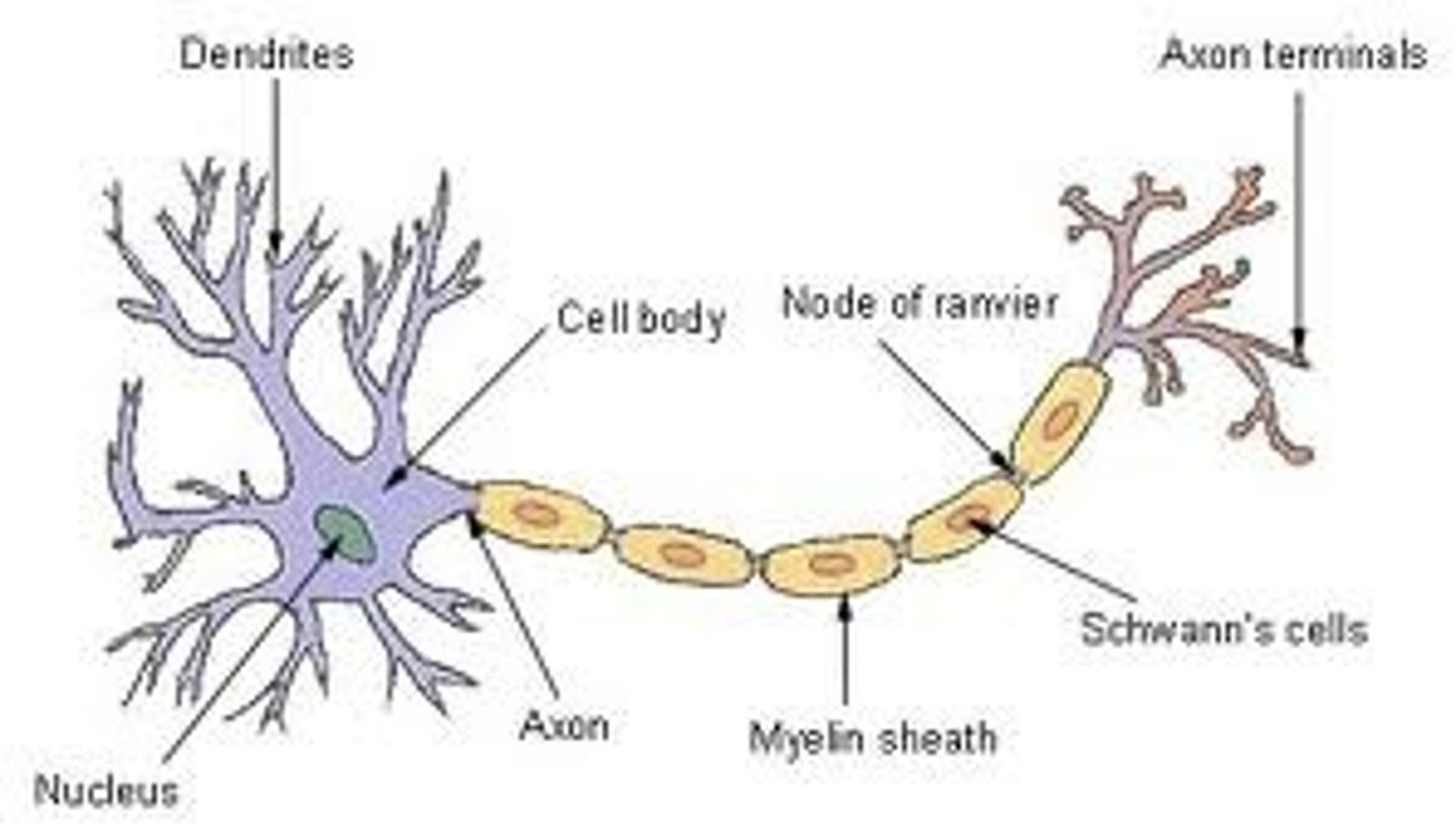
main characteristics of a neuron
- dendrites
- cell body or soma
- axon
- presynaptic terminal
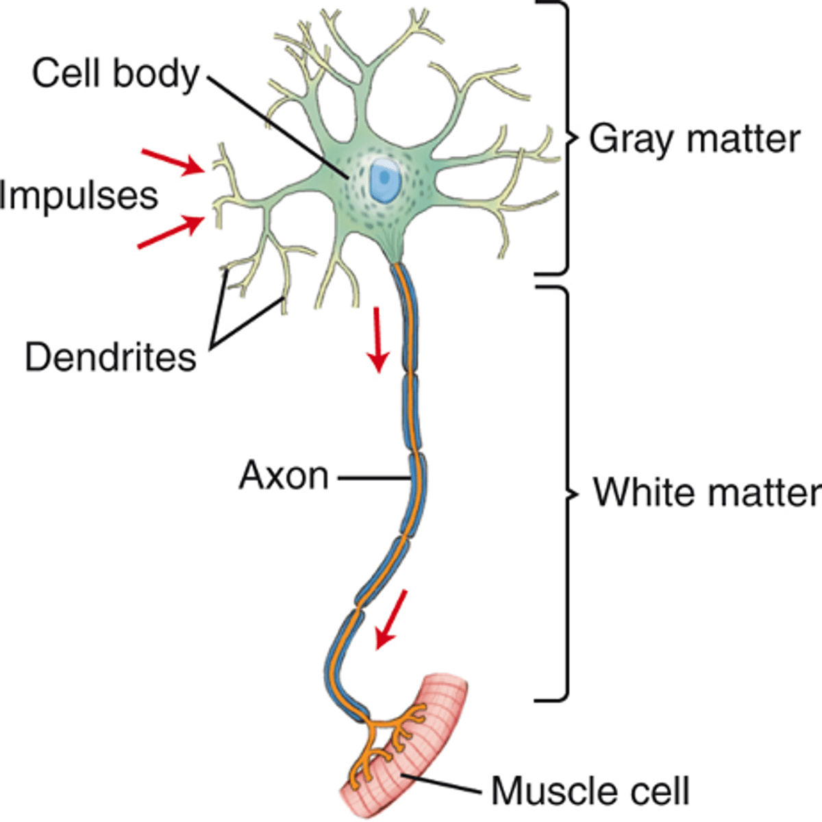
dendrites structure/function
- fibers with enlarged surface w/ synaptic receptors that receive information from other neurons via synapses
- receive electrical signals and send them to the cell body
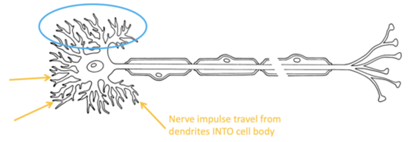
cell body or soma structure/function
- composed of large, round nucleus surrounded by cytoplasm
- contains the cytoplasm and all cytoplasmic organelles EXCEPT centrioles, hence neurons are generally amitotic
- generate and transmit action potentials AWAY from the cell body
- contains well-developed rough ER (called Nissl Body or Chrmatophilic substance) as well as prominent nucleoli
- cell body is referred to as biosynthetic region of a neuron since neurotransmitters are synthesized in the cell body
presynaptic terminal structure/function
- at the presynaptic terminal, the axon releases chemicals that cross through the junction from one neuron and the next
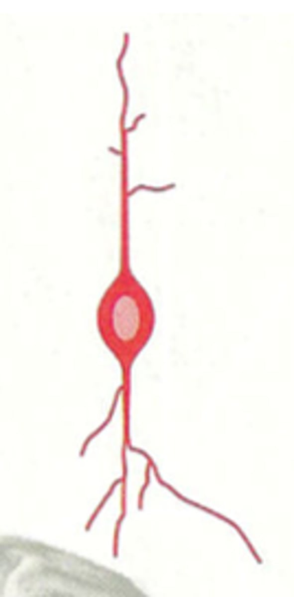
example of bipolar neuron: von-economo neurons (spindle neurons) structure/functions
- found only in a few species, mostly humans, in very particular parts of the brain (also found in primates, whales, dolphins, elephants)
- kind of bipolar neurons, longer than average
- allow for long distance communication between long distance neurons in the brain
- responsible for empathy and socialization: judgement, morality, higher order cognition
- drastically affected by frontotemporal dementia -> lose empathy and other personality traits
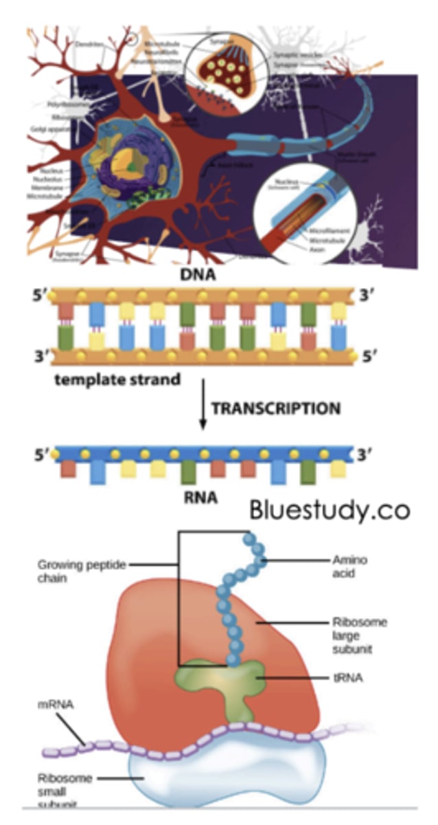
the neuron as a factory
- final product of neurons -> proteins
- genotype: organism's genetic code (or set of genes)
- phenotype: organism's observable characteristics
- the nucleus is the cell's executive office, where the genes for making proteins are stored, copied and sent to the factory floor:
~1. chromosomes are constantly changing shape so that the genes can be expressed (DNA uncoils to expose a gene)
~2. then the gene information is transcribed -> RNA
~3. the RNA leaves the nucleus and comes in contact with ribosomes, where its genetic code is read and translated into a specific amino acid chain, which forms the protein
- new proteins are created in response to environmental factors (ex. stress)
the neuron as a factory -> COMT proteins; COMT proteins + cocaine
- variation is associated with variation in the COMT gene: degraded dopamine -> more impulsive
- specifically, the val-val genotype, linked to greater COMT activity and lower levels of prefrontal dopamine, is associated with greater impulsivity (more COMT protein -> more impulsive)
- these findings raise the intriguing possibility that an inhibitor of COMT (tolcapone) may reduce impulsive behavior
COMT + cocaine:
- raises dopamine levels
- COMT protein levels rise in response to degrade dopamine
- COMT protein levels remain high, degrading dopamine levels to below their base state:
- COMT removes/inactivates dopamine from synapse space -> results in withdrawal symptoms + can lead to addiction
the neuron as a factory -> MAO proteins
- degrades neurotransmitters, specifically serotonin
- less MAO results in too much serotonin -> can lead to psychopathy, increases confidence
glia structure/functions
Glia = Supporting Cells
- provide firmness and structure for the brain (role played by connective tissue cells in other parts of the body)
- smaller but more numerous than neurons
Glial Functions
- Guidance: help with migration of neurons
- Form myelin: oligodendrocytes in CNS (central nervous system) and Schwann in PNS (peripheral nervous system)
- synapse formation and maintenance of synapses depends on signals from astrocytes
- supply oxygen and nutrients, remove dead neurons
- removal of K+ by astrocytes to keep right K+ concentration -> removes extra potassium to prevent toxicity

types of glia
Macroglia:
- Astrocytes
- Oligodendrocytes
Microglia
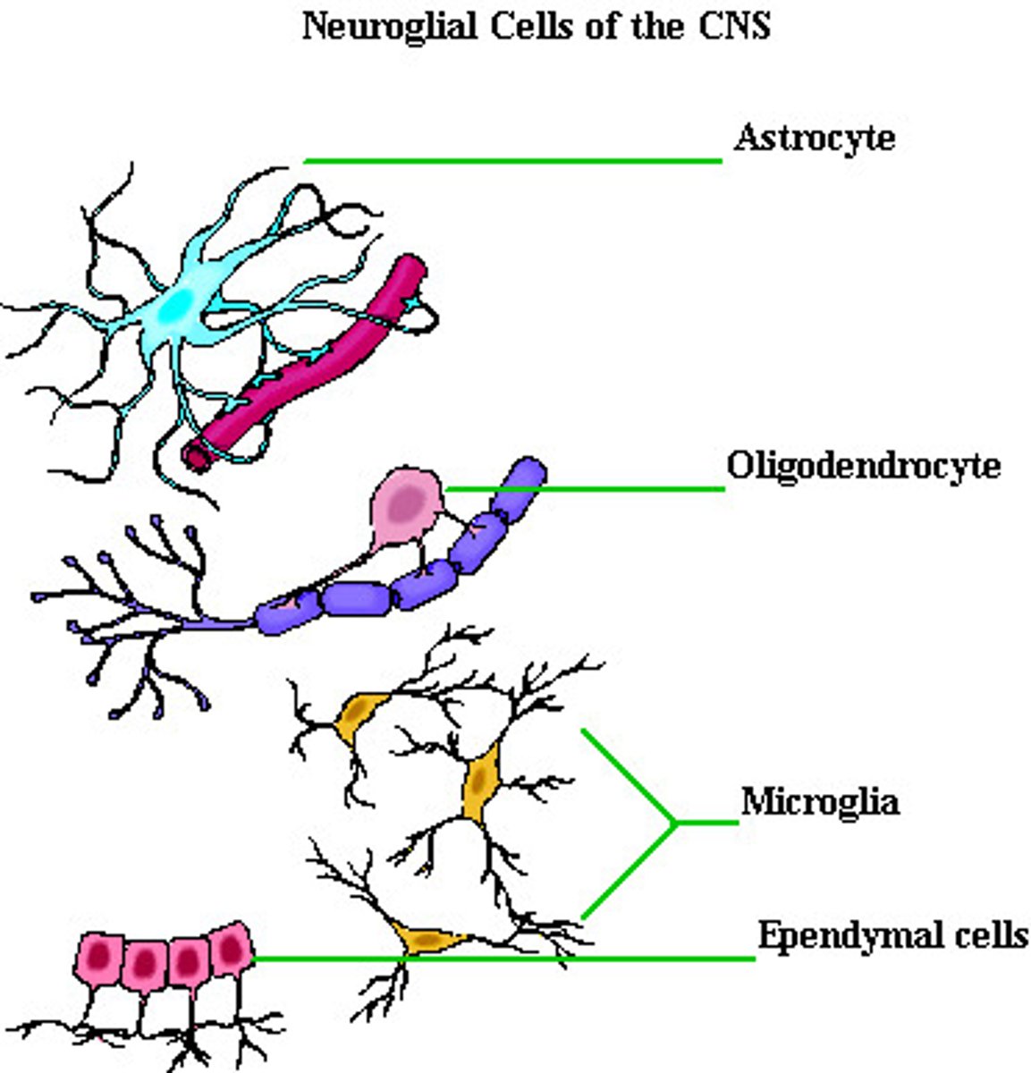
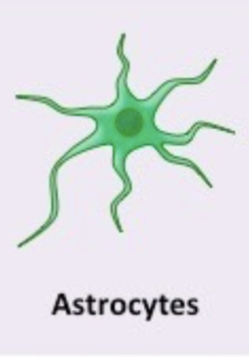
astrocytes structure/functions
Structure:
- type of macroglia
- star shaped cell
Functions:
- nutritive function together with microglias
- removes dead neurons after brain injury + forms glial scars
- prohibits recovery of sensitivity and other brain functions/reconnection of axons
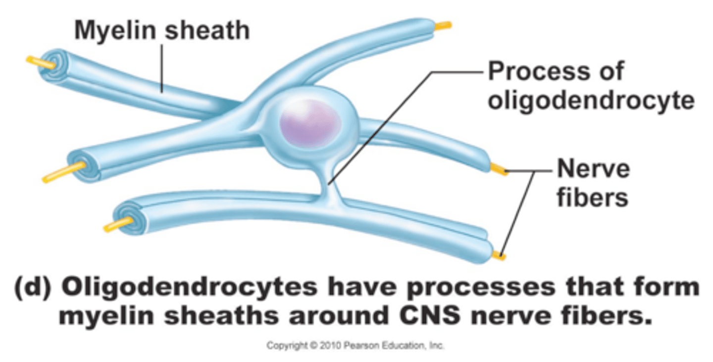
oligodendrocytes structure/function
Structure:
- smaller cells
Function:
- contribute the myelin sheath to the axon in the CNS and to schwann cells in the PNS
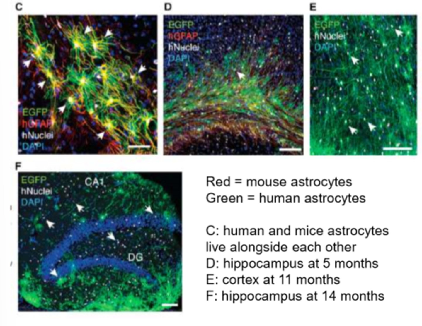
Ted talk by Steve Goldman
Astrocytes as Key Players in Human Intelligence:
- Astrocytes are star-shaped glial cells that outnumber neurons in the brain and play a critical role in supporting neural activity.
- Human astrocytes are larger and more complex than those in rodents, with more fibers and connections, contributing to the greater processing power of the human brain.
- When human astrocytes were transplanted into mice, the mice demonstrated enhanced cognitive abilities, such as improved learning and memory.
Glial Cells and Mental Health
- Astrocytes regulate synaptic activity, neurotransmitter balance, and brain plasticity, meaning they are essential for healthy brain function and mental well-being.
- Dysfunction in astrocytes has been linked to neurological and psychiatric disorders, including schizophrenia, depression, and neurodegenerative diseases like Alzheimer’s.
- The study of glial cells may open up new therapeutic avenues for treating mental health conditions.
Evolutionary Differences Between Humans and Rodents
- Compared to rodents, human astrocytes are more intricate, with more connections per neuron, which enhances information processing and cognitive flexibility.
-Rodents have less complex astrocytes with fewer fibers, which may explain why their cognitive abilities are more limited compared to humans.
Potential for Future Research and Therapy
- Understanding glial cell function can reshape how we approach brain disorders by targeting astrocytes as potential therapeutic agents.
- Research on glial transplantation (introducing human glia into animal models) may help develop treatments for cognitive and psychiatric disorders.
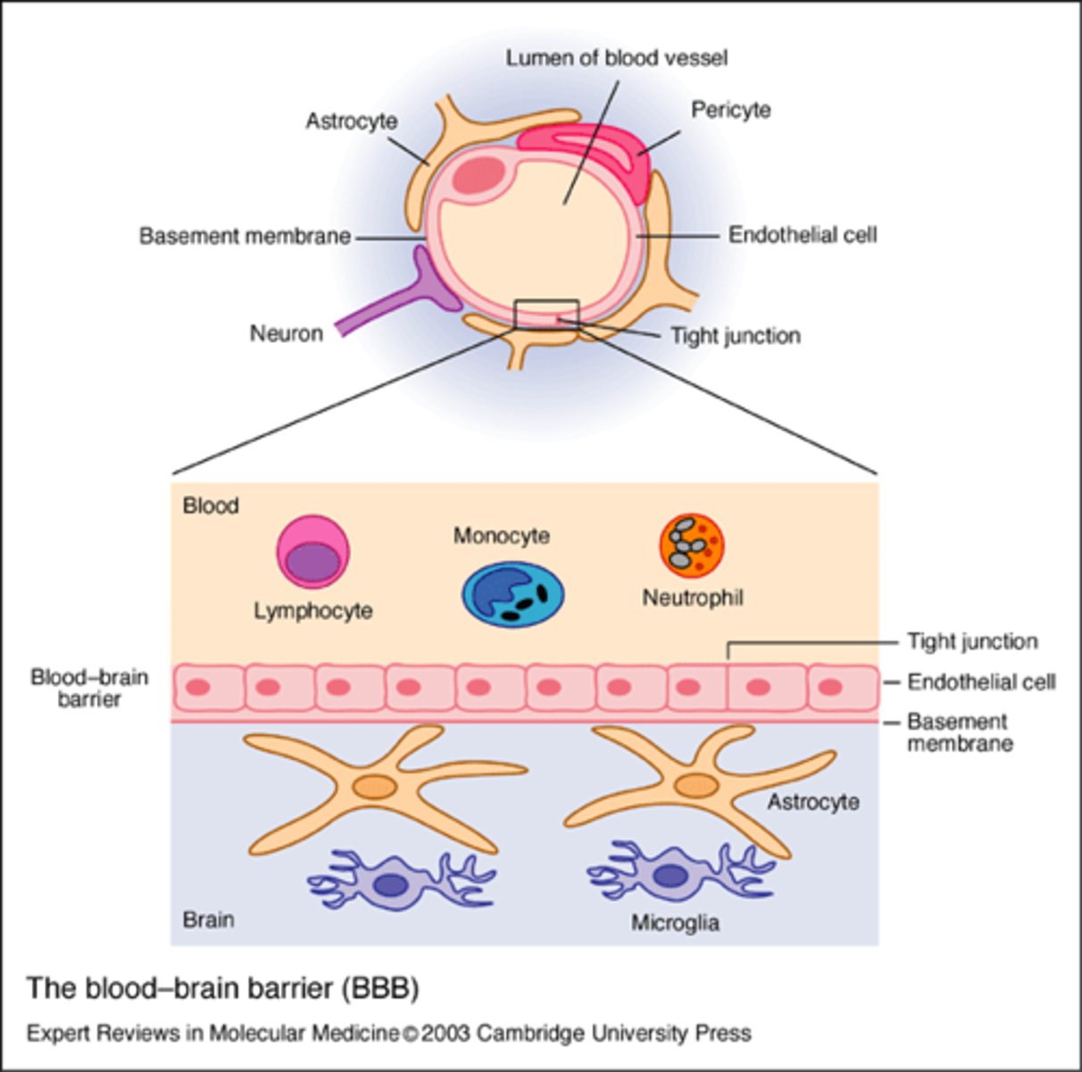
blood-brain barrier + its function
- made of endothelial cells
- the brain is protected against surging fluctuations in the content of many constituents of the blood
- there is not a single barrier; there are many different systems that exist for excluding substances from blood to the brain (i.e. morphological and functional characteristics of brain capillaries; they differ from those of other organs)
action potential
- neurons communicate with each other through electrical or chemical signals that travel down their axons
- electrical -> action potential
electrolytes involved in action potential
- sodium, potassium, calcium
- neurons cannot survive without the cell membrane
- cell membrane is key for electrical signal
- more potassium ions inside cell than outside -> more negative (-) inside cell
- more sodium ions outside cell than inside -> more positive (+) outside cell
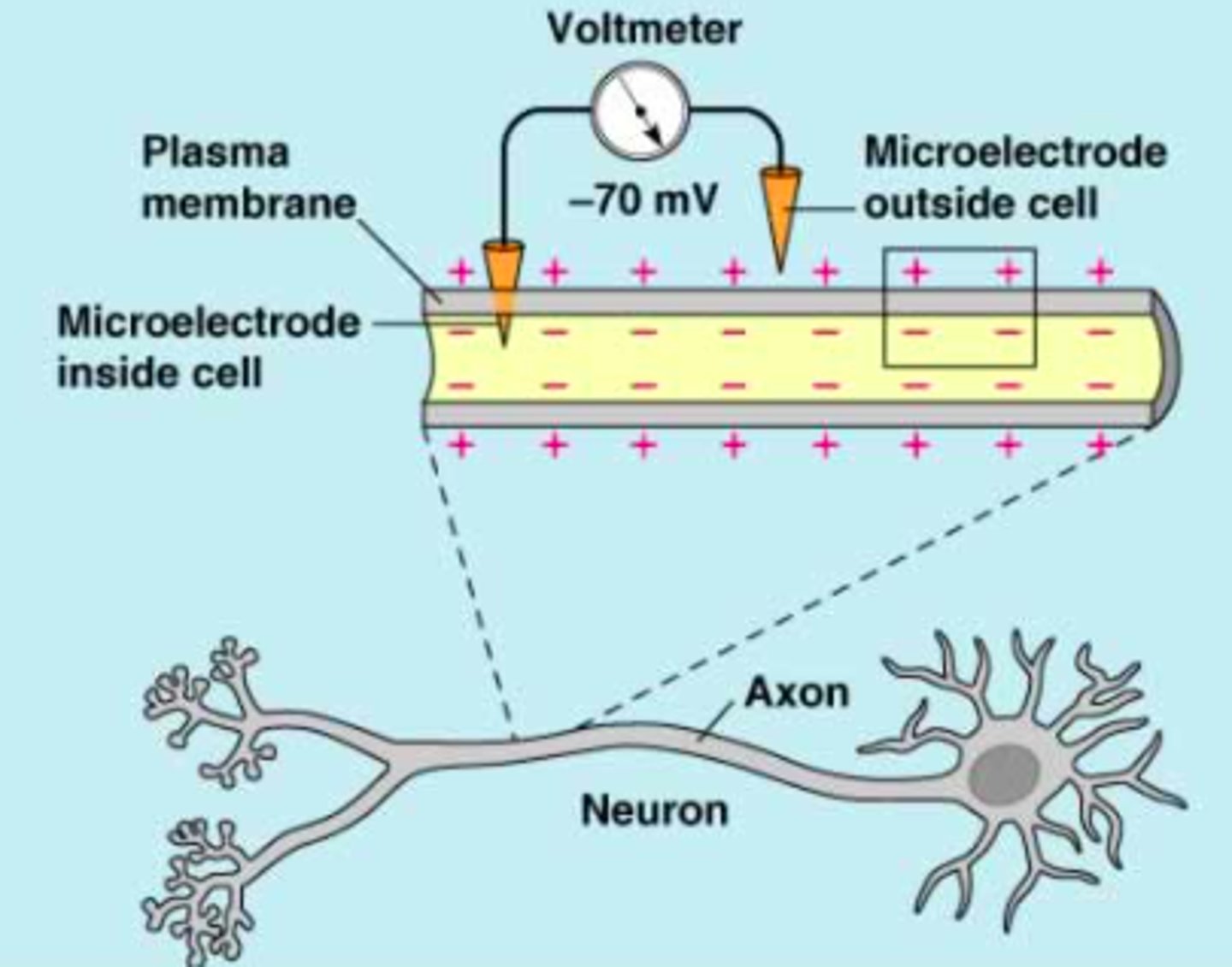
resting potential of the neuron
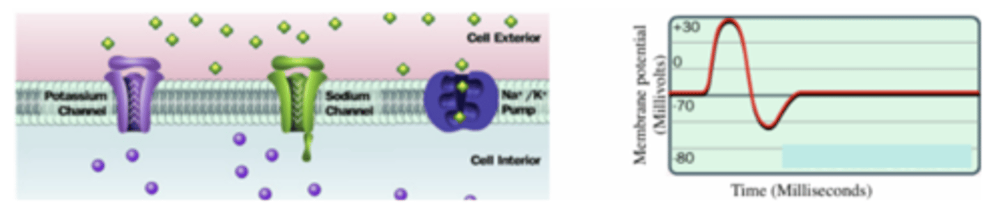
- there are proteins in the membrane (skin) of the neuron through which chemicals can pass (channels or gates)
- polarization: electrical gradient (different electrical charge between inside and outside of neuron)
Resting Potential = -70 mV (millivolts)
- inside is more negative than outside
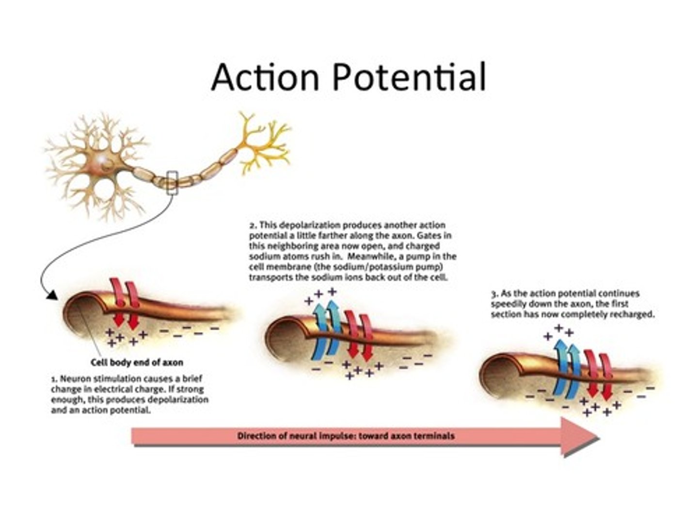
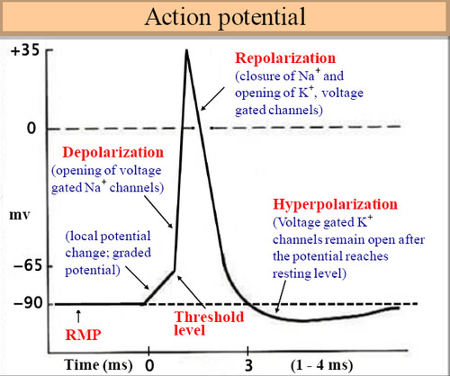
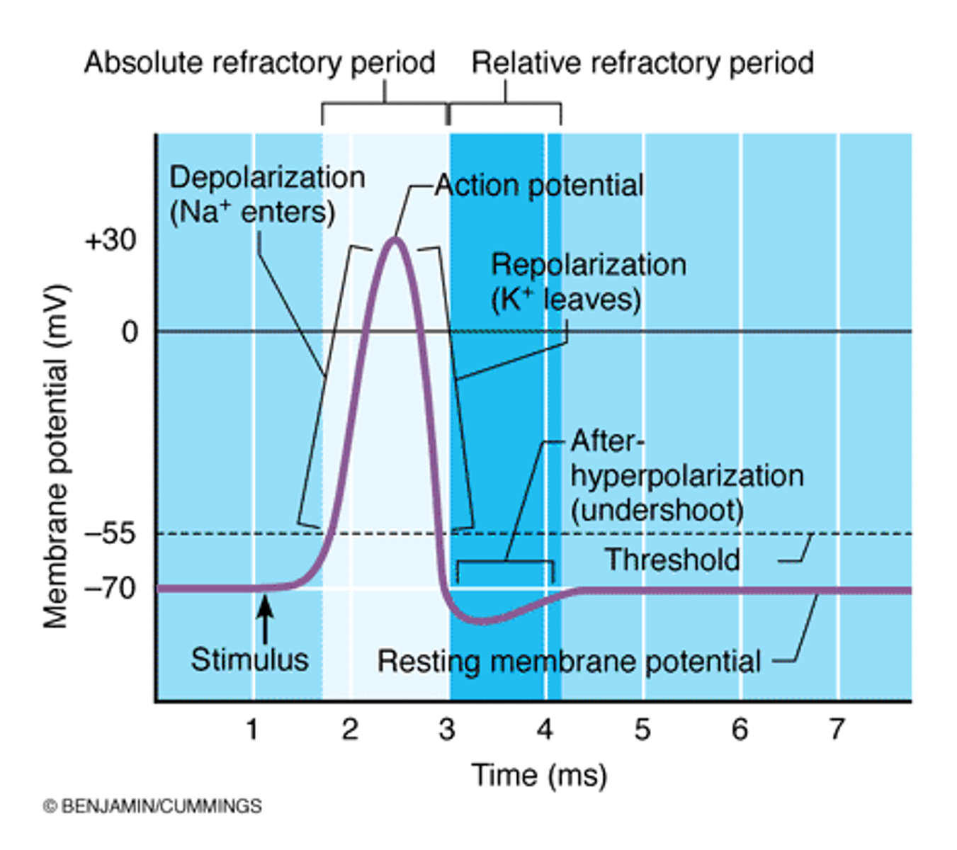
stages of action potential (4)
1. resting state
2. stimulation -> depolarization -> generation of nerve impulse
3. repolarization
4. hyperpolarization - returning to resting state

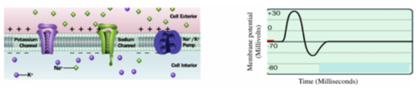
resting state stage of acting potential
1st state of action potential
- the neuron is polarized
- high Na+ concentration outside cell, high K+ concentration inside cell (maintained by active sodium-potassium pumps)
- all gated Na+ and K+ channels are closed
- the cytoplasmic surface of the plasma membrane is more negative than its extracellular surface
the charge separation = resting membrane potential (-70 mV)
stimulation (depolarization) stage of action potential
stimulation -> depolarization
- the neuron is activated by a stimulus of appropriate intensity (threshold stimulus: around -55 mV)
- sodium gates open in the membrane and Na+ ions moves into the cell (sodium influx), down their concentration gradient
- potassium gates are closed
- the interior of the cell becomes less negative, and eventually becomes positive (+) (depolarization)
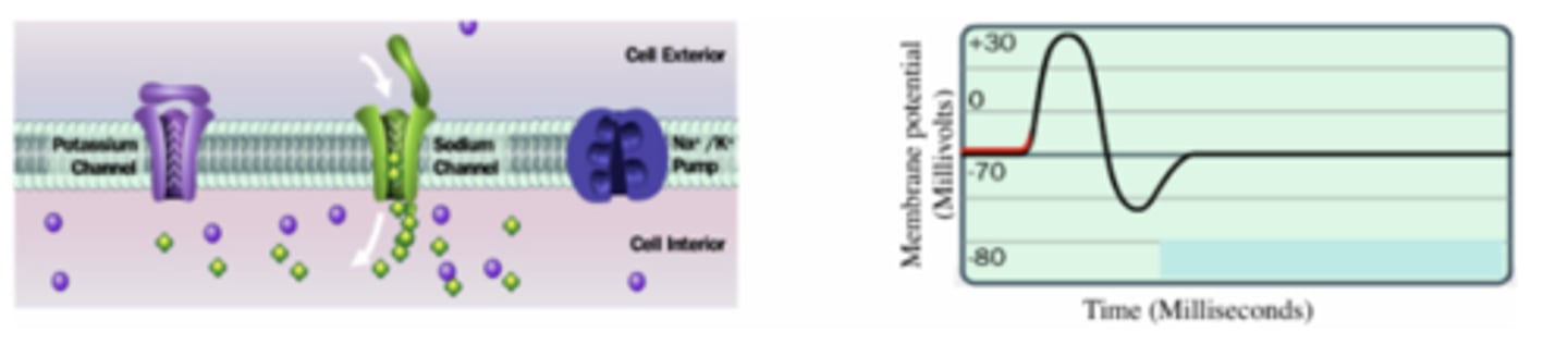
generation of a nerve impulse
- depolarization occurs when the charge across the plasma membrane reverses from a negative to a positive charge
- when the threshold potential (-55mV) is reached -> an action potential is generated
- a transmitted action potential = a nerve impulse
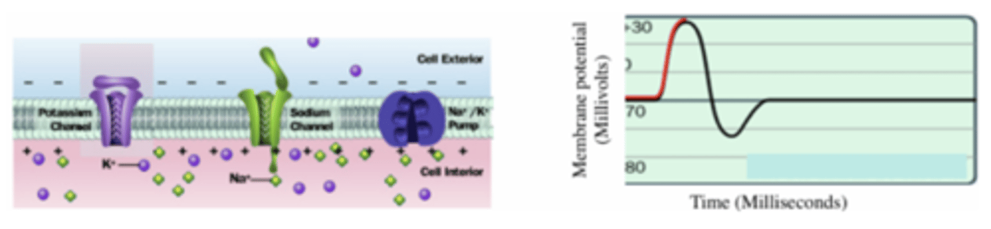
repolarization stage of action potential
- after depolarization (~ +30 mV) -> potassium gates open, K+ ions move out of the cell, down their concentration gradient (potassium efflux)
- sodium channels close -> results in reversal of membrane potential toward a negative (-) membrane potential
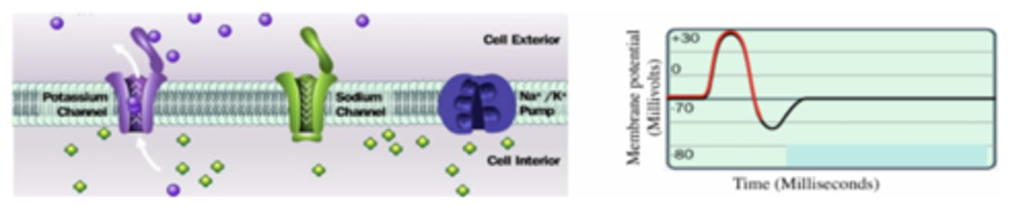
hyperpolarization stage of action potential (returning to resting state)
-the sodium-potassium pump reestablishes the concentration gradient of the resting membrane potential so that the neuron can fire again
- more potassium efflux occurs past the resting membrane potential -> temporarily driving the membrane potential below the RMP
refractory periods
absolute refractory period and relative refractory period
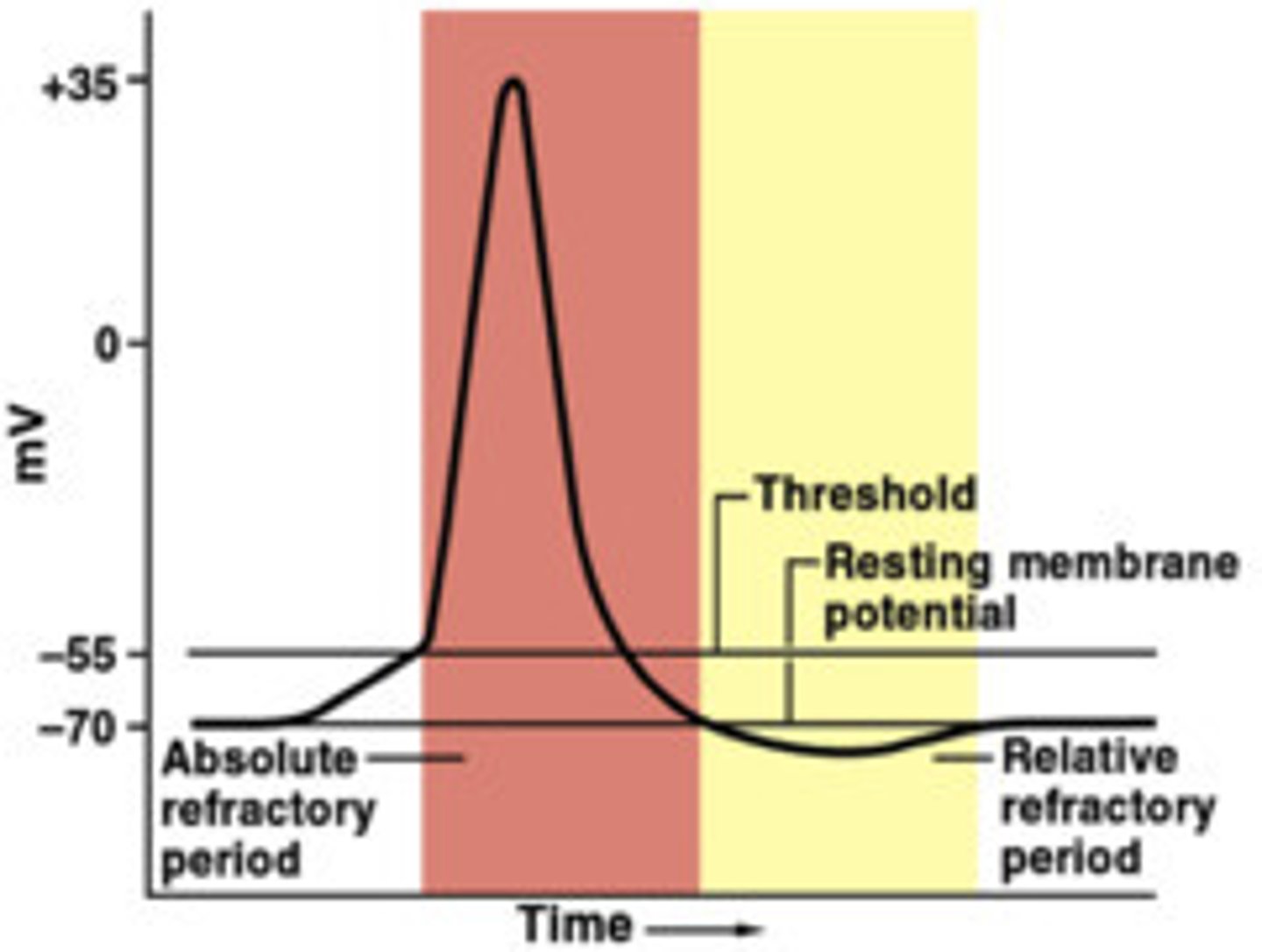
absolute refractory period
-occurs when the sodium gates are still open (depolarization phase) and the neuron is unable to initiate a new action potential since all Na+ channels are open
relative refractory
- when the neuron is undergoing repolarization (and therefore the sodium gates are closed) an exceptionally strong stimulus can cause initiation of another action potential by opening Na+ channels and allowing sodium influx

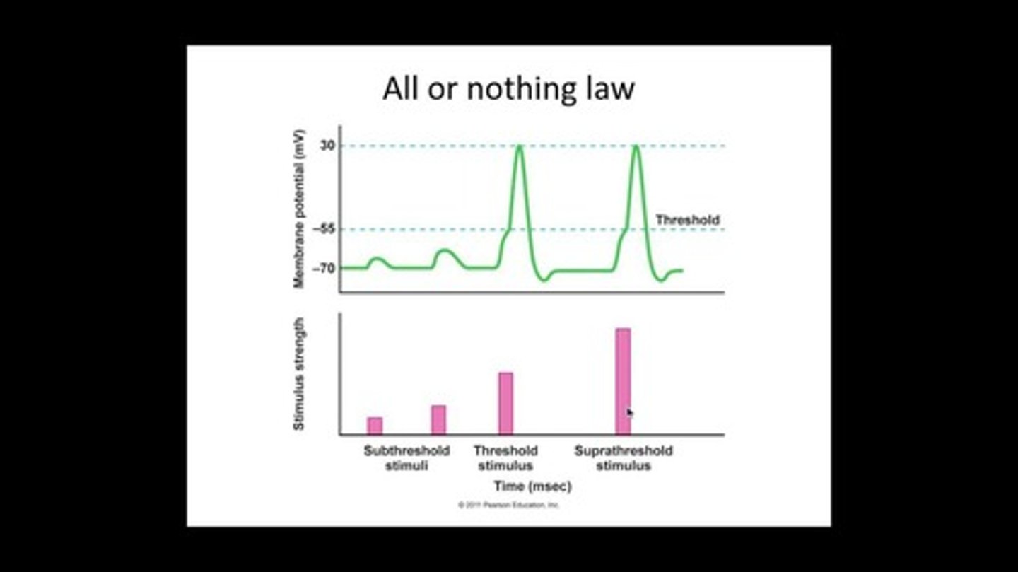
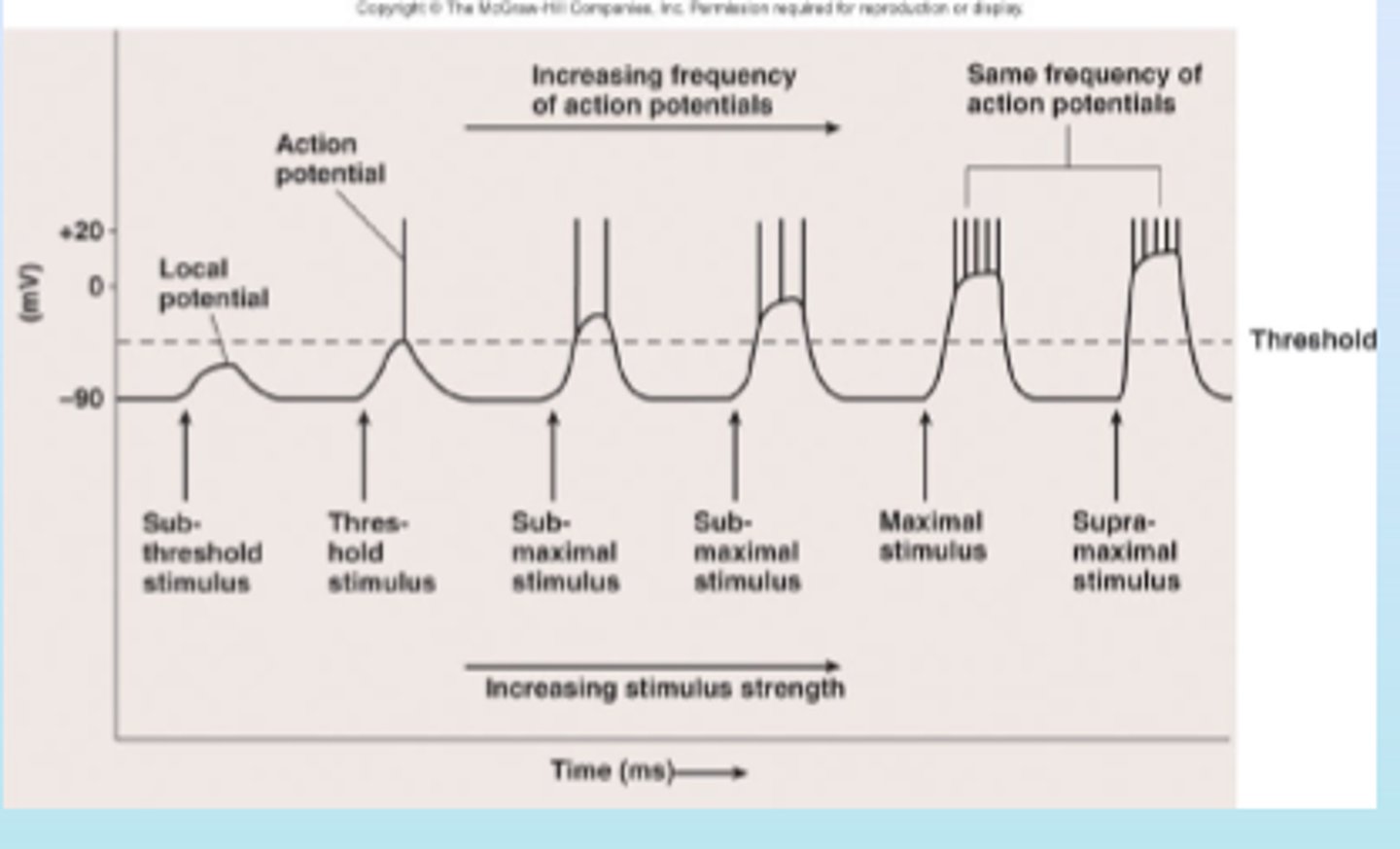
characteristics of action potentials
- all-or-non phenomenon
- self propagating
all-or-none phenomenon
- an action potential will be generated if depolarization reaches a threshold potential
- all action potentials have same shape and same amplitude (+30 mV) regardless of stimulus strength
self-propagating action potential
once generated by the axon, the action potential is propagated down the axon to the axon terminals; a propagated or transmitted action potential is called a nerve impulse
all action potentials traced have the same shape and the SAME amplitude (+30mV) regardless of stimulus strength
- the difference between a stronger stimulus that causes the generation of an action potential and a weaker stimulus that causes the generation of an action potential is that the stronger causes the impulse to be generated at a higher frequency than the weaker stimulus
myelin sheath
- whitish fatty material (fats and protein) covering most long nerve fibers (myelinated fibers)
- concentric rings of the plasma membrane
- insulates fibers and increases the speed of transmission of nerve impulse
- in the CNS (central nervous system), axons are myelinated by oligodendrocytes
- in the PNS (peripheral nervous system) -> axons are myelinated by schwann cells
myelin sheath functions
Protection:
- physical protection against trauma
Electrical Insulation:
- to prevent interference from neighboring axons in a nerve (if in the PNS) or tract (if in the CNS)
Increase in rate of impulse transmission:
- using saltatory conduction occurring only at the nodes of ranvier
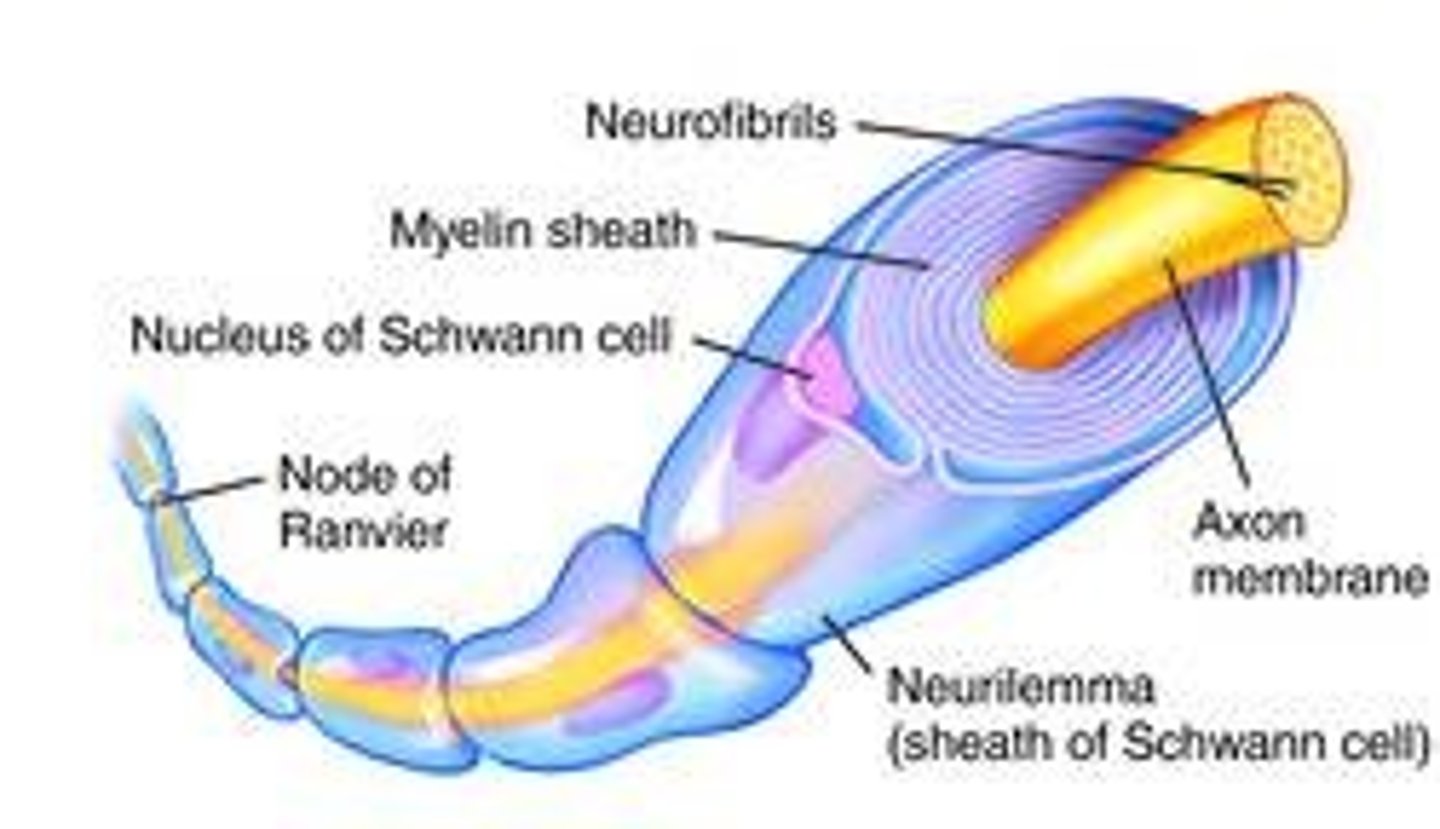
nodes of ranvier
- at these nodes, sodium ions enter the axon, push the positive charges to the next node to regenerate a new action potential (saltatory conduction)
- action potential cannot be regenerated at parts of axons with myelin
- axons with no myelin -> slower action potential
- if axon was completely covered with myelin -> signal would not regenerate and would eventually dissipate
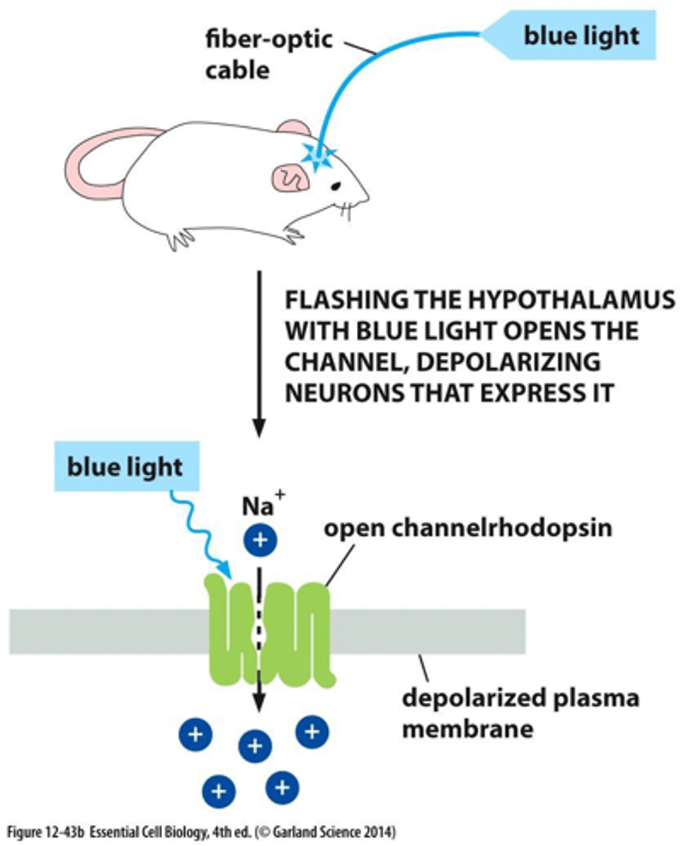
optogenetics
- a treatment that uses a combination of light stimulation and genetics to manipulate the activity of individual neurons
- insert light-sensitive proteins into a type of neuron -> proteins will activate or inhibit the neurons
- insert an optical fiber to shine light
- could potentially work in humans, but is invasive and likely unethical
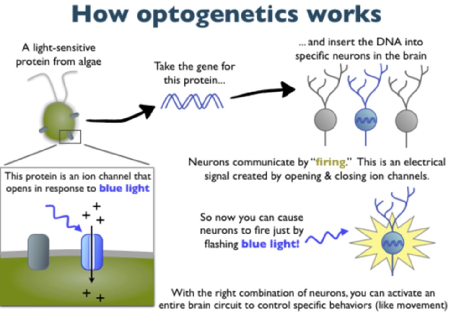
why are optogenetics revolutionary?
- you can target a very specific types of neurons
example:
- protein found in algae responds to light
- get DNA from protein -> insert DNA into specific neurons you want to work with -> use flashing blue light, neuron will activate when light turns on
brain stimulation
stimulation = increase or decrease activity in the brain
- electrophysiology = techniques that are used to try to read the electrical signals of the brain
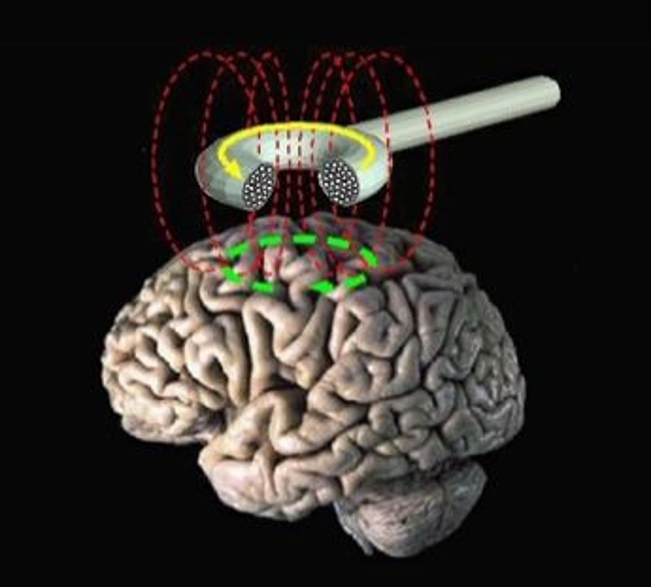
brain stimulation: transcranial magnetic stimulation (TMS)
transcranial magnetic stimulation (TMS)- non-invasive technique
- coiled (device) placed on top of scale + sends magnetic signals to brain -> controls electrical activity of neurons
- magnetic stimulation to a portion of the scalp
- activates or inactivates neurons in a narrow area blow the magnet
- good temporal resolution
- temporary brain activity
limitations
- a little painful, discomfort
- only for brain cortex (most outer layer of the brain) -> does not work on deeper areas of the brain (subcortical areas)
- difficult to target very specific areas of then brain (poor spatial resolution)
- transient cognitive changes (temporary, still a safe technique to use)
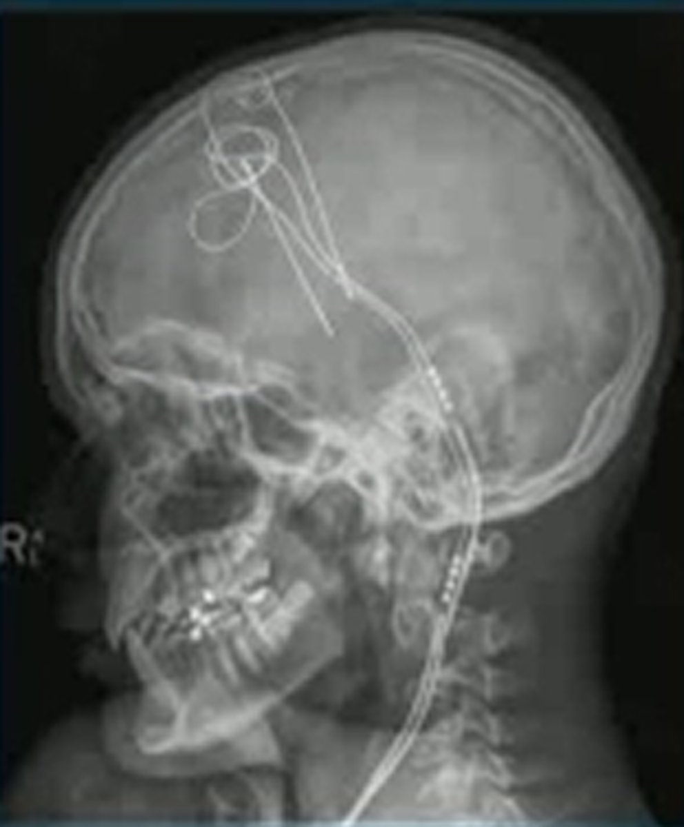
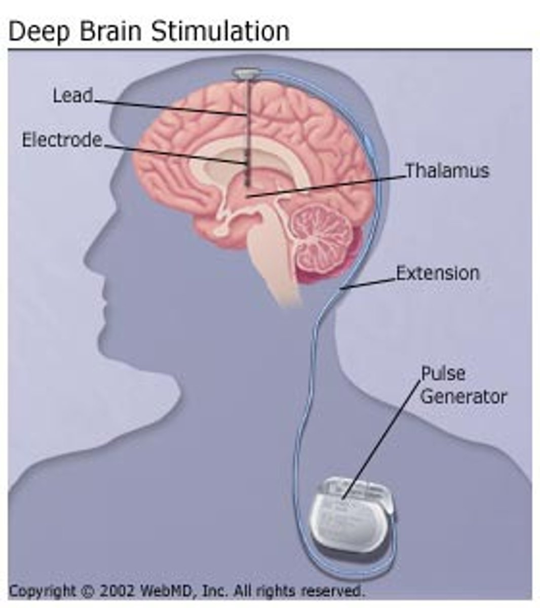
brain stimulation: deep brain stimulation
- electrical stimulation of the brain
- only used in patients when medication is not working (i.e. parkinson's diseases, depression, OCD... )
- there is a neurotransmitter implanted under the skin that will send electrical impulses to the brain -> very invasive (brain surgery)
electrical recording techniques
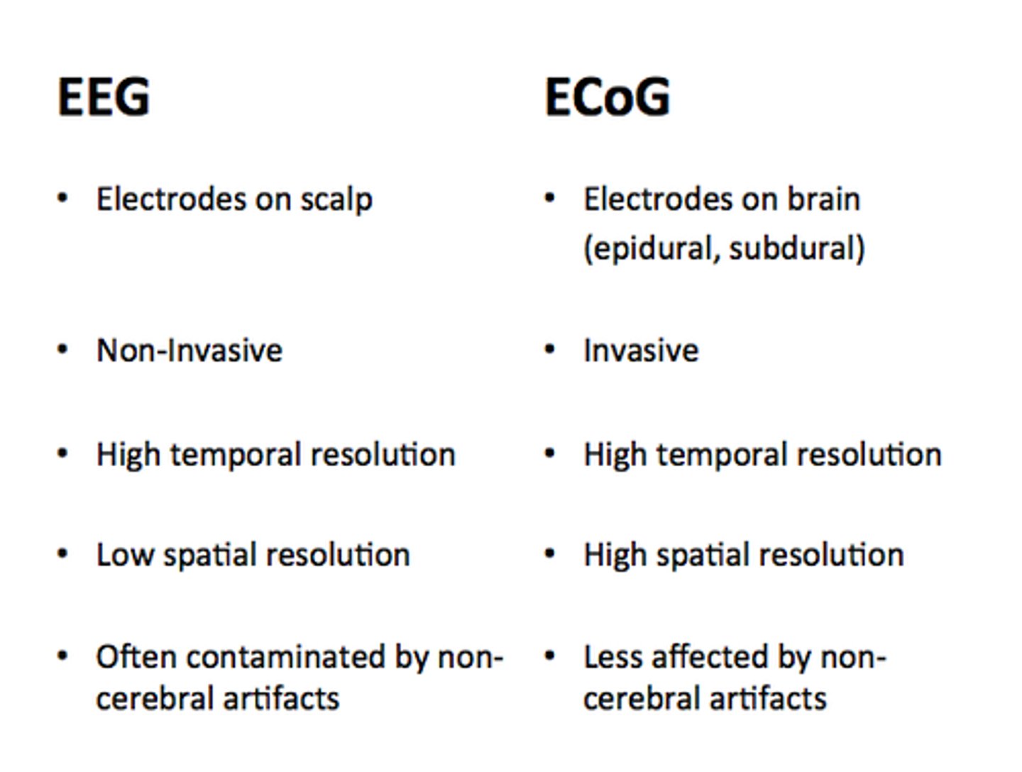
electroencephalography (EEG)
intracranial EEG (iEEG)
magnetoencephalography (MEG)
electrocorticography (ECOG)
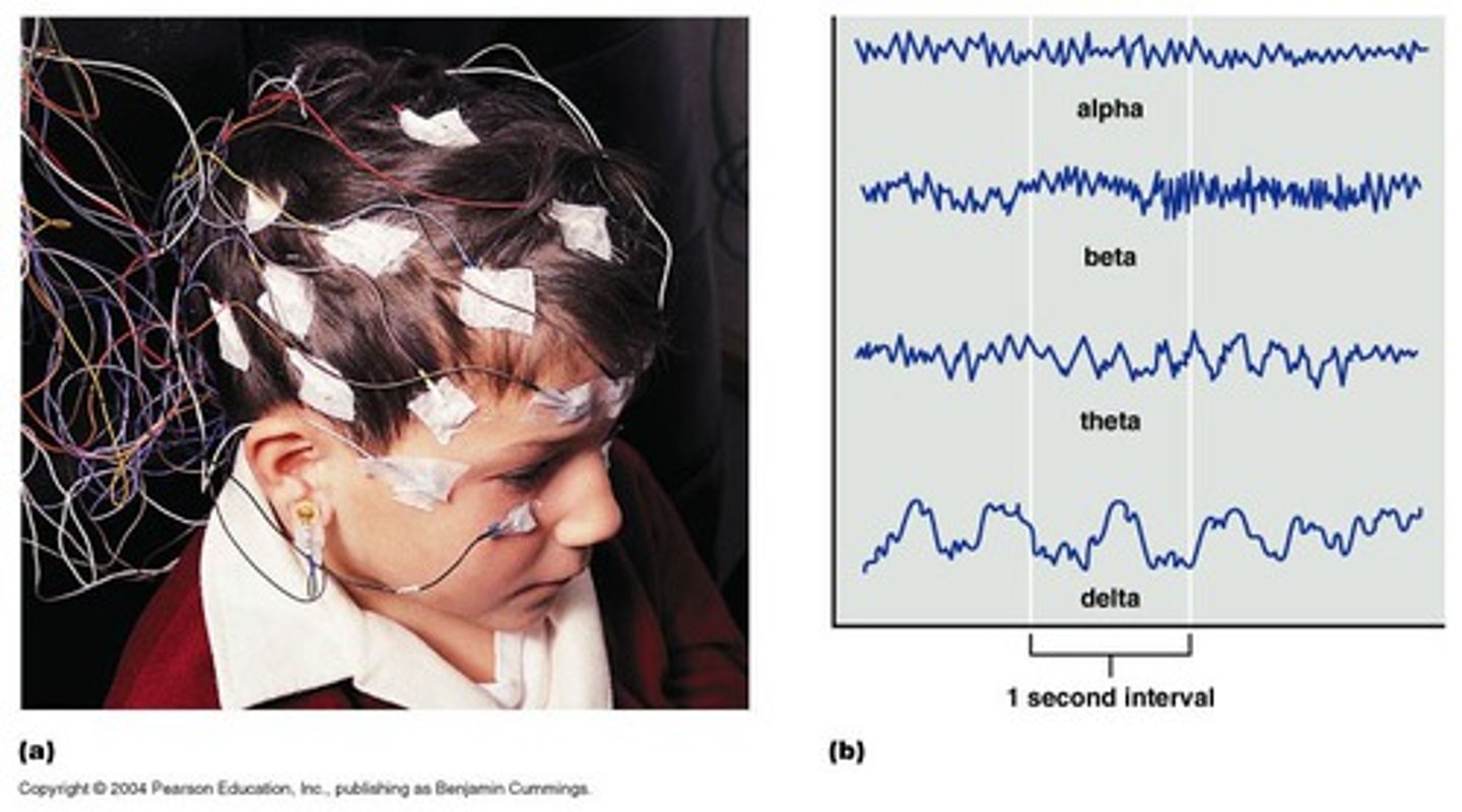
electroencephalography (EEG)
- when many millions of neurons are active at the same time, the current generated is strong enough to be detected on the surface of the scalp with sensitive electrodes
- cap with electrodes
- studies electrical activity of the brain
- each electrode measures activity from many different parts of the brain -> limitation because you cannot measure very specific parts of the brain
bad spatial resolution
very good temporal resolution (milliseconds):
- allows us to very quickly see how brain activity changes
- can show changes in consciousness (ex. sleep studies, epilepsy, comas, attention)
intracranial EEG (iEEG)/ECOG
- one single electrode inside the brain
- good spatial and temporal resolution: can accurately pinpoint both the location ("spatial") and the precise time ("temporal) of brain activity

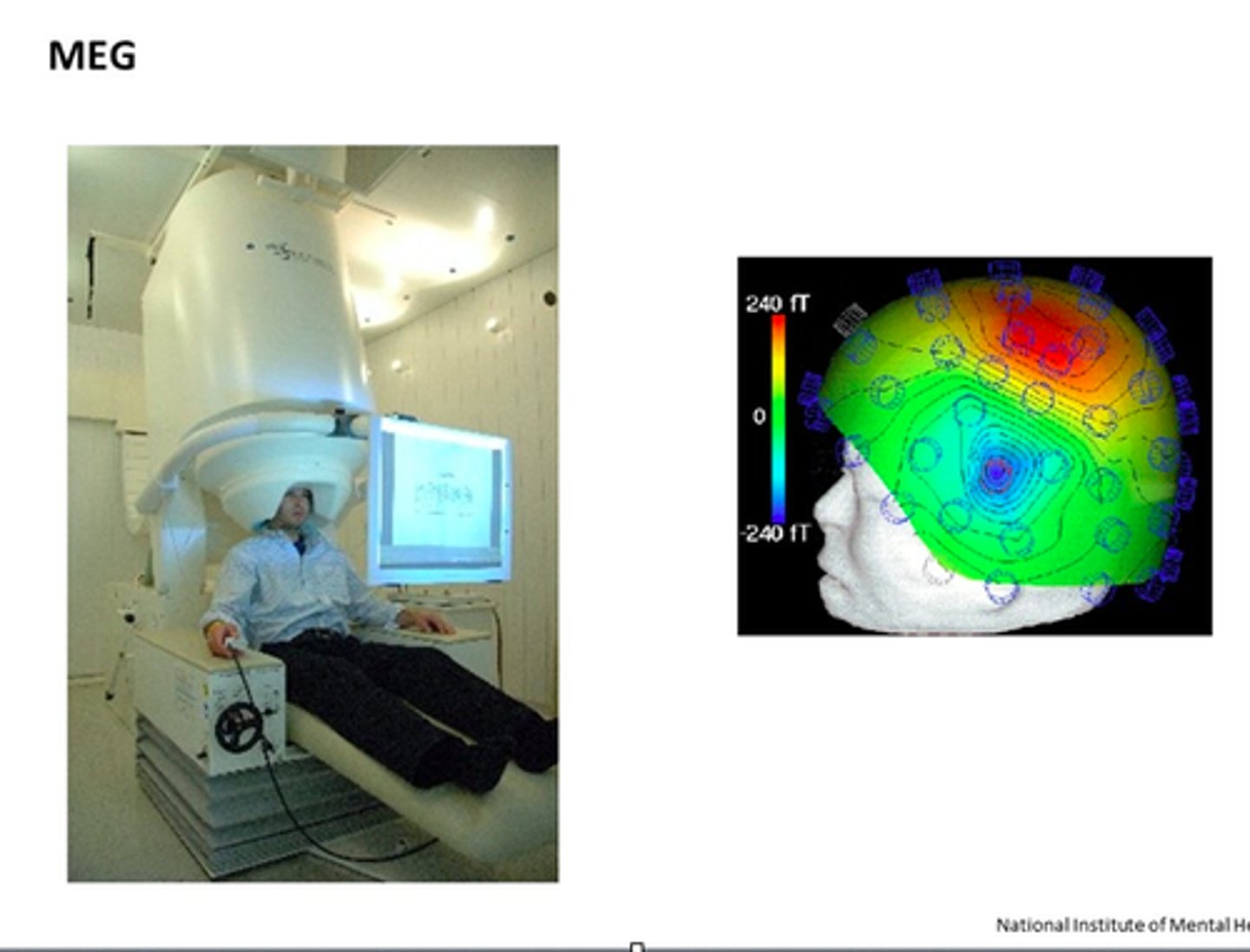
magnetoencephalography (MEG)
- similar to EEG, it measures the magnetic field generated by brain activity
- same limitation -> cannot measure specific parts of the brain
- a little cleaner than EEG because electrical signals would have to travel through the bone/scalp to be read
bad spatial resolution (slightly better than EEG) -> more expensive
very good time resolution (milliseconds):
- allows us to very quickly see how brain activity changes
- can show changes in consciousness (ex. sleep studies, epilepsy, comas, attention)
electrocorticography (ECOG)
- electrodes are placed directly on the brain
- good spatial and temporal resolution: can accurately pinpoint both the location ("spatial") and the precise time ("temporal) of brain activity
- better spatial resolution than EEG

neuroimaging
- the use of various techniques to provide pictures of the structure and function of the living brain
- neuroimaging is an indirect measure
- has good spatial resolution and bad temporal resolution
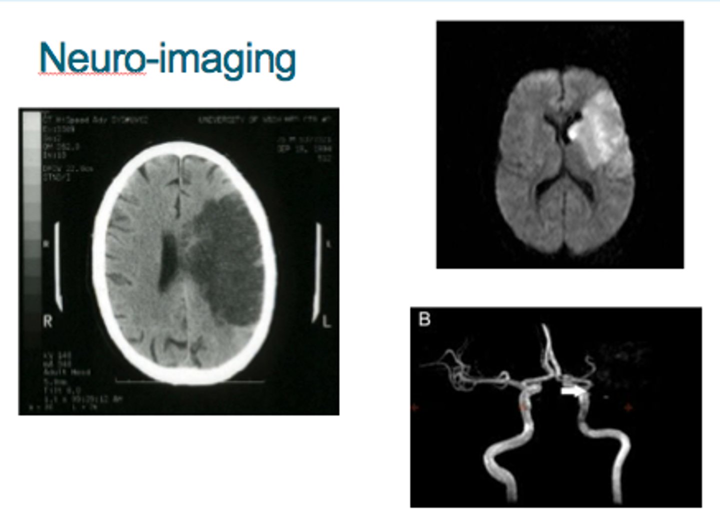
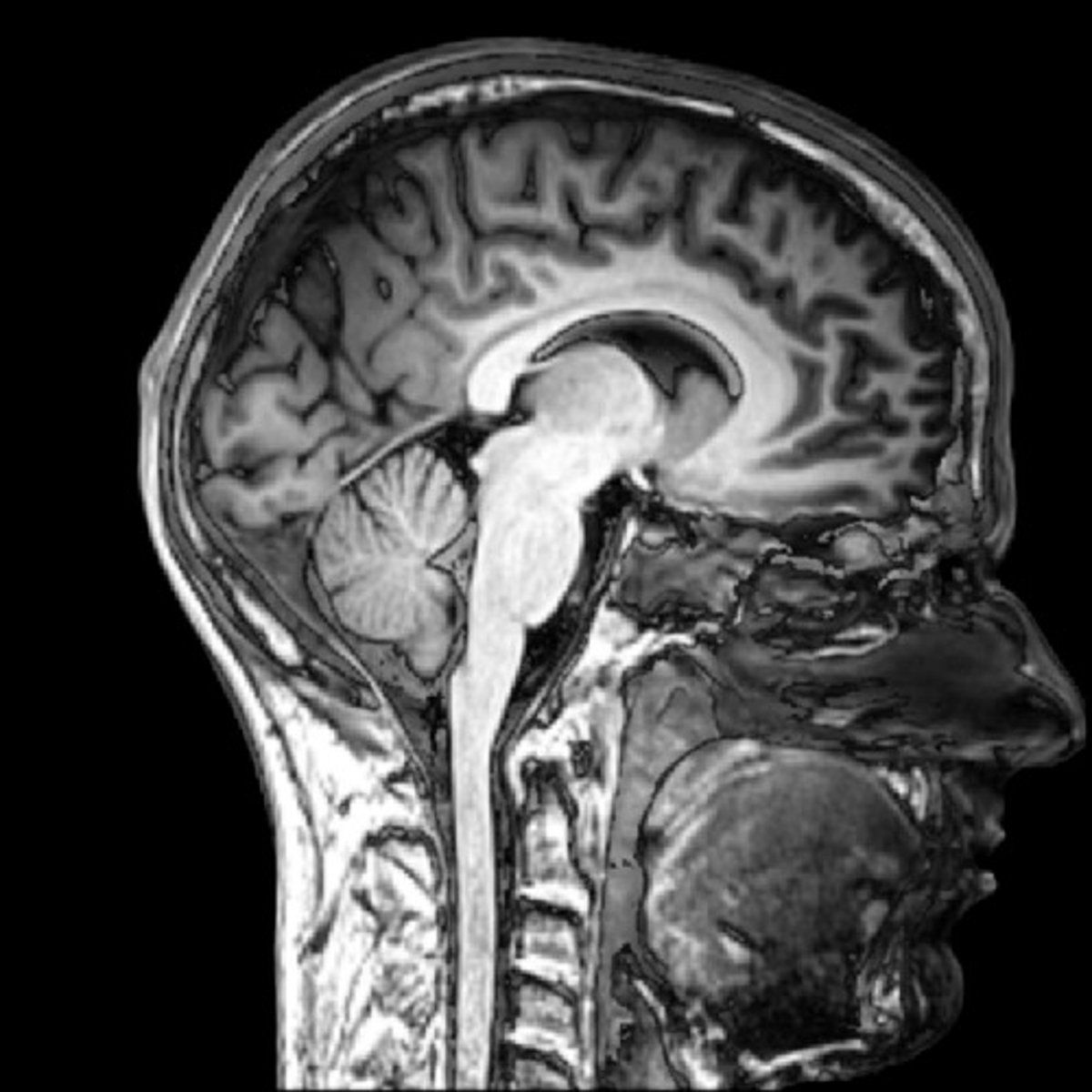
neuroimaging: Magnetic Resonance Imaging (MRI)
-structural image of the brain (anatomy)
- doesn't use x-rays (CT), instead uses strong magnetic fields
- takes picture of the brain
- good spatial resolution
- bad temporal resolution
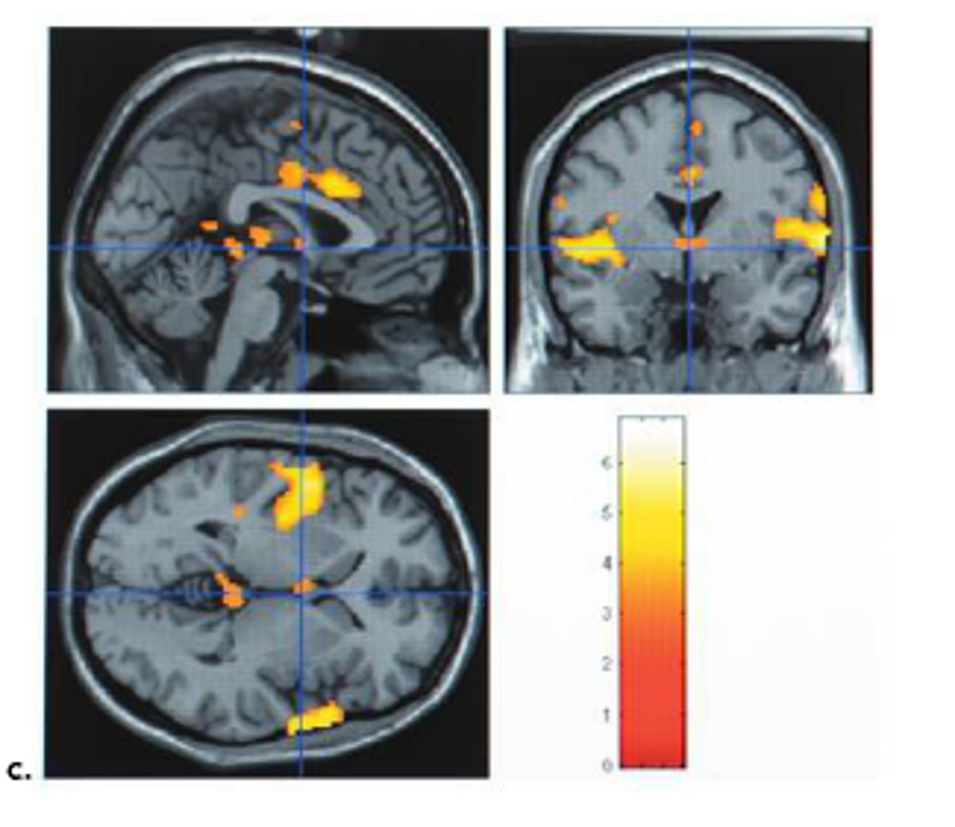
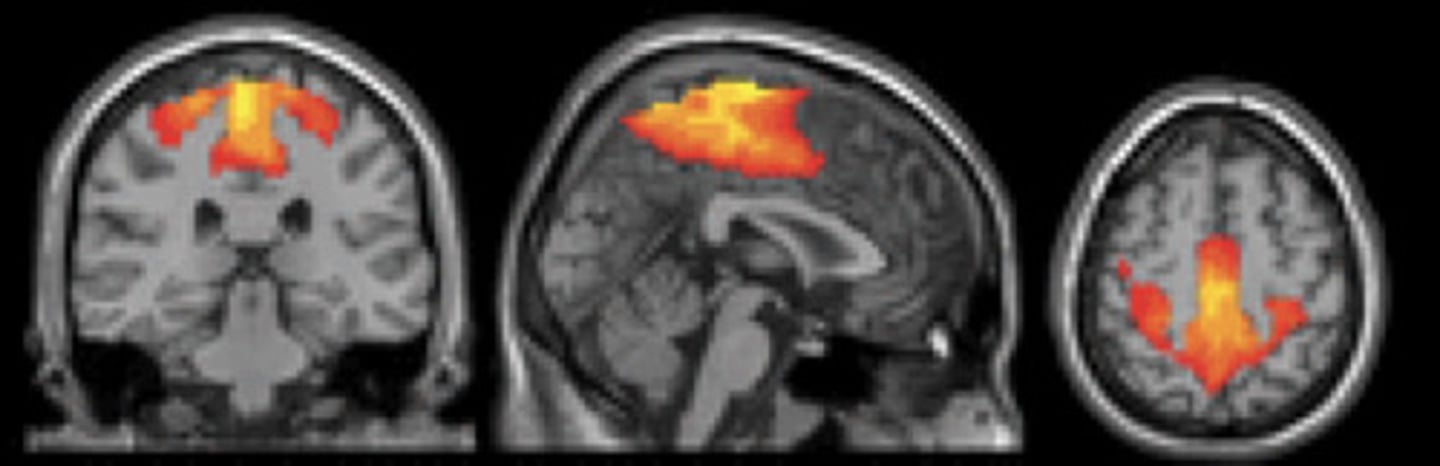
neuroimaging: Functional Magnetic Resonance Imaging (fMRI)
-used to study brain function
- measures the oxygen content in the blood flowing through each region of the brain (relies on the magnetic properties of the atoms)
- good spatial resolution
- bad temporal resolution (i.e. ~ 2 seconds)
- you can do EEG and MRI at the same time -> but signal is noisy
neuroimaging: resting-state fMRI
- fMRI study of brain connectivity while the person is not performing a specific task
- class activity: default mode network (autopilot)
Based on sentence, what technique are they using:
Schizophrenic subjects show aberrant ... activation of dorsolateral prefrontal cortex and basal ganglia during working memory performance.
fMRI
deep area = fMRI
Based on sentence, what technique are they using:
Sleep in outpatients with generalized anxiety: a preliminary comparison with depressed outpatients
EEG/MEG
Epilepsy or sleep = EEG or iEEG/ECOG
Based on sentence, what technique are they using:
Applied to left dorsolateral prefrontal cortex disrupts verbal working memory performance in humans
TMS
Stimulating outer cortex → TMS
Based on sentence, what technique are they using:
Loss of consciousness during induction with thipental, etomidate, midazolam, or sevoflurane
EEG
Epilepsy, changes in consciousness or sleep = EEG or iEEG/ECOG
Based on sentence, what technique are they using:
Imbalance between left and right dorsolateral prefrontal cortex in major depression is linked to negative emotional judgement
fMRI (specific area of brain)
Based on sentence, what technique are they using:
Subthalamic nucleus: A key structure for emotional component synchronization in humans
intracranial EEG (iEEG)
default mode brain network + resting-state fMRI
The default mode network is a large-scale brain network that was first identified as the network that is consistently active when the brain is not engaged in a task, as measured through resting-state functional MRI (fMRI; Raichle et al., 2001; Shulman et al., 1997).
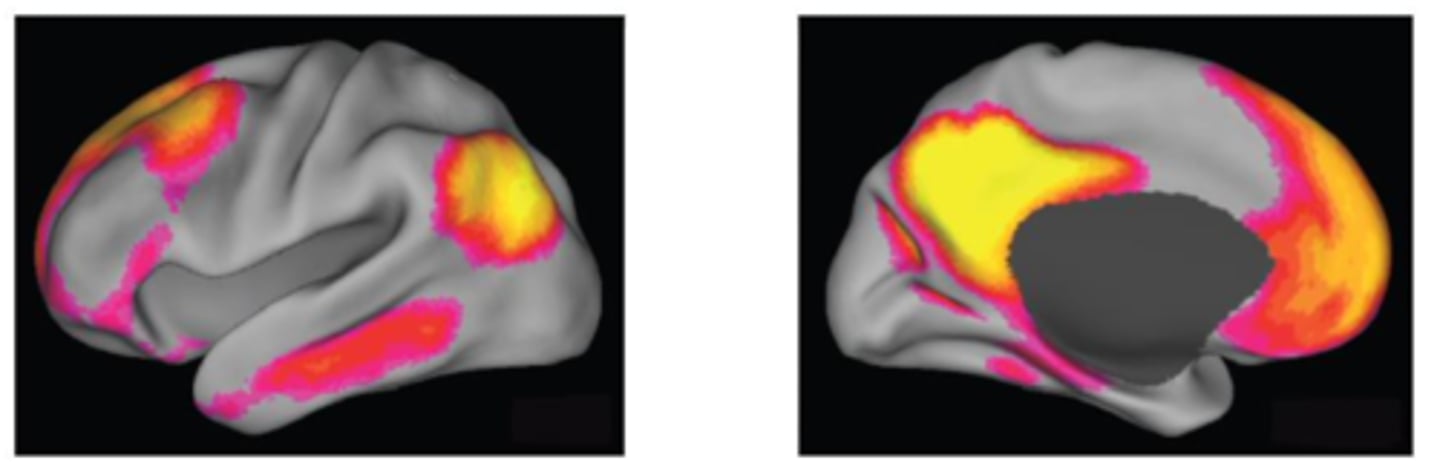
default mode network (DMN)
- active when the brain is at rest, involved in introspection and daydreaming.

central executive network (CEN)
- coordinates attention, working memory, and decision-making
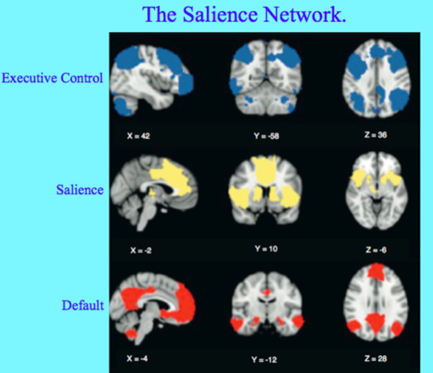
salience network
- detects and responds to important stimuli

sensorimotor network
- controls movement and perception
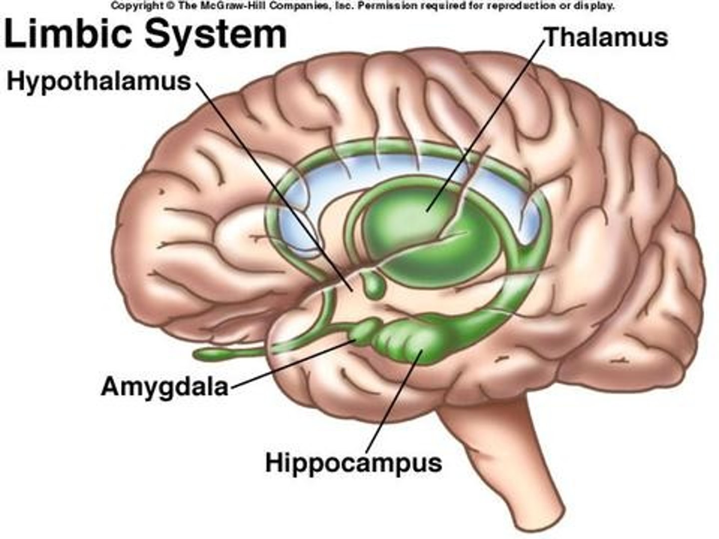
limbic network
- regulates emotions and motivation
what does electrocorticography (ECOG) provide that EEG cannot?
- high spatial resolution (EEG has poor spatial resolution)
- less contaminated by non-cerebral artifacts