Brain Imaging & Cross Sections
1/62
There's no tags or description
Looks like no tags are added yet.
Name | Mastery | Learn | Test | Matching | Spaced | Call with Kai |
|---|
No analytics yet
Send a link to your students to track their progress
63 Terms
CT Image Acquisition
Computed Tomography, a bunch of x-rays showing structues
CT Radiation/Magnets
Yes radiation (x-ray), no magnets
CT Advantages
quicker and comfier than MRI, less sensitive to movement, quieter
CT Disadvantages
not as finely detailed, worse spatial resolution, hard to see lesions
MRI Image Acquisition
Magnetic Resonance Imagine, spins protons from applying a magnetic field around them, shows structures
MRI Radiation/Magents
No radiation, Yes magnets
MRI Advantages
High spatial resolution, easy to compare slices since thinner
MRI Disadvantages
Long time to record, confined space, louder, sensitive to movement
MRI T1
CSF dark, white matter light, lesions dark
MRI T2
CSF bright, white matter dark grey, lesions bright
MRI Flair
CSF dark, white matter dark grey, lesions bright
most common to read lesions
PET Image Acquisition
positron emission tomography, radioactive tracer injected into patient and detected by gamma ray detectors, shows metabolic activity
PET Radiation/Magnets
Yes radiation (positron), no magnets
Reading PET scans
Green = low activity, red = high activity
PET advantages
different tracers image different things
PET disadvantages
poor spatial resolution when used alone
18FDG Tracer
most commonly used, shows glucose metabolism
SPECT Image Acquistion
Single Photon Emission Computed Tomography, gamma-emitting radioisotopes used as tracers, show activity
SPECT Radiation/Magnets
Yes radiation (gamma), no magnets
SPECT advantages
can use different tracers, can detect problems earlier than MRI, CT, X-rays
SPECT Disadvantages
radiation exposure
PET v SPECT
Pet uses positron-emitting radioisotopes, SPECT uses gamma-emitting radioisotopes & a 2 panel machine that spins around the patient
Surface EEG
electroencephalogram, uses electrodes to indirectly (on surface of skull) measure electrical activity of brain
Surface EEG Advantages
less invasive
Surface EEG disadvantages
lower accuracy since skull, skin, and hair obstruct signals, lots of pre- and post-processing
Intracranial EEG
electroencephalogram reserved for epileptic patients, electrodes directly on brain to measure electrical activity
Intracranial EEG Advantages
Increased accuracy since directly only brain
Intracranial EEG Disadvantages
Invasive, lots of pre- and post-processing
MEG Image Acquisition
Magnetoencephalogram, uses magnets to measure electrical activity in the brain, based on right hand rule
MEG Output
look for synced activity that correlated with a change
MEG Advantages
less invasive, “mobile” options available
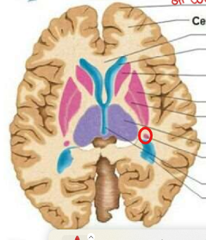
Axial Section View
cut to let us see top & bottom
Axial Section Advantages
see left and right differences to compare hemispheres
Axial Section - what is visible
basal ganglia, ventricles, anterior-posterior white matter tracts
Axial Section - Head of Caudate Nucleus
Axial Section - Putamen
Axial Section - Globus Pallidus
Axial Section - Thalamus
Axial Section - Tail of Caudate Nucleus
Coronal Section View
cut to divide into anterior/posterior slices
Coronal Section Advantages
see left and right differences, whole lobes, subcortical structures, and brainstem
Coronal Section - what is visible
Medial-lateral white tracts, corpus callosum, lobes, deep nuclei, brainstem
Sagittal Section View
cuts to let us see left and right
Sagittal Section - what is viewable
rostro-caudal size, structure, anterior-posterior structures and white-matter tracts, cerebellar and brainstem
Sagittal Section Disadvantages
not able to view left and right differences
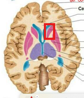
Axial Section - Caudate Nucleus (Scans)
Seen in red
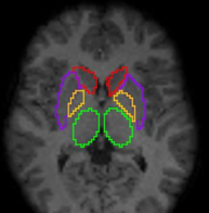
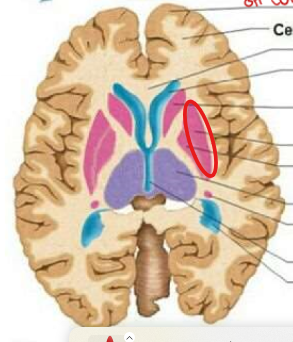
Axial Section - Putamen (Scan)
seen in purple

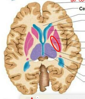
Axial Section - Globus Pallidus (Scans)
seen in yellow

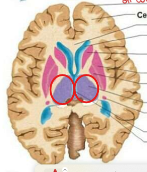
Axial Section - Thalamus (Scans)
seen in green

Coronal Section - Caudate Nucleus
seen in red

Coronal Section - Putamen
seen in purple
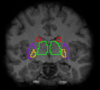
Coronal Section - Globus Pallidus
seen in yellow

Coronal Section - Thalamus
seen in green

Sagittal Section - Caudate Nucleus
seen in red
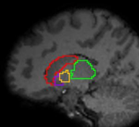
Sagittal Section - Putamen
seen in purple

Sagittal Section - Globus Pallidus
seen in yellow

Sagittal Section - thalamus
seen in green

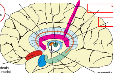
Sagittal Section - corpus callosum
seen in magenta
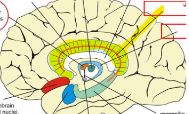
Sagittal Section - Cingulum
highlighted in yellow
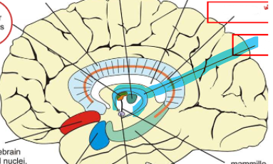
Sagittal Section - fornix
highlighted in blue
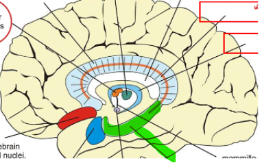
Sagittal Section - hippocampus
highlighted in green
Layers of Grey Matter
not uniform, certain things clustered in certain areas
Cerebral Cortex Grey Matter Layers
molecular layer (superficial), external pyramidal layer, multiform cell layer (deep)